. 2012;7(8):e42080.
doi: 10.1371/journal.pone.0042080. Epub 2012 Aug 6.

Aging of the microenvironment influences clonality in hematopoiesis

Affiliations

Aging of the microenvironment influences clonality in hematopoiesis

Virag Vas et al. PLoS One. 2012.

Abstract

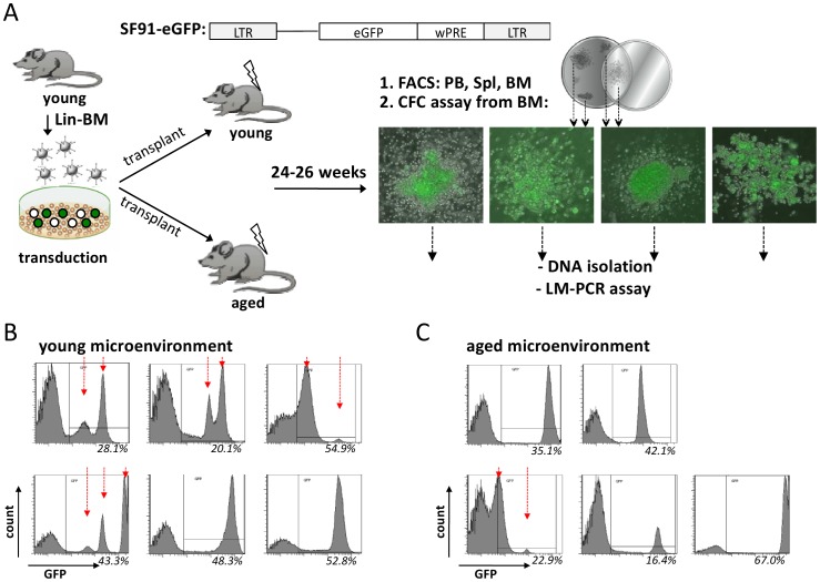
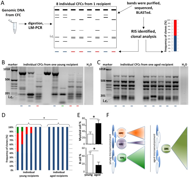
The mechanisms of the age-associated exponential increase in the incidence of leukemia are not known in detail. Leukemia as well as aging are initiated and regulated in multi-factorial fashion by cell-intrinsic and extrinsic factors. The role of aging of the microenvironment for leukemia initiation/progression has not been investigated in great detail so far. Clonality in hematopoiesis is tightly linked to the initiation of leukemia. Based on a retroviral-insertion mutagenesis approach to generate primitive hematopoietic cells with an intrinsic potential for clonal expansion, we determined clonality of transduced hematopoietic progenitor cells (HPCs) exposed to a young or aged microenvironment in vivo. While HPCs displayed primarily oligo-clonality within a young microenvironment, aged animals transplanted with identical pool of cells displayed reduced clonality within transduced HPCs. Our data show that an aged niche exerts a distinct selection pressure on dominant HPC-clones thus facilitating the transition to mono-clonality, which might be one underlying cause for the increased age-associated incidence of leukemia.


Conflict of interest statement

Competing Interests: The authors have declared that no competing interests exist.

Figures

Figure 1
Figure 1. The clonal composition of the hematopoiesis is different in old compared to young microenvironment. (A)
Schematic representation of the gammaretroviral SF91/IRES-eGFP vector used for insertional mutagenesis and the experimental setup (LTR: long terminal repeat with strong enhancer element, wPRE: woodchuck hepatitis virus posttranscriptional regulatory element). Lineage depleted (lin-) BM cells from young C57BL/6 mice were pre-stimulated with a cytokine cocktail and transduced. Cells were subsequently transplanted in equal amounts into young and aged recipient mice. (The graft contained 51.6%, 30.7%, 32.4% GFP+ cells in the 3 transduction/transplantation.) 24–26 week post-transplantation recipients were sacrificed and PB, spleen and BM analyzed by flow cytometry for lineage differentiation markers as well as the number of primitive L-S+K+ cells. At the same time CFC-assays on BM cells were performed and DNA from GFP+ colonies (representative pictures) isolated for LM-PCR. GFP expression among BM cells of young (B) and aged (C) recipient mice. (young mice n = 6, aged mice n = 5, from 3 independent experiments).
Figure 2
Figure 2. Coexistence of balanced HSC populations is characteristic for young microenvironment whereas an aged microenvironment favors the expansion of single dominant hematopoietic clone.
(A) Schematic representation of the retroviral insertional mutagenesis screen. DNA was extracted from individual CFCs (average: 7.3±1.8 GFP+CFCs/mouse were isolated) and digested with four-cutter enzymes. After LM-PCR, the recovered bands were isolated and sequenced. The sequences were aligned to the mouse genome with NCBI/BLAST program. The RISs were identified and the closest genes (within a ±100 kb window) were listed. Based on the LM-PCR band-pattern and the identified RISs, clonal analyses were performed. (B) Representative agarose gel analysis of LM-PCR performed on methylcellulose colonies isolated from BM of one representative young transplanted mouse. Clonality was identified based on the band pattern and DNA sequence information on individual bands. Distinct colors represent distinct clones. (C) Insertion sites recovered by LM-PCR from CFCs of one representative aged recipient mouse. The red arrows depict distinct GFP peaks. i.c.: internal control bands represent PCR products amplified from the viral genome. (D) Distribution of clonality within the GFP+ CFC population of young and aged recipient mice determined by the LM-PCR pattern and sequence information. (E) Flow cytometric analyses of PB of young and aged recipient mice (same mice were used in clonal analysis and RISs identification). Myeloid cells were identified based on Gr1 and Mac1 expression and B220 was used as B cell marker. (young mice n = 6, aged mice n = 5, from 3 independent experiments), * = p<0.05 (F) Proposed model for clonality of hematopoiesis in young and aged microenvironment.

Similar articles

Cited by

References

    1. Iwasaki H, Suda T (2009) Cancer stem cells and their niche. Cancer Sci 100: 1166–1172. - PMC - PubMed
    1. Raaijmakers MHGP (2011) Niche contributions to oncogenesis: emerging concepts and implications for the hematopoietic system. Haematologica 96: 1041–1048. - PMC - PubMed
    1. Henry CJ, Marusyk A, Degregori J (2011) Aging-associated changes in hematopoiesis and leukemogenesis: what’s the connection? Aging (Albany NY) 3: 643–656. - PMC - PubMed
    1. Dick JE, Lapidot T (2005) Biology of normal and acute myeloid leukemia stem cells. Int J Hematol 82: 389–396. - PubMed
    1. Geiger H, Rudolph KL (2009) Aging in the lympho-hematopoietic stem cell compartment. Trends Immunol 30: 360–365. - PubMed

Publication types