. 2012;7(8):e42106.
doi: 10.1371/journal.pone.0042106. Epub 2012 Aug 3.

Binding of the heterogeneous ribonucleoprotein K (hnRNP K) to the Epstein-Barr virus nuclear antigen 2 (EBNA2) enhances viral LMP2A expression

Affiliations

Binding of the heterogeneous ribonucleoprotein K (hnRNP K) to the Epstein-Barr virus nuclear antigen 2 (EBNA2) enhances viral LMP2A expression

Henrik Gross et al. PLoS One. 2012.

Abstract

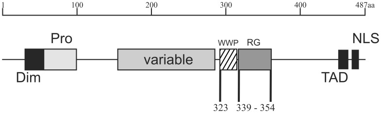
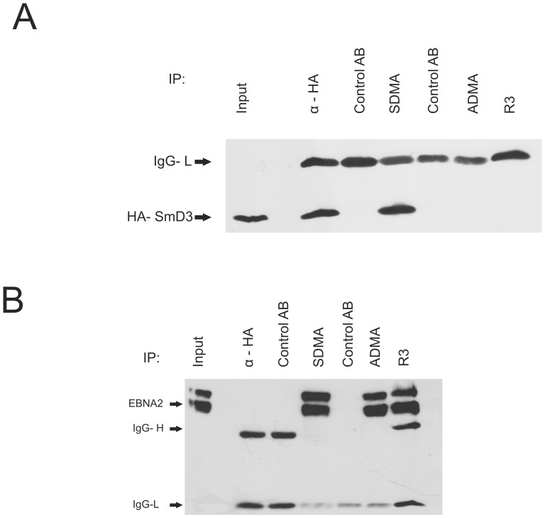
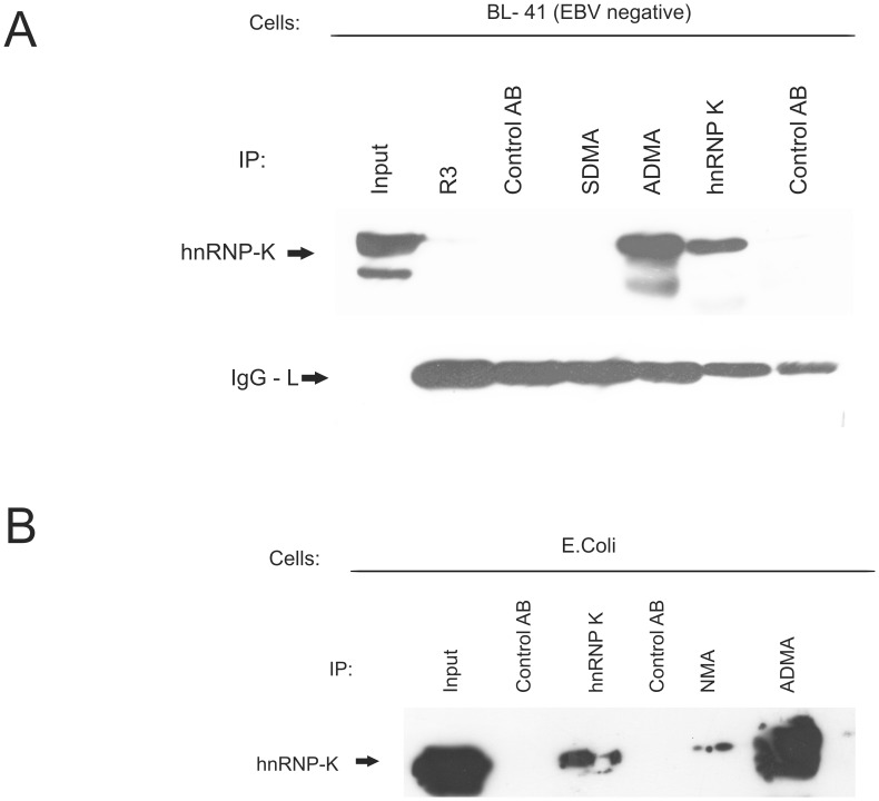
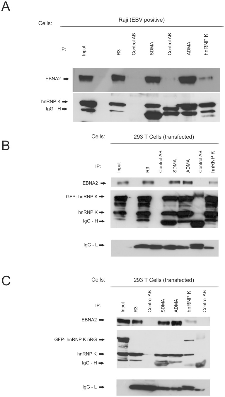
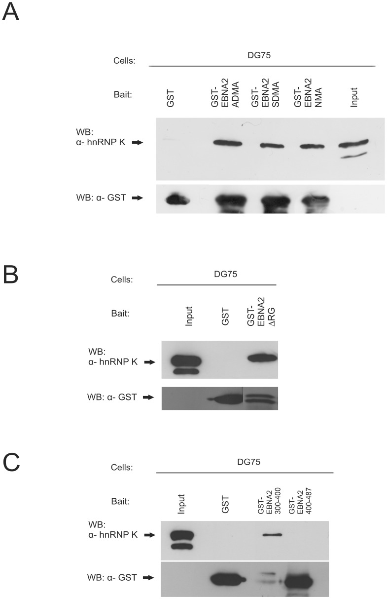
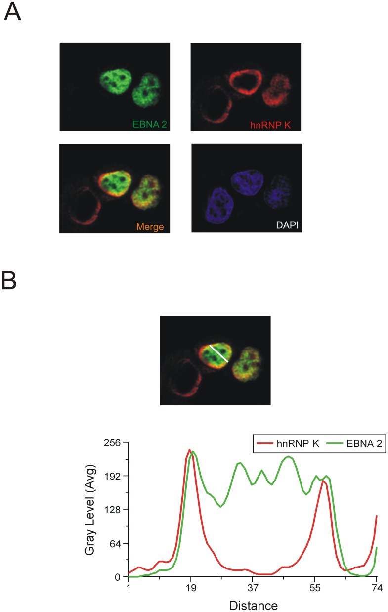
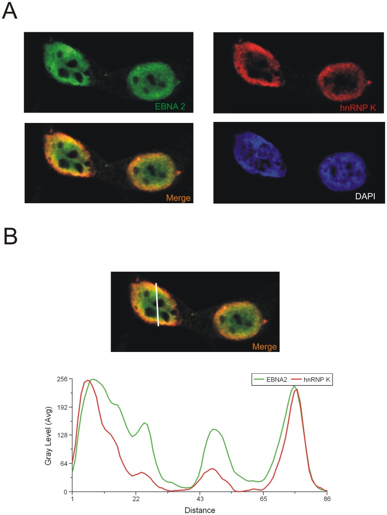
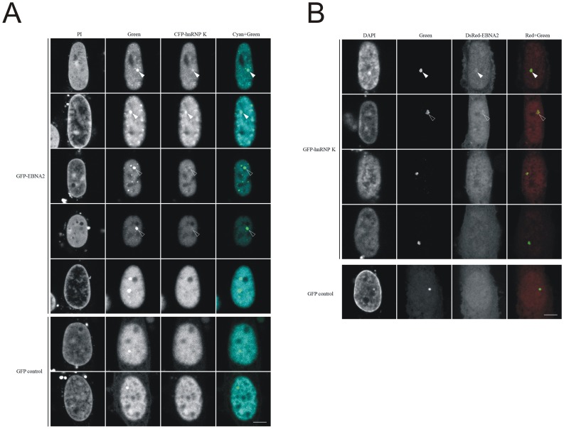
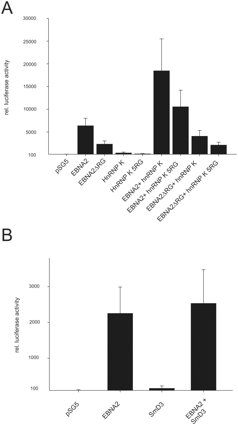
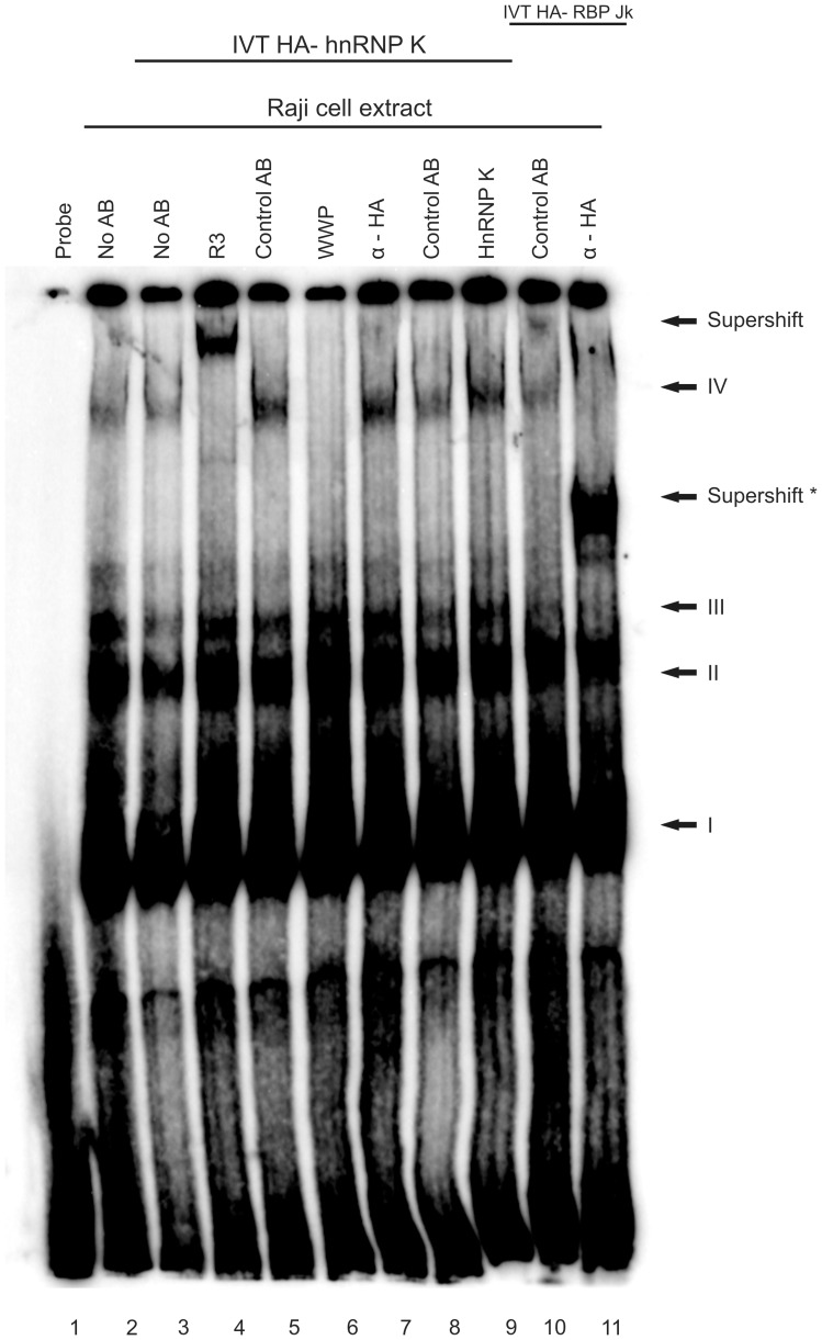
The Epstein-Barr Virus (EBV) -encoded EBNA2 protein, which is essential for the in vitro transformation of B-lymphocytes, interferes with cellular processes by binding to proteins via conserved sequence motifs. Its Arginine-Glycine (RG) repeat element contains either symmetrically or asymmetrically di-methylated arginine residues (SDMA and ADMA, respectively). EBNA2 binds via its SDMA-modified RG-repeat to the survival motor neurons protein (SMN) and via the ADMA-RG-repeat to the NP9 protein of the human endogenous retrovirus K (HERV-K (HML-2) Type 1). The hypothesis of this work was that the methylated RG-repeat mimics an epitope shared with cellular proteins that is used for interaction with target structures. With monoclonal antibodies against the modified RG-repeat, we indeed identified cellular homologues that apparently have the same surface structure as methylated EBNA2. With the SDMA-specific antibodies, we precipitated the Sm protein D3 (SmD3) which, like EBNA2, binds via its SDMA-modified RG-repeat to SMN. With the ADMA-specific antibodies, we precipitated the heterogeneous ribonucleoprotein K (hnRNP K). Specific binding of the ADMA- antibody to hnRNP K was demonstrated using E. coli expressed/ADMA-methylated hnRNP K. In addition, we show that EBNA2 and hnRNP K form a complex in EBV- infected B-cells. Finally, hnRNP K, when co-expressed with EBNA2, strongly enhances viral latent membrane protein 2A (LMP2A) expression by an unknown mechanism as we did not detect a direct association of hnRNP K with DNA-bound EBNA2 in gel shift experiments. Our data support the notion that the methylated surface of EBNA2 mimics the surface structure of cellular proteins to interfere with or co-opt their functional properties.


Conflict of interest statement

Competing Interests: The authors have declared that no competing interests exist.

Figures

Figure 1
Figure 1. Schematic representation of the Epstein-Barr virus nuclear antigen 2 (EBNA2).
EBNA2 of the standard B95.8 strain (accession number: AJ507799) of EBV consists of 487 amino acids (aa) present in an A-type virus. The N-terminal dimerization domain (“Dim”) is located next to a poly-Proline stretch (“Pro”). The variable region (“variable”) differs between the A-type viruses and B-type viruses. B-type viruses have a reduced in vitro transformation potential. The binding site for RBPjκ (“WWP”) is located around a Trp-Trp-Pro motif at aa 323–325. The adjacent Arginine-Glycine repeat (“RG”) between aa 339–354 confers binding to the survival of motor neurons (SMN) protein and represents the second nuclear localization signal (“NLS”) in addition to the canonical NLS found at the extreme C-terminus between aa 468–487. The C-terminal acidic transactivation domain (“TAD”) between aa 424–468 interacts with various basal transcription factors.
Figure 2
Figure 2. SmD3 is precipitated by the SDMA- EBNA2 specific antibody.
(A) Monoclonal antibodies (mAbs) directed against the SDMA- and ADMA- containing Arginine-Glycine (RG)-repeat of EBNA2 were tested by precipitation using extracts of HEK 293-T cells expressing EBNA2-wt and HA- SmD3. For each antibody, an appropriate isotype control was tested in parallel to exclude unspecific binding to the protein G Sepharose used for precipitation. Precipitated HA- SmD3 protein was visualised using the HA -specific mAb 3F10. The position of HA- SmD3 is indicated by an arrow. (B) Immunoprecipitation of EBNA2 from transiently transfected cells. HEK 293-T cells expressing EBNA2-wt and HA- SmD3 were precipitated with monoclonal antibodies directed against the SDMA- and ADMA- containing Arginine-Glycine (RG)-repeat of EBNA2 using appropriate isotype control antibodies. Precipitated EBNA2 protein was visualised using the EBNA2 mAb R3. The position of EBNA2 is indicated by an arrow.
Figure 3
Figure 3. hnRNP K is precipitated by the ADMA- specific antibody.
(A) Immunoprecipitation of hnRNP K from BL- 41 cells. EBV negative BL- 41 cells were precipitated with monoclonal antibodies directed against the SDMA- and ADMA- containing Arginine-Glycine (RG)-repeat of EBNA2 and an hnRNP K specific antibody using appropriate isotype control antibodies. The EBNA2 specific mAb R3 served as a negative control. The position of hnRNP K is indicated by an arrow. (B) ADMA- modified hnRNP K is precipitated by the ADMA- specific antibody. Soluble extract containing ADMA- hnRNP K methylated in E. coli with the type I methyltransferase PRMT1 was subjected to immunoprecipitation using either a hnRNP K- specific mAb, the ADMA- specific mAb and the NMA- specific mAb. Precipitated hnRNP K was visualised using the hnRNP K mAb.
Figure 4
Figure 4. EBNA2 is co-precipitated with wild- type hnRNP K but not with the methylation deficient hnRNP K 5RG mutant.
(A) Co-immunoprecipitation of EBNA2 and hnRNP K from EBV positive Raji cells. Raji cells expressing EBNA2 were precipitated with monoclonal antibodies directed against the SDMA- and ADMA- containing Arginine-Glycine (RG)-repeat of EBNA2, an EBNA2 specific mAB (R3) and an hnRNP K specific mAB (D6). The positions of EBNA2 and hnRNP K are indicated by arrows. (B) Co-immunoprecipitation of EBNA2 and GFP - hnRNP K from transfected 293T cells. The cells were precipitated with monoclonal antibodies directed against the SDMA- and ADMA- containing Arginine-Glycine (RG)-repeat of EBNA2, an EBNA2 specific mAB (R3) and an hnRNP K specific mAB (D6). The positions of EBNA2 and hnRNP K are indicated by arrows. (C) No co-immunoprecipitation of EBNA2 and GFP - hnRNP K 5RG is observed from transfected 293T cells. The cells were precipitated with monoclonal antibodies directed against the SDMA- and ADMA- containing Arginine-Glycine (RG)-repeat of EBNA2, an EBNA2 specific mAB (R3) and an hnRNP K specific mAB (D6). The positions of EBNA2 and hnRNP K are indicated by arrows.
Figure 5
Figure 5. hnRNP K binds to the amino acids 300 – 400 of EBNA2 regardless of the methylation or presence of the RG- repeat.
(A) In vitro methylated (SDMA and ADMA) and unmethylated (NMA) GST- EBNA2 fusion protein containing amino acids 300–400 of EBNA2 and GST alone were coupled to glutathione sepharose and were incubated with DG75 cell extract treated with methylation inhibitor AdOX. Precipitated hnRNP K was visualised using the hnRNP K mAb D-6. (B) GST- EBNA2ΔRG fusion protein containing amino acids 300–400 without the RG- Repeat of EBNA2 and GST alone were coupled to glutathione sepharose and were incubated with DG75 cell extract. Precipitated hnRNP K was visualised using the hnRNP K mAb D-6. (C) GST- EBNA2 aa400–487 fusion protein containing amino acids 400- 487 of EBNA2, GST- EBNA2 aa300–400 and GST alone were coupled to glutathione sepharose and were incubated with DG75 cell extract. Precipitated hnRNP K was visualised using the hnRNP K mAb D-6.
Figure 6
Figure 6. hnRNP K and EBNA2 co-localize in transiently transfected cells.
(A) HeLa cells transfected with EGFP- EBNA2 were analysed by confocal laser scanning microscopy. Endogenous hnRNP K was detected using the monoclonal D-6 antibody and an Alexa 647 coupled anti mouse antibody. The signals for hnRNP K (red) or EBNA2 (green) are shown. The merged signals show co-localisation of hnRNP K and EBNA2, resulting in a yellow color. Also shown is the DAPI staining of DNA. The fluorescence profiles of hnRNP K and EBNA2 (B) at a co-localisation hotspot (indicated by the line, left picture - lower lane) were analysed with the Leica MMAF software. The signals for hnRNP K and EBNA2 show the same progression of intensity at the inner nuclear membrane.
Figure 7
Figure 7. hnRNP K and EBNA2 co-localize in EBV positive cells.
(A) 293-EBV cells were analysed by confocal laser scanning microscopy. Endogenous hnRNP K was detected using the monoclonal D-6 antibody and an Alexa 647 coupled anti mouse antibody. Endogenous EBNA2 expressed from the viral episome was detected using the monoclonal R3 antibody and an TRITC coupled anti rat antibody. The signals for hnRNP K (red) or EBNA2 (green) are shown. The merged signals show co-localisation of hnRNP K and EBNA2, resulting in a yellow color. Also shown is the DAPI staining of DNA. The fluorescence profiles of hnRNP K and EBNA2 (B) at a co-localization hotspot (indicated by the line, left picture - lower lane) were analysed with the Leica MMAF software. The signals for hnRNP K and EBNA2 show the same progression of intensity at the inner nuclear membrane.
Figure 8
Figure 8. hnRNP K interacts with EBNA2 in a cell based interaction system.
Cells containing a lac operator (lacO) array inserted in the genome were transfected with expression vectors for a lac repressor fused with a GFP binding protein (GBP) and the indicated fluorescent fusion proteins as indicated. For comparison and orientation the nucleus was stained with PI or DAPI. The GFP fusion proteins are captured at the lac operator array by the LacI-GBP and the co-localization of cyan or red fusion proteins visualized. Clear and weak interactions at lacO array spots are marked with filled and open arrow tips, respectively. The displayed cells represent the different patterns observed in several independent experiments. GFP expression vectors were used as negative control. Scale bar is 5 µm.
Figure 9
Figure 9. hnRNP K but not SmD3 enhances the EBNA2-mediated promoter activation at the viral LMP2a promoter.
(A) A LMP2a promoter luciferase construct was co- transfected into DG75 cells with EBNA2, EBNA2-ΔRG, hnRNP K, and hnRNP K-5RG expression constructs in the indicated combinations. The luciferase value (RLU) obtained with empty pSG5 and the reporter construct was set to 100%. The graph represents the values obtained from 5 independent experiments carried out in duplicate. (B) A LMP2a promoter luciferase construct was co- transfected into DG75 cells with EBNA2 and SmD3 expression constructs in the indicated combinations. The luciferase value (RLU) obtained with empty pSG5 and the reporter construct was set to 100%. The graph represents the values obtained from 4 independent experiments carried out in duplicate.
Figure 10
Figure 10. hnRNP K is not present in EBNA2-containing DNA complexes.
EBNA2-containing Raji cell extract was incubated with in vitro translated hnRNP K and RBP- Jk and antibodies as indicated above and then assayed in a gel shift assay. R3 recognizes EBNA2 regardless of its methylation status and induces a “super- shift” indicated by the upper arrow, the mAb 6C8 directed against the “WWP”-repeat of EBNA2 destroys the EBNA2/RBPjκ-complex IV. Control antibodies corresponded to the respective IgG-subtype of each antibody. To efficiently separate the high molecular weight complexes, the electrophoresis was carried out for an extended time. Therefore, uncomplexed 32P-labelled probe ran out of the gel. The position of the RBPjκ-containing complexes I-IV as described in the text are indicated; the arrow (“Supershift”) points at the EBNA2-containing complex IV that is supershifted by R3 but not by the hnRNP K specific D6 antibody or the HA- specific antibody. The arrow (“Supershift *”) indicates the RBP- Jk containing complex which is supershifted by the HA- specific antibody and served as an internal control.

Similar articles

Cited by

References

    1. Crawford DH (2001) Biology and disease associations of Epstein-Barr virus. Philos Trans R Soc Lond B Biol Sci 356: 461–473. - PMC - PubMed
    1. Kieff E, Rickinson AE (2007) Epstein-Barr Virus and its replication. In: Knipe D, Griffin DE, Lamb RA, Strauss SE, Howley PM, et al., editors. editors. Fields Virology. 5 ed. Philadelphia: Lippincott-Raven. 2603–2654.
    1. Bornkamm GW, Hammerschmidt W (2001) Molecular virology of Epstein-Barr virus. Philos Trans R Soc Lond B Biol Sci 356: 437–459. - PMC - PubMed
    1. Zimber Strobl U, Kremmer E, Grässer F, Marschall G, Laux G, et al. (1993) The Epstein-Barr virus nuclear antigen 2 interacts with an EBNA2 responsive cis-element of the terminal protein 1 gene promoter. EMBO J 12: 167–175. - PMC - PubMed
    1. Ling PD, Hayward SD (1995) Contribution of conserved amino acids in mediating the interaction between EBNA2 and CBF1/RBPJk. J Virol 69: 1944–1950. - PMC - PubMed

Publication types

MeSH terms