

Quantitative model of cell cycle arrest and cellular senescence in primary human fibroblasts
- PMID: 22879912
- PMCID: PMC3413708
- DOI: 10.1371/journal.pone.0042150
Quantitative model of cell cycle arrest and cellular senescence in primary human fibroblasts
Abstract
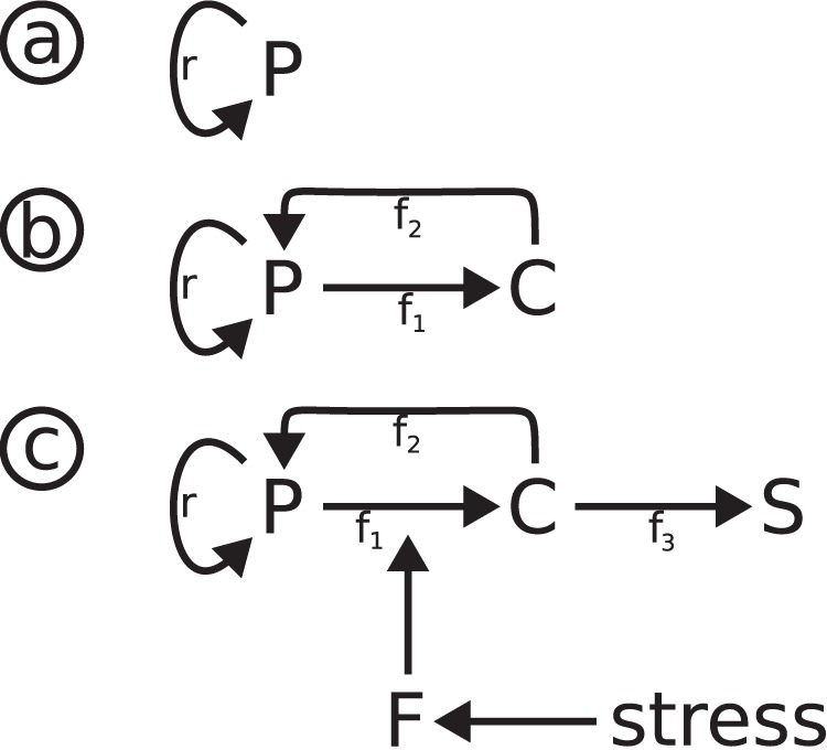
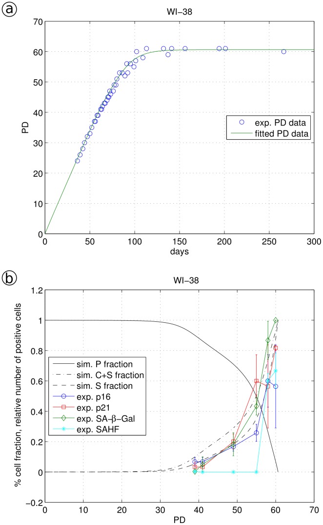
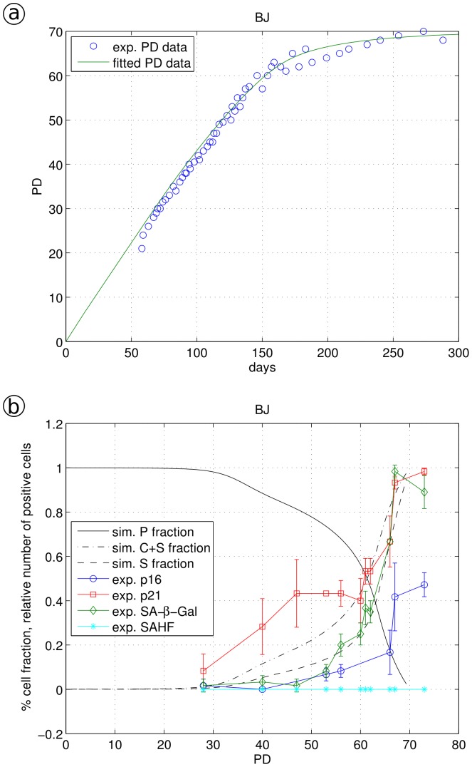
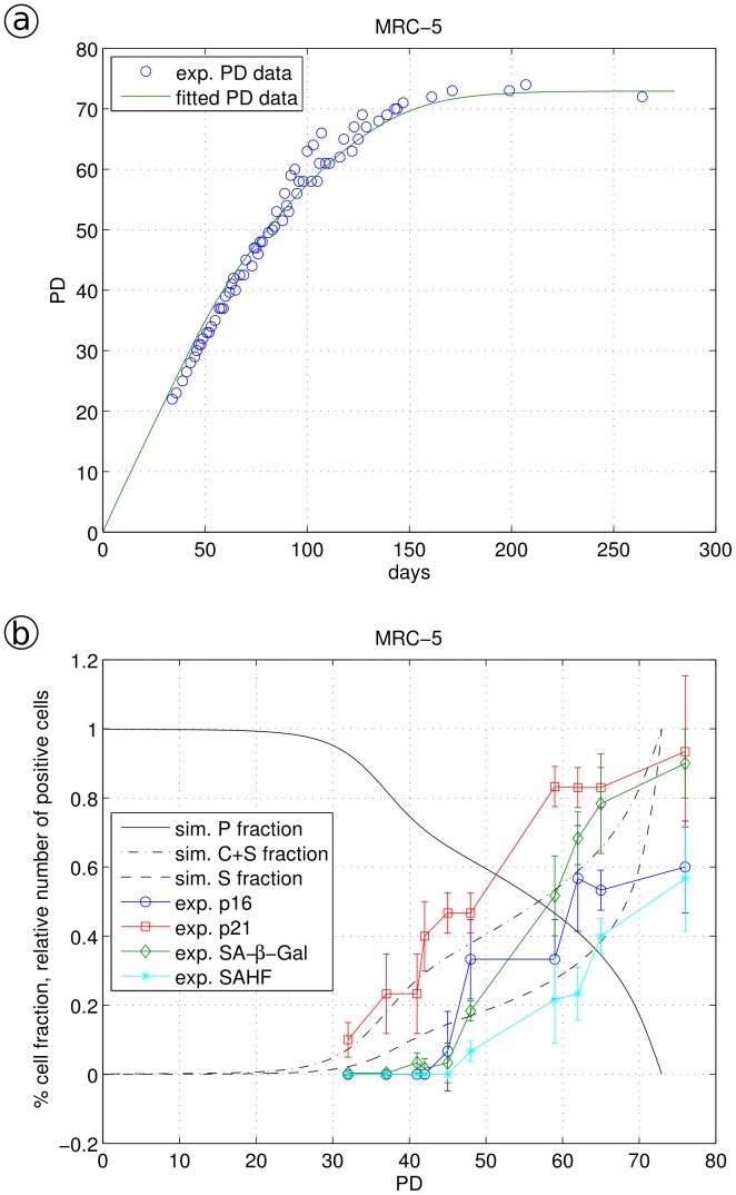
Primary human fibroblasts in tissue culture undergo a limited number of cell divisions before entering a non-replicative "senescent" state. At early population doublings (PD), fibroblasts are proliferation-competent displaying exponential growth. During further cell passaging, an increasing number of cells become cell cycle arrested and finally senescent. This transition from proliferating to senescent cells is driven by a number of endogenous and exogenous stress factors. Here, we have developed a new quantitative model for the stepwise transition from proliferating human fibroblasts (P) via reversibly cell cycle arrested (C) to irreversibly arrested senescent cells (S). In this model, the transition from P to C and to S is driven by a stress function γ and a cellular stress response function F which describes the time-delayed cellular response to experimentally induced irradiation stress. The application of this model based on senescence marker quantification at the single-cell level allowed to discriminate between the cellular states P, C, and S and delivers the transition rates between the P, C and S states for different human fibroblast cell types. Model-derived quantification unexpectedly revealed significant differences in the stress response of different fibroblast cell lines. Evaluating marker specificity, we found that SA-β-Gal is a good quantitative marker for cellular senescence in WI-38 and BJ cells, however much less so in MRC-5 cells. Furthermore we found that WI-38 cells are more sensitive to stress than BJ and MRC-5 cells. Thus, the explicit separation of stress induction from the cellular stress response, and the differentiation between three cellular states P, C and S allows for the first time to quantitatively assess the response of primary human fibroblasts towards endogenous and exogenous stress during cellular ageing.
Conflict of interest statement
Figures
Similar articles
-
Raman and Infrared Spectroscopy Distinguishing Replicative Senescent from Proliferating Primary Human Fibroblast Cells by Detecting Spectral Differences Mainly Due to Biomolecular Alterations.Anal Chem. 2017 Mar 7;89(5):2937-2947. doi: 10.1021/acs.analchem.6b04264. Epub 2017 Feb 14. Anal Chem. 2017. PMID: 28192961
-
Rapamycin Protects Skin Fibroblasts from Ultraviolet B-Induced Photoaging by Suppressing the Production of Reactive Oxygen Species.Cell Physiol Biochem. 2018;46(5):1849-1860. doi: 10.1159/000489369. Epub 2018 Apr 25. Cell Physiol Biochem. 2018. PMID: 29705807
-
A dynamical systems model for the measurement of cellular senescence.J R Soc Interface. 2019 Oct 31;16(159):20190311. doi: 10.1098/rsif.2019.0311. Epub 2019 Oct 9. J R Soc Interface. 2019. PMID: 31594522 Free PMC article.
-
Replicative senescence of human fibroblast-like cells in culture.Physiol Rev. 1993 Jul;73(3):617-38. doi: 10.1152/physrev.1993.73.3.617. Physiol Rev. 1993. PMID: 8332640 Review.
-
Cellular and molecular mechanisms of stress-induced premature senescence (SIPS) of human diploid fibroblasts and melanocytes.Exp Gerontol. 2000 Oct;35(8):927-45. doi: 10.1016/s0531-5565(00)00180-7. Exp Gerontol. 2000. PMID: 11121681 Review.
Cited by
-
Doxorubicin-sensitive and -resistant colorectal cancer spheroid models: assessing tumor microenvironment features for therapeutic modulation.Front Cell Dev Biol. 2023 Dec 22;11:1310397. doi: 10.3389/fcell.2023.1310397. eCollection 2023. Front Cell Dev Biol. 2023. PMID: 38188017 Free PMC article.
-
Hormetic effect of rotenone in primary human fibroblasts.Immun Ageing. 2015 Sep 16;12:11. doi: 10.1186/s12979-015-0038-8. eCollection 2015. Immun Ageing. 2015. PMID: 26380578 Free PMC article.
-
The impact of the spatial heterogeneity of resistant cells and fibroblasts on treatment response.PLoS Comput Biol. 2022 Mar 9;18(3):e1009919. doi: 10.1371/journal.pcbi.1009919. eCollection 2022 Mar. PLoS Comput Biol. 2022. PMID: 35263336 Free PMC article.
-
A mathematical model of tumor growth and its response to single irradiation.Theor Biol Med Model. 2016 Feb 27;13:6. doi: 10.1186/s12976-016-0032-7. Theor Biol Med Model. 2016. PMID: 26921069 Free PMC article.
-
Similarities in Gene Expression Profiles during In Vitro Aging of Primary Human Embryonic Lung and Foreskin Fibroblasts.Biomed Res Int. 2015;2015:731938. doi: 10.1155/2015/731938. Epub 2015 Aug 3. Biomed Res Int. 2015. PMID: 26339636 Free PMC article.
References
Publication types
MeSH terms
Substances
LinkOut - more resources
Full Text Sources
Other Literature Sources
Molecular Biology Databases
