. 2012;7(8):e42291.
doi: 10.1371/journal.pone.0042291. Epub 2012 Aug 3.

c-Abl is an upstream regulator of acid sphingomyelinase in apoptosis induced by inhibition of integrins αvβ3 and αvβ5

Affiliations

c-Abl is an upstream regulator of acid sphingomyelinase in apoptosis induced by inhibition of integrins αvβ3 and αvβ5

Xiuhai Ren et al. PLoS One. 2012.

Abstract

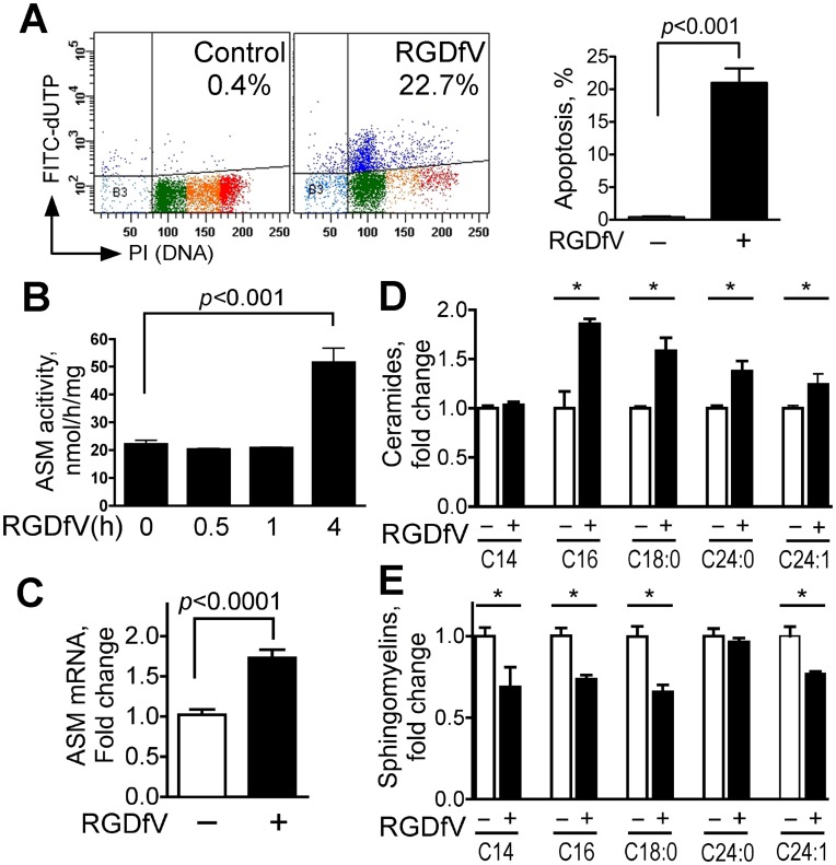
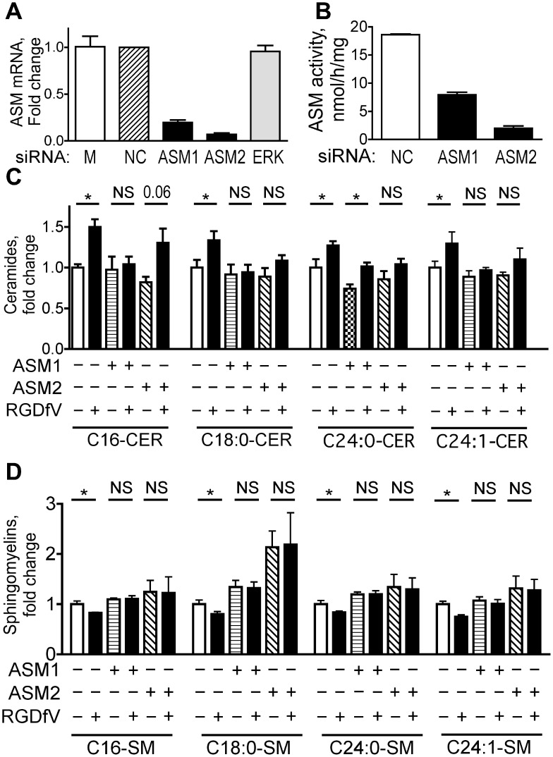
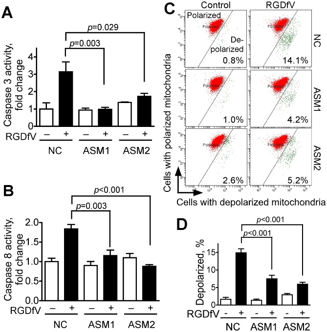
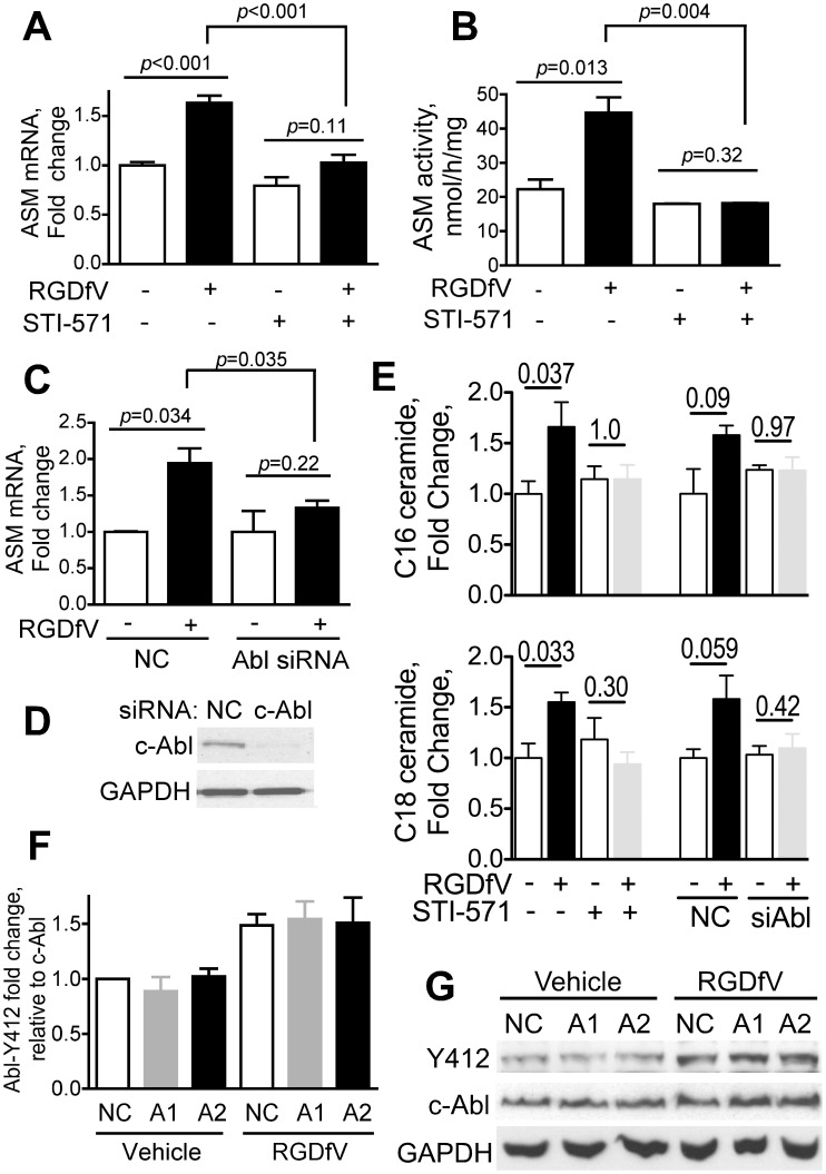
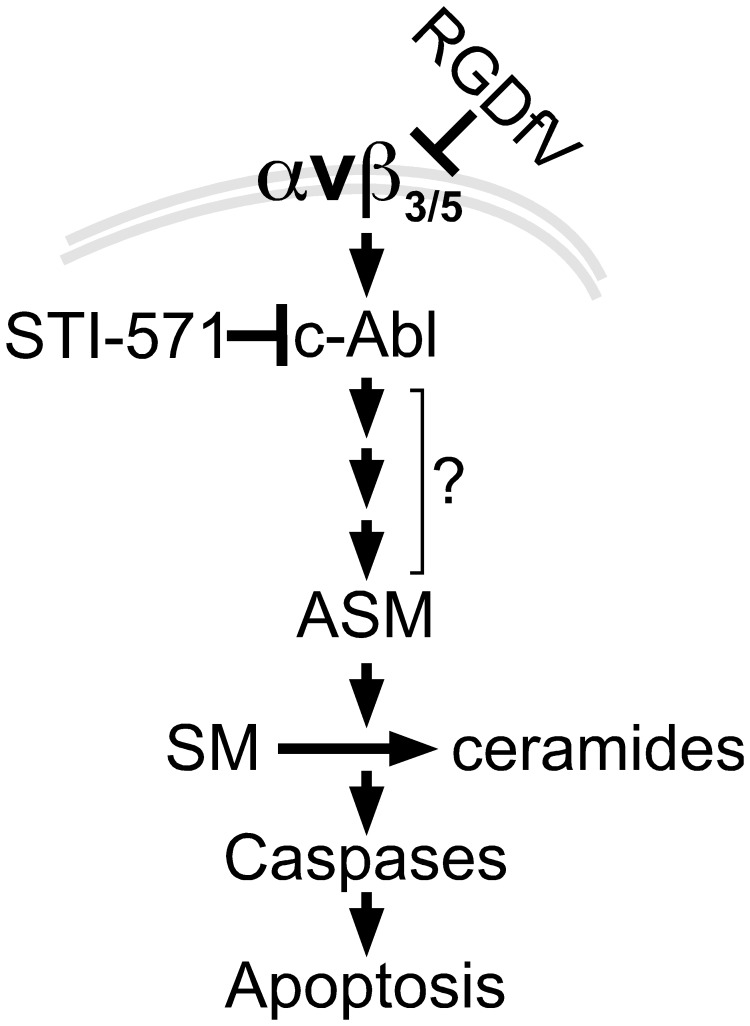
Inhibition of integrins αvβ3/αvβ5 by the cyclic function-blocking peptide, RGDfV (Arg-Gly-Asp-Phe-Val) can induce apoptosis in both normal cells and tumor cells. We show that RGDfV induced apoptosis in ECV-304 carcinoma cells, increased activity and mRNA expression of acid sphingomyelinase (ASM), and increased ceramides C(16), C(18:0), C(24:0) and C(24:1) while decreasing the corresponding sphingomyelins. siRNA to ASM decreased RGDfV-induced apoptosis as measured by TUNEL, PARP cleavage, mitochondrial depolarization, and caspase-3 and caspase-8 activities, as well as by annexinV in a 3D collagen model. These findings indicate a causal role for ASM in RGDfV-induced apoptosis in ECV-304. We have shown that c-Abl, a non-receptor tyrosine kinase, also mediates RGDfV-induced apoptosis. However, c-Abl, has not been previously linked to ASM in any system. Here we show that STI-571 (imatinib, inhibitor of c-Abl) inhibited RGDfV-induced ASM activity. Furthermore, STI-571 and c-Abl-siRNA both inhibited RGDfV-induced increase in ASM mRNA, but ASM-siRNA did not affect c-Abl phosphorylation or expression, supporting that c-Abl regulates the RGDfV-induced increase in ASM expression. These studies implicate ASM as a mediator of apoptosis induced by inhibition of integrins αvβ3/αvβ5, and for the first time place c-Abl as an upstream regulator of ASM expression and activity.


Conflict of interest statement

Competing Interests: The authors have declared that no competing interests exist.

Figures

Figure 1
Figure 1. RGDfV increases ASM mRNA, ASM activity and ceramide levels and decreases sphingomyelins.
A) Apoptosis (Apo-Direct kit) in ECV-304 cells treated with vehicle or RGDfV (5 µg/ml; 48 hrs). Left panel: representative flow cytometry plots showing percentage of apoptotic cells (FITC-dUTP positive cells in the two upper quadrants; dark blue dots). Color dots in the lower quadrants represent position in the cell cycle of the non-apoptotic cells based on DNA content (propidium iodide). Light blue is sub G0, green is G1, orange is S-phase and red is G2/M. Right panel: mean±SEM apoptotic (dark blue) cells of 24 independent experiments. B) ASM activity (Echelon Biosciences kit) in extracts from ECV-304 treated with vehicle or RGDfV (5 µg/ml). Shown are mean±SEM of three independent experiments. C) Real time quantitative RT-PCR for ASM from ECV-304 cells treated with vehicle or RGDfV (24 hrs). Shown are mean±SEM ASM mRNA normalized to GAPDH, depicted as fold-increase compared to baseline from 16 independent experiments performed in 1–4 replicates each. D–E) Change in ceramide (D) and sphingomyelin (E) content in extracts from ECV-304 cells incubated with vehicle or RGDfV (5 µg/ml; 24 hrs) and analyzed by mass spectrometry. Bars represent mean±SEM fold change from control in one of two experiments with similar results, each of which was performed in 3 biological triplicates and analyzed in duplicates.
Figure 2
Figure 2. ASM knockdown attenuates the RGDfV-induced changes in ceramides and sphingomyelins.
ECV-304 cells were transfected with siRNA as described in Methods. A) Messenger RNA extracted from HBMEC 48 hrs after transfection with ASM1, ASM2, ERK, or control siRNA was analyzed for ASM expression by qRT-PCR (n = 6; p<0.001 for ASM1, p<0.001 for ASM2 and p = 0.54 for ERK by unpaired t-test for each siRNA compared to non-silencing control). B) ASM activity in extracts of ECV-304 72 hrs after ASM-siRNA transfection compared to NC controls (Echelon Biosciences kit). Shown are mean±SEM; n = 3; p<0.001 for ASM1 and for ASM2, by unpaired t-test. C–D) Change in ceramide (panel C) and sphingomyelin (panel D) in extracts of ECV-304 cells after NC, ASM siRNA #1 or ASM siRNA #2, that were then incubated with vehicle or RGDfV (5 µg/ml; 24 hrs) and analyzed by mass spectrometry. Bars represent mean±SEM fold change from control in one of two experiments with similar results, each of which was performed in total of 3 biological triplicates. Absolute lipid values are provided in Table 1.
Figure 3
Figure 3. ASM knockdown inhibits RGDfV-induced apoptosis in 2D and 3D conditions.
A–B) Apoptosis was assessed by flow cytometry (Apo-Direct kit) in siRNA-treated (48 hrs) cells incubated with RGDfV or vehicle (5µg/ml; additional 48 hrs). A) Representative experiment of three with similar results; the percent apoptotic cells indicated is sum of both upper quadrants. Color dots are as detailed in Fig. 1A. B) Mean±SEM of three experiments as shown in panel A. C–D) PARP cleavage (western blotting) in lysates of cells treated with siRNA (48 hrs) and incubated with RGDfV (5µg/ml) or vehicle for additional 24 hrs. Panel D provides densitometry of PARP cleavage showing mean±SEM of three similar experiments. E–F) AnnexinV-FITC staining of ECV-304 (5×10⁴ cells/ml) cultured in 3D-collagen, treated with siRNA to ASM or non-silencing control (NC) and incubated 24 hrs with RGDfV (5 µg/ml) or vehicle. E) Representative fields (top panels: light microscopy, bottom panels: fluorescence) in presence of RGDfV. Control panels without RGDfV had only rare AnnexinV-positive cells and are not shown. F) Effect of ASM siRNA on percent of AnnexinV-positive cells relative to total cells in the same fields. Total cells counted on bright field images per sample were between 300–800 cells. Original magnification: 400x. p<0.001. NC: negative control (non-specific non-silencing siRNA); ASM1: siRNA#1 to ASM; ASM2: siRNA#2 to ASM.
Figure 4
Figure 4. ASM knockdown inhibits RGDfV-induced activation of caspase-3 and caspase-8 and diminishes RGDfV-induced disruption of mitochondrial membrane potential.
ECV-304 cells transfected 48 hrs with non-specific non-silencing negative control siRNA, ASM1-siRNA or ASM2-siRNA were treated with vehicle or RGDfV (5 µg/ml; 24 hrs). A–B) Lysates analyzed using the ApoTarget caspase-3/CPP32 (A) or caspase-8/FLICE (B) colorimetric protease assays. Bars represent mean±SEM; n = 6 for each condition. C–D) Loss of mitochondrial membrane potential (ΔΨm) was measured by flow cytometry using the JC-1 mitochondrial probe. Panel C shows representative flow cytometry plots. Red: mitochondria with normal polarization, green: depolarized mitochondria. Values in the panels represent percentage of cells with depolarized mitochondrial membrane. Panel D shows percent of cells with depolarized mitochondrial membrane potential from 3 independent experiments, each performed in triplicate (means ± SEM).
Figure 5
Figure 5. Inhibition of c-Abl abrogates the RGDfV-induced increase in ASM mRNA and ASM activity.
A–B) ECV-304 cells were treated with STI-571 (c-Abl inhibitor; 10 µM) or vehicle starting 2 hrs before adding RGDfV (5 µg/ml) or vehicle for additional 24 hrs. (A) ASM mRNA (real time qRT-PCR; mean±SEM of 4 independent experiments performed in 2 replicates), (B) ASM activity (Echelon Biosciences kit; mean±SEM of 2 independent experiments performed in 4 replicates). C–D) ASM mRNA (real time qRT-PCR relative to GAPDH) of ECV-304 transfected with non-specific non-silencing negative control siRNA (NC) or c-Abl siRNA (48 hrs) and incubated with RGDfV (5 µg/ml) or vehicle for 24 hrs. Shown are mean±SEM, n = 4. Representative efficacy of c-Abl siRNA knockdown (western blot) is shown in Panel D. E) Change in ceramide content in extracts from cells incubated with STI-571 or siRNA-c-Abl and their controls with/without RGDfV as in panels A–D was analyzed by mass spectrometry. Bars represent mean±SEM fold change in ceramides C16 and C18 from four (STI-571) and three (siRNA, either non-silencing controls NC, or c-Abl) independent experiments. Changes in other ceramides were not significant. P-values were calculated using paired t-tests. F–G) Phospho-c-Abl (Y412) and total c-Abl (western blotting) in lysates of ECV-304 transfected with NC, ASM1 or ASM2 siRNA as in Fig. 3 and treated 24 hrs with RGDfV (5 µg/ml) or vehicle. GAPDH served as loading control. Panel G shows a typical experiment and panel F shows mean±SEM of phospho-c-Abl (Y412) relative to total c-Abl from densitometry of three experiments. ASM mRNA level in these experiments was 28±1.2% of the non-silencing control samples for siRNA-ASM1 and 11.4±0.9% for siRNA-ASM2.
Figure 6
Figure 6. Model to link RGDfV-induced integrin αvβ3/αvβ5 inhibition, c-Abl, ASM and apoptosis.
Our data support a model in which inhibition of integrin αvβ3/αvβ5 signaling by RGDfV activates c-Abl (26). c-Abl in turn increases expression of ASM, which results in increased ASM activity and changes in sphingolipids, including increase in ceramides and decrease in sphingomyelins. The consequence of the increase in ASM expression and activity is apoptosis. Since ASM mediates the apoptosis, but it is not known which sphingolipid(s) is/are the mediator(s) of the apoptosis downstream of ASM, the downstream arrows are placed below ASM rather than below specific sphingolipids. Since downregulation of ASM expression leads to inhibition of caspase 3 and caspase 8 activity, caspases are placed downstream of ASM. Inhibition of c-Abl blocked the increase in ASM mRNA and activity and prevented the apoptosis, placing c-Abl upstream of ASM. It is likely that the interaction between c-Abl and ASM is not direct and that other yet-unknown mediators (denoted by the three small arrows and question mark) exist within this pathway. Not shown are possible initial upstream regulation of ASM by caspase 8 and other intermediate effectors.

Similar articles

Cited by

References

    1. Hynes RO (1992) Integrins: versatility, modulation, and signaling in cell adhesion. Cell 69: 11–25. - PubMed
    1. Desgrosellier JS, Cheresh DA (2010) Integrins in cancer: biological implications and therapeutic opportunities. Nat Rev Cancer 10: 9–22. - PMC - PubMed
    1. Lorger M, Krueger JS, O’Neal M, Staflin K, Felding-Habermann B (2009) Activation of tumor cell integrin αvβ3 controls angiogenesis and metastatic growth in the brain. Proc Natl Acad Sci U S A 106: 10666–10671. - PMC - PubMed
    1. Felding-Habermann B, Mueller BM, Romerdahl CA, Cheresh DA (1992) Involvement of integrin αV gene expression in human melanoma tumorigenicity. J Clin Invest 89: 2018–2022. - PMC - PubMed
    1. Bello L, Francolini M, Marthyn P, Zhang J, Carroll RS, et al. (2001) αvβ3 and αvβ5 integrin expression in glioma periphery. Neurosurgery 49: 380–389; discussion 390. - PubMed

Publication types

MeSH terms