. 2012;7(8):e42316.
doi: 10.1371/journal.pone.0042316. Epub 2012 Aug 3.

TGFβ-stimulated microRNA-21 utilizes PTEN to orchestrate AKT/mTORC1 signaling for mesangial cell hypertrophy and matrix expansion

Affiliations

TGFβ-stimulated microRNA-21 utilizes PTEN to orchestrate AKT/mTORC1 signaling for mesangial cell hypertrophy and matrix expansion

Nirmalya Dey et al. PLoS One. 2012.

Abstract

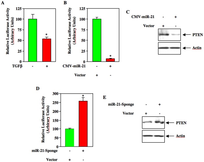
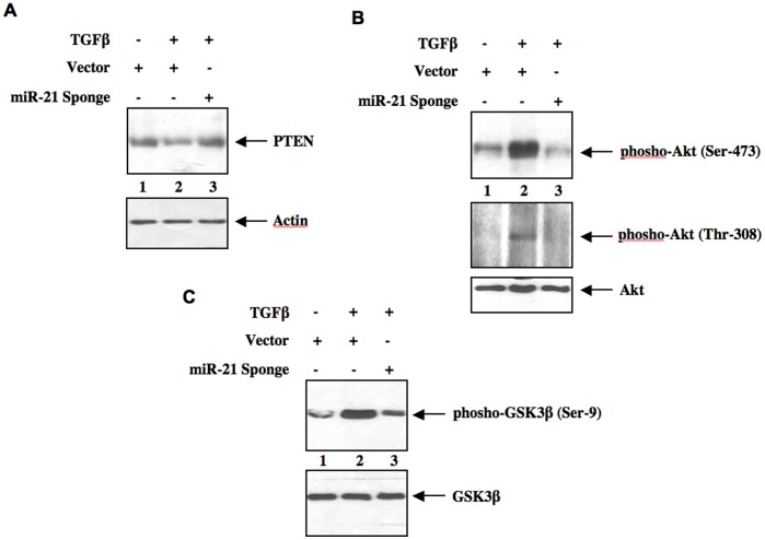
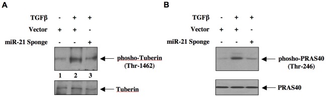
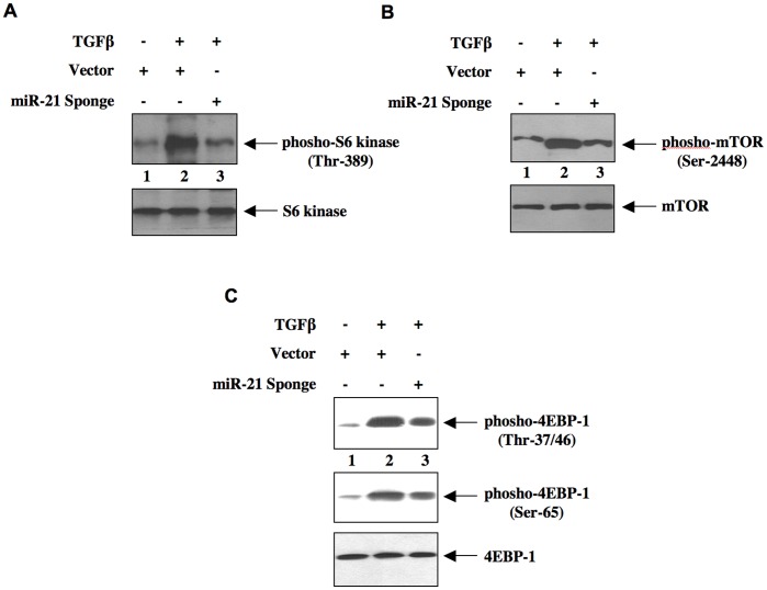
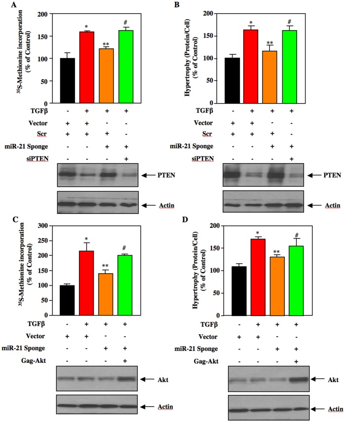
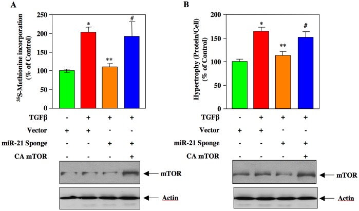
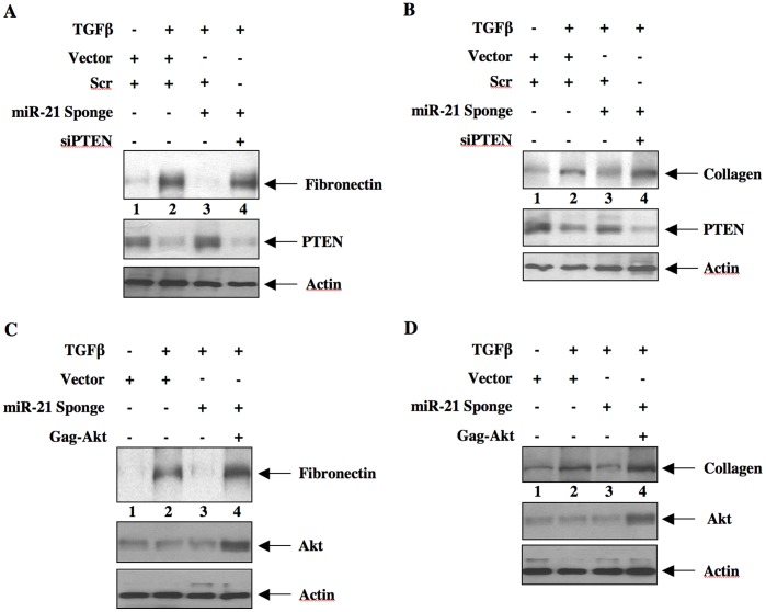
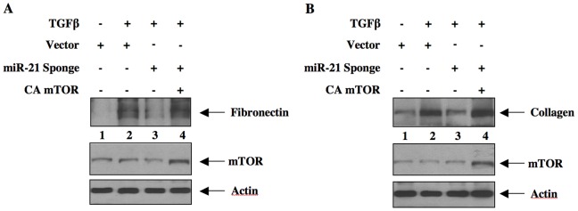
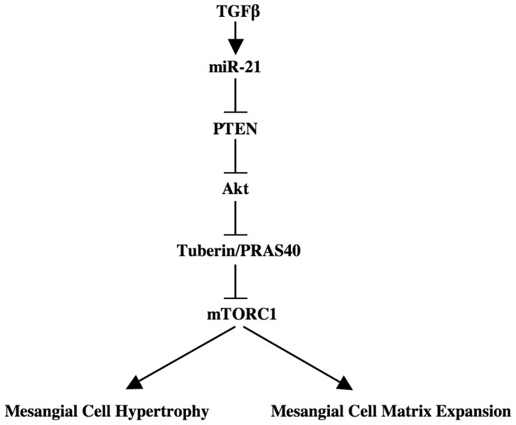
Transforming growth factor-β (TGFβ) promotes glomerular hypertrophy and matrix expansion, leading to glomerulosclerosis. MicroRNAs are well suited to promote fibrosis because they can repress gene expression, which negatively regulate the fibrotic process. Recent cellular and animal studies have revealed enhanced expression of microRNA, miR-21, in renal cells in response to TGFβ. Specific miR-21 targets downstream of TGFβ receptor activation that control cell hypertrophy and matrix protein expression have not been studied. Using 3'UTR-driven luciferase reporter, we identified the tumor suppressor protein PTEN as a target of TGFβ-stimulated miR-21 in glomerular mesangial cells. Expression of miR-21 Sponge, which quenches endogenous miR-21 levels, reversed TGFβ-induced suppression of PTEN. Additionally, miR-21 Sponge inhibited TGFβ-stimulated phosphorylation of Akt kinase, resulting in attenuation of phosphorylation of its substrate GSK3β. Tuberin and PRAS40, two other Akt substrates, and endogenous inhibitors of mTORC1, regulate mesangial cell hypertrophy. Neutralization of endogenous miR-21 abrogated TGFβ-stimulated phosphorylation of tuberin and PRAS40, leading to inhibition of phosphorylation of S6 kinase, mTOR and 4EBP-1. Moreover, downregulation of miR-21 significantly suppressed TGFβ-induced protein synthesis and hypertrophy, which were reversed by siRNA-targeted inhibition of PTEN expression. Similarly, expression of constitutively active Akt kinase reversed the miR-21 Sponge-mediated inhibition of TGFβ-induced protein synthesis and hypertrophy. Furthermore, expression of constitutively active mTORC1 prevented the miR-21 Sponge-induced suppression of mesangial cell protein synthesis and hypertrophy by TGFβ. Finally, we show that miR-21 Sponge inhibited TGFβ-stimulated fibronectin and collagen expression. Suppression of PTEN expression and expression of both constitutively active Akt kinase and mTORC1 independently reversed this miR-21-mediated inhibition of TGFβ-induced fibronectin and collagen expression. Our results uncover an essential role of TGFβ-induced expression of miR-21, which targets PTEN to initiate a non-canonical signaling circuit involving Akt/mTORC1 axis for mesangial cell hypertrophy and matrix protein synthesis.


Conflict of interest statement

Competing Interests: The authors have declared that no competing interests exist.

Figures

Figure 1
Figure 1. TGFβ-stimulated miR-21 targets PTEN 3′UTR to inhibit PTEN expression.
(A) Human glomerular mesangial cells were transfected with PTEN 3′UTR-containing luciferase (PTEN 3′UTR-Luc) reporter plasmid PTEN 3′UTR-Luc. Transfected cells were serum-starved for 16 hours followed by incubation with 2 ng/ml TGFβ for 24 hours. The cell lysates were assayed for luciferase activity as described in the Materials and Methods , , . Mean ± SE of six measurements is shown. *p = 0.018 vs control. (B and C) Mesangial cells were cotransfected with PTEN 3′UTR-Luc and CMV-miR-21 (expressing mature miR-21). The cell lysates were assayed for luciferase activity as described (panel B) , , . Mean ± SE of triplicate measurements is shown; *p = 0.003 vs vector. For panel C, the cell lysates were immunoblotted with PTEN and actin antibodies. (D and E) Mesangial cells were transfected with PTEN 3′UTR-Luc plus miR-21 Sponge. For panel D the cell lysates were assayed for luciferase activity as described , , . Mean ± SE of 12 measurements is shown; *p = 0.0001 vs vector. For panel E, the cell lysates were immunoblotted with PTEN and actin antibodies.
Figure 2
Figure 2. miR-21 targets PTEN to regulate activation of Akt kinase in response to TGFβ.
Mesangial cells were transfected with miR-21 Sponge or vector followed by incubation with 2 ng/ml TGFβ for 24 hours. Cell lysates were immunoblotted with PTEN, actin (panel A), phospho-Akt (Ser-473), phospho-Akt (Thr-308), Akt (panel B), phospho-GSK3β and GSK3β (panel C) antibodies as indicated.
Figure 3
Figure 3. miR-21 Sponge inhibits TGFβ-stimulated phosphorylations of tuberin and PRAS40.
Glomerular mesangial cells were transfected with miR-21 Sponge or vector. The serum-starved cells were incubated with 2 ng/ml TGFβ for 24 hours. The cell lysates were immunoblotted with phospho-tuberin (Thr-1462), tuberin (panel A), phospho-PRAS40 (Thr-246) and PRAS40 (panel B) antibodies as indicated.
Figure 4
Figure 4. miR-21 Sponge blocks mTORC1 activity in response to TGFβ.
Mesangial cells were transfected with miR-21 Sponge or vector. The serum-starved cells were incubated with 2 ng/ml TGFβ for 24 hours. The cell lysates were immunoblotted with phospho-S6 kinase (Thr-389), S6 kinase (panel A), phospho-mTOR (Ser-2448), mTOR (panel B), phospho-4EBP-1 (Thr-34/46), phospho-4EBP-1 (Ser-65) and 4EBP-1 (panel C) antibodies as indicated.
Figure 5
Figure 5. miR-21/PTEN/Akt axis regulates mesangial cell protein synthesis and hypertrophy in response to TGFβ.
Mesangial cells were cotransfected with miR-21 Sponge and siRNAs targeting PTEN mRNA (siPTEN) or scrambled RNA (Scr) (panels A and B). Mesangial cells were cotransfected with miR-21 Sponge and constitutively active Gag-Akt as indicated (panels C and D). The transfected cells were starved for 16 hours prior to incubation with 2 ng/ml TGFβ for 24 hours. Protein synthesis (panels A and C) and hypertrophy (panels B and D) were determined as described in the Materials and Methods , , . Mean ± SE of 3 measurements is shown. For panel A, *p<0.01 vs control; **p<0.01 vs TGFβ; #p<0.05 vs miR-21 Sponge plus TGFβ. For panel B, *p<0.05 vs control; **p<0.05 vs TGFβ; #p<0.05 vs miR-21 Sponge plus TGFβ. For panel C, *p<0.05 vs control; **p<0.05 vs TGFβ; #p<0.05 vs miR-21 Sponge plus TGFβ. For panel D, *p<0.01 vs control; **p<0.05 vs TGFβ; #p<0.05 vs miR-21 Sponge plus TGFβ. Bottom panels show expression of PTEN and Akt in representative samples. Actin expression was used as a control for immunoblotting.
Figure 6
Figure 6. Expression of constitutively active mTORC1 blocks the inhibitory effect of miR-21 Sponge on TGFβ-induced mesangial cell protein synthesis and hypertrophy.
Glomerular mesangial cells were cotransfected with miR-21 Sponge and CA mTOR as indicated. The cells were incubated with 2 ng/ml TGFβ for 24 hours. Protein synthesis (panel A) and hypertrophy (panel B) were determined as described , , . For panel A, mean ± SE of triplicate measurements is shown; *p<0.05 vs control; **p<0.05 vs TGFβ; #p<0.05 vs miR-21 Sponge in the presence of TGFβ. For panel B, mean ± SE of triplicate measurements is shown; p<0.01 vs control; **p<0.01 vs TGFβ; #p<0.05 vs miR-21 Sponge in the presence of TGFβ. Bottom panels show expression of mTOR and actin in the representative samples.
Figure 7
Figure 7. miR-21/PTEN/Akt axis regulates mesangial cell matrix protein expression in response to TGFβ.
Mesangial cells were transfected with miR-21 Sponge and siPTEN or scrambled RNA as indicated in panels A and B. Similarly, mesangial cells were transfected with miR-21 Sponge and Gag-Akt as indicated in panels C and D. The transfected cells were incubated with 2 ng/ml TGFβ for 24 hours. The cell lysates were immunoblotted with fibronectin, PTEN, actin (panel A), collagen I (α2), PTEN, actin (panel B), fibronectin, Akt, actin (panel C) and collagen I (α2), Akt, actin (panel D) antibodies as indicated.
Figure 8
Figure 8. Constitutively active mTORC1 prevents the inhibition of miR-21 Sponge on TGFβ-stimulated fibronectin and collagen expression.
Glomerular mesangial cells were transfected with miR-21 Sponge and CA mTOR plasmids as indicated. The cells were incubated with TGFβ for 24 hours. The cell lysates were immunoblotted with fibronectin (panel A) and collagen I (α2) (panel B) antibodies. Immunoblots of mTOR and actin are shown at the bottom.
Figure 9
Figure 9. Schematic showing the results described in the paper.
TGFβ-stimulated miR-21 decreases PTEN to activate Akt-dependent mTORC1, leading to hypertrophy of mesangial cells and matrix protein expression.

Similar articles

Cited by

References

    1. Schnaper HW, Hayashida T, Hubchak SC, Poncelet AC (2003) TGF-beta signal transduction and mesangial cell fibrogenesis. Am J Physiol Renal Physiol 284: F243–252. - PubMed
    1. Eddy AA, Neilson EG (2006) Chronic kidney disease progression. J Am Soc Nephrol 17: 2964–2966. - PubMed
    1. Bottinger EP (2007) TGF-beta in renal injury and disease. Semin Nephrol 27: 309–320. - PubMed
    1. Iwano M, Kubo A, Nishino T, Sato H, Nishioka H, et al. (1996) Quantification of glomerular TGF-beta 1 mRNA in patients with diabetes mellitus. Kidney Int 49: 1120–1126. - PubMed
    1. Kopp JB, Factor VM, Mozes M, Nagy P, Sanderson N, et al. (1996) Transgenic mice with increased plasma levels of TGF-beta 1 develop progressive renal disease. Lab Invest 74: 991–1003. - PubMed

Publication types

MeSH terms