. 2012;7(8):e42349.
doi: 10.1371/journal.pone.0042349. Epub 2012 Aug 7.

fussel (fuss)--A negative regulator of BMP signaling in Drosophila melanogaster

Affiliations

fussel (fuss)--A negative regulator of BMP signaling in Drosophila melanogaster

Susanne Fischer et al. PLoS One. 2012.

Abstract

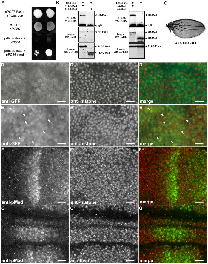
The TGF-β/BMP signaling cascades control a wide range of developmental and physiological functions in vertebrates and invertebrates. In Drosophila melanogaster, members of this pathway can be divided into a Bone Morphogenic Protein (BMP) and an Activin-ß (Act-ß) branch, where Decapentaplegic (Dpp), a member of the BMP family has been most intensively studied. They differ in ligands, receptors and transmitting proteins, but also share some components, such as the Co-Smad Medea (Med). The essential role of Med is to form a complex with one of the two activating Smads, mothers against decapentaplegic (Mad) or dSmad, and to translocate together to the nucleus where they can function as transcriptional regulators of downstream target genes. This signaling cascade underlies different mechanisms of negative regulation, which can be exerted by inhibitory Smads, such as daughters against decapentaplegic (dad), but also by the Ski-Sno family. In this work we identified and functionally analyzed a new member of the Ski/Sno-family, fussel (fuss), the Drosophila homolog of the human functional suppressing element 15 (fussel-15). fuss codes for two differentially spliced transcripts with a neuronal expression pattern. The proteins are characterized by a Ski-Sno and a SAND homology domain. Overexpression studies and genetic interaction experiments clearly reveal an interaction of fuss with members of the BMP pathway, leading to a strong repression of BMP-signaling. The protein interacts directly with Medea and seems to reprogram the Smad pathway through its influence upon the formation of functional Mad/Medea complexes. This leads amongst others to a repression of downstream target genes of the Dpp pathway, such as optomotor blind (omb). Taken together we could show that fuss exerts a pivotal role as an antagonist of BMP signaling in Drosophila melanogaster.


Conflict of interest statement

Competing Interests: The authors have declared that no competing interests exist.

Figures

Figure 1
Figure 1. Genomic organisation of the fussel locus CG11093 and its transcription pattern.
(A) 20 kb of the reverse complemented 102F4 cytological region of chromosome IV and the transcripts fussB (RefSeq NM_001169358.1) and fussC (RefSeq NM_001169359.1) are shown. The position of the Tc1-2- family transposon and the size of the two alternate exons are indicated. In-situ hybridisation of fuss shows expression in (B) two segmental clusters of cells in stage 14 embryos and (C) cells in the mediolateral (arrow) and SE- (arrowhead) and Tv-neuron-region (open arrowheads) in L3 brains.
Figure 2
Figure 2. Fussel-Proteins and their relationship to the Ski-Sno/CORL/DACH-family.
(A) Comparison of FussB and FussC proteins to Drosophila dSnoN-, human Fussel 15 and 18 and human Ski-Sno proteins. Shaded boxes show the relative positions of structural features as indicated in the legend; the scale-bar represents aminoacids. (B) Unrooted phylogenetic tree of the Ski-Sno/CORL/DACH- family. Branch length reflects phylogenetic divergence and the scale bar indicates the number of amino acid substitutions per site. Bootstrap values are given to indicate statistical significance at each node. (C) Multiple Sequence Alignment of the Ski-Sno-homology (upper alignment) and SMAD4- binding domains (lower alignment) of Drosophila and human Ski-Sno/CORL-proteins. Shading and the Clustal consensus indicates similar or identical amino acids. Height of bars reflects the quality of conservation while the symbols are denoting the conservation type: stars mark identical or conserved residues in all sequences while colons and dots indicate conserved or semi-conserved substitutions. Red arrows mark the zinc binding domain.
Figure 3
Figure 3. Ectopic expression of fussel in the wing disc reduces wing size, leads to loss of veins, loss of campaniform sensilla and interferes with the expression of BMP target genes.
(A) Control wing of a male fly from the A9-Gal line. Longitudinal (L2 to L5) and cross veins (a-cv, p-cv) are indicated. (B) A9-Gal4; UAS-fussC. The wing is smaller, arrows indicate the truncation of L2, L5 and the p-cv. (C) A9-Gal4; UAS-fussC/UAS-fussC. Expression of two copies of UAS-fussC enhances the observed phenotype. (D) A9-Gal4; UAS-fussB. Expression of one copy of fussB leads to a reduction of wing size and a severe disruption of the overall wing structure. (E) nub-Gal4; UAS-fussC. The L2 and L5 veins are truncated. (F) A9-Gal4; UAS-dSno. Expression of dSno leads to a reduction of wing size and a loss of the L4 vein. (G) Mis-expression of fuss leads to loss of campaniform sensilla. (G’) Medial part of the L3 vein of a male A9-Gal4 fly. Three campaniform sensilla are marked with arrowheads (G”) A9-Gal4; UAS-fussC. Distal to the p-cv, all campaniform sensilla are lost. (H) fuss represses omb expression. Micrographs of X- Gal- stained female L3- wing discs: (H’) omb-lacZ; UAS-fussB; (H”) omb-lacZ/A9-Gal4; UAS-fussB/+. (I) The blistered (dSRF) domain in male L3-wing discs is disrupted by fuss expression. Confocal scans of (I’) A9-Gal4; (I”) A9-Gal4; UAS-fussB. (J) Relative expression of omb, salm and ecr1b in actin-Gal4/fussC L3-larvae compared to actin-Gal4 controls. Asterisks indicate the level of statistical significance (t-test **p<0.01, ***p<0.001). (K) fuss disrupts the pattern of activated Mad but does not inhibit its phosphorylation. Confocal scans of anti-phospho-SMAD1/5 stained L3-wing discs: (K’) A9-Gal4; (K”) A9-Gal4; UAS-fussB. Scale bars represent 500 µm (A), 100 µm(H’) and 50 µm(I’, K’).
Figure 4
Figure 4. Genetic interaction of fussel with members of the BMP pathway.
(A) A9-Gal4/y; UAS-saxA. Overexpression of constitutively active sax causes growth of additional vein material between L3–L5 (B) A9-Gal4/y; UAS-saxA; UAS-fussC. Coexpression of fussC ameliorates vein overgrowth. (C) A9-Gal4/y; UAS-mad. Overexpression of mad transforms most of the intervein tissue into vein tissue and eventually results in a blistered wing. (D) A9-Gal4/y; UAS-mad; UAS-fussC. Coexpression of fussC ameliorates blistering and considerably improves vein pattering. (E) A9-Gal4/y; UAS-med. Overexpression of med causes distinct overgrowth and dublication of wing veins. (F) A9-Gal4/y; UAS-med; UAS-fussC. Coexpression of fussC rescues the vein overgrowth and restores the fussC-phenotype.
Figure 5
Figure 5. Formation of a Fuss/Med protein complex and its translocation into the nucleus.
(A) Yeast Two Hybrid experiment showing physical interaction of Fuss and Med: pPC97-Fos + pPC86-Jun: interaction control; pCL1+ pPC86: Gal4- growth control and empty prey vector; pdbLeu-Fuss + pPC86: negative control; pdbLeu-Fuss + pPC86-Med: positive interaction of Fuss and Med. (B) Coimmunoprecipitations: in lysates from S2-cells co-expressing HA-Fuss together with FLAG-Med or FLAG-Mad, HA-Fuss co-precipitates with FLAG-Med but not with FLAG-Mad. In lysates from S2-cells co-expressing FLAG-Fuss with HA-Med or HA-Mad, HA-Med co-precipitates with FLAG-Fuss and HA-Mad does not. (C) Wing of a male fly of the genotype A9-Gal4; UAS-fuss-GFP. Overexpression of fuss-GFP leads to truncations of L2 and L5 veins and a loss of the p-cv. (D–E”) Upon co-expression of Med, Fuss-GFP is partially translocated into the nucleus while the cytoplasmic fraction of the protein is reduced. Confocal scans of L3 wing discs stained with anti-GFP (D, E) and anti-Histone (D’, E’) antibodies. (D–D”) A9-Gal4; UAS-fuss-GFP. The Fuss-GFP fusion protein is mainly localised in the cytoplasm. (E) A9-Gal4; UAS-fuss-GFP; UAS-Med. Arrows emphasize some of the cells that clearly show nuclear localisation of the Fuss-GFP fusion protein. (F–G”) Fuss does not inhibit the nuclear translocation of pMad. Confocal scans of L3 wing discs stained with anti-phospho-SMAD1/5 (F, G) and anti-Histone (F’, G’) antibodies. (F–F”) A9-Gal4. (G–G”) A9-Gal4; UAS-fussB. All Scale bars: 10 µm.

References

    1. Padgett RW, St Johnston RD, Gelbart WM (1987) A transcript from a Drosophila pattern gene predicts a protein homologous to the transforming growth factor-beta family. Nature 325: 81–84. - PubMed
    1. Wozney JM, Rosen V, Celeste AJ, Mitsock LM, Whitters MJ, et al. (1988) Novel regulators of bone formation: molecular clones and activities. Science 242: 1528–1534. - PubMed
    1. Twombly V, Blackman RK, Jin H, Graff JM, Padgett RW, et al. (1996) The TGF-beta signaling pathway is essential for Drosophila oogenesis. Development 122: 1555–1565. - PubMed
    1. Shravage BV, Altmann G, Technau M, Roth S (2007) The role of Dpp and its inhibitors during eggshell patterning in Drosophila . Development 134: 2261–2271. - PubMed
    1. O’Connor MB, Umulis D, Othmer HG, Blair SS (2006) Shaping BMP morphogen gradients in the Drosophila embryo and pupal wing. Development 133: 183–193. - PMC - PubMed

Publication types

MeSH terms