. 2012;7(8):e42351.
doi: 10.1371/journal.pone.0042351. Epub 2012 Aug 7.

Understanding the basis of drug resistance of the mutants of αβ-tubulin dimer via molecular dynamics simulations

Affiliations

Understanding the basis of drug resistance of the mutants of αβ-tubulin dimer via molecular dynamics simulations

Kathiresan Natarajan et al. PLoS One. 2012.

Abstract

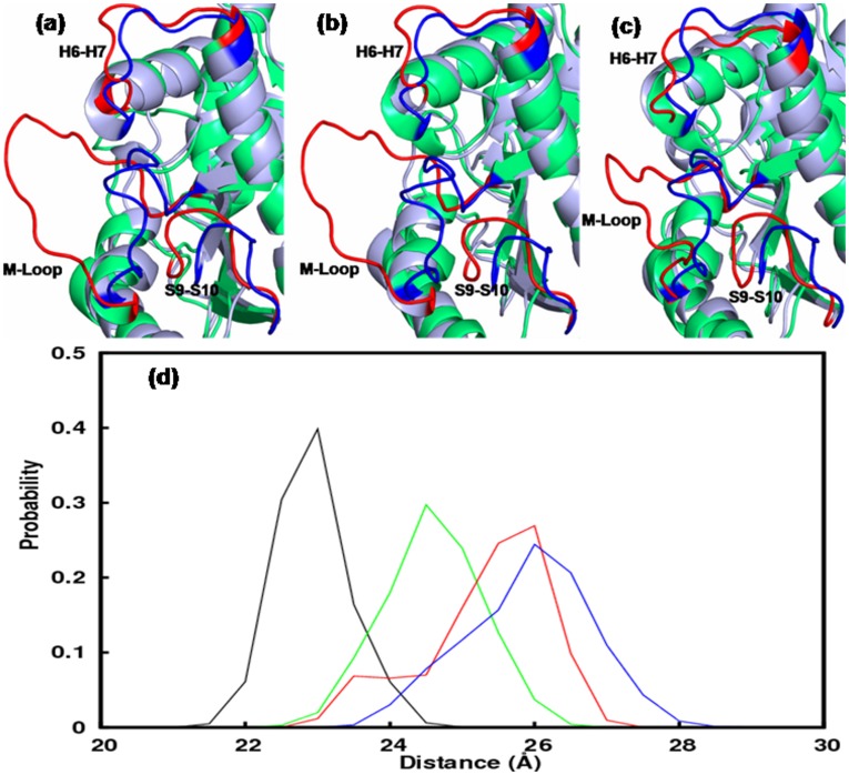
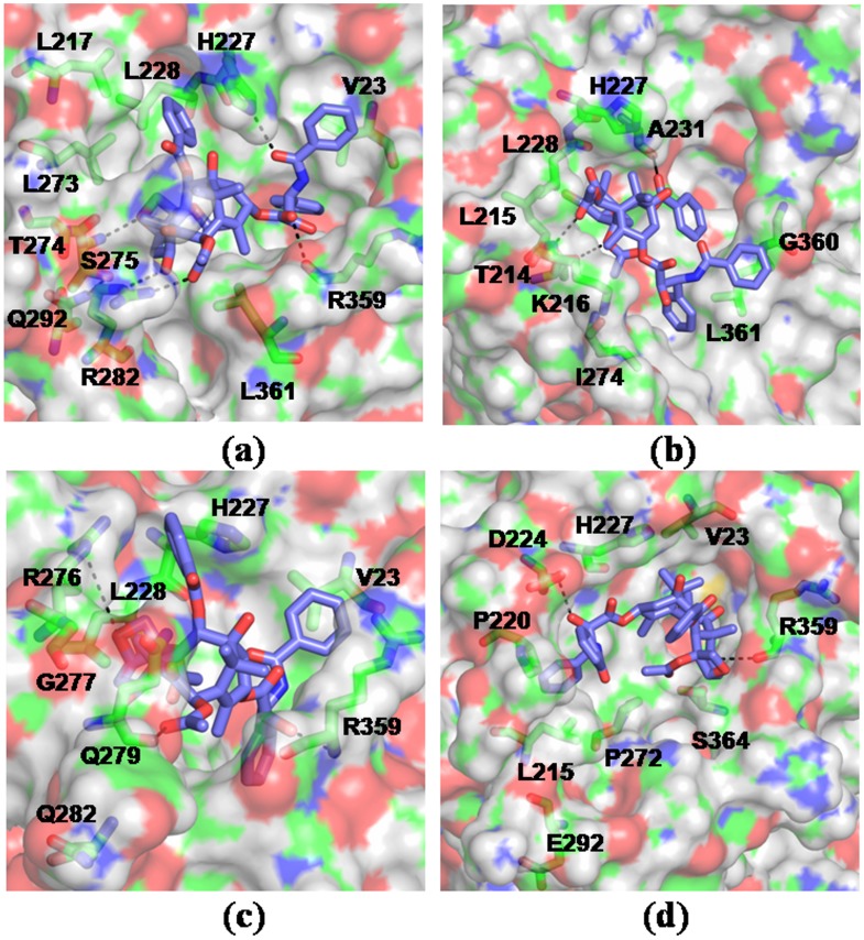
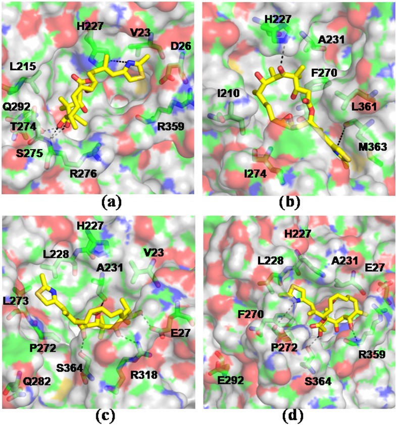
The vital role of tubulin dimer in cell division makes it an attractive drug target. Drugs that target tubulin showed significant clinical success in treating various cancers. However, the efficacy of these drugs is attenuated by the emergence of tubulin mutants that are unsusceptible to several classes of tubulin binding drugs. The molecular basis of drug resistance of the tubulin mutants is yet to be unraveled. Here, we employ molecular dynamics simulations, protein-ligand docking, and MMPB(GB)SA analyses to examine the binding of anticancer drugs, taxol and epothilone to the reported point mutants of tubulin--T274I, R282Q, and Q292E. Results suggest that the mutations significantly alter the tubulin structure and dynamics, thereby weaken the interactions and binding of the drugs, primarily by modifying the M loop conformation and enlarging the pocket volume. Interestingly, these mutations also affect the tubulin distal sites that are associated with microtubule building processes.


Conflict of interest statement

Competing Interests: The authors have declared that no competing interests exist.

Figures

Figure 1
Figure 1. Tubulin-drug interactions.
(a) Crystal structure of taxol bound αβ-tubulin dimer (1JFF). The mutated residues are highlighted in yellow and the taxol/epothilone (ice blue) binding site is noted. A few functionally important loops, such as M, H6–H7, S9–S10 are labeled. The protein residues that involve in direct interactions with the drugs are shown in (b) and (c). Taxol (violet) and epothilone (yellow) are shown in licorice representations.
Figure 2
Figure 2. Structural Changes in tubulin mutants relative to WT.
a) Time-averaged structures of the β-subunit of tubulin mutants superposed on the time-averaged structure of the β-subunit of WT tubulin. Secondary structural elements which underwent the most significant conformational changes are highlighted. b) Solvent accessible surface area of the β-subunit of WT tubulin and the mutants as a function of time. Color scheme: Wild-type (black), T274I mutation (blue), R282Q mutation (red), Q292E mutation (green).
Figure 3
Figure 3. Residue-level displacements and fluctuations.
Difference in average Cα-RMSD of β-tubulin residues of tubulin mutants and the WT (left column) and comparison of average Cα-RMSF of β-tubulin residues of mutants and the WT (right column). Color scheme is similar to Fig. 2.
Figure 4
Figure 4. Correlations of the motions of various regions in β-tubulin.
Two dimensional cross-correlation maps of the β-subunit of WT and mutated tubulins. Red patches indicate the positively correlated motions, whereas blue patches indicate anti-correlated motion. The maps have been calculated for the Cα aoms from the final 10 ns MD data. Very similar patterns were obtained when the maps were generated on other sets of 10 ns data.
Figure 5
Figure 5. Local changes due to mutations in β-tubulin.
The conformations of the drug binding loops, M, H6–H7, S9–S10, in mutants and the WT: a) T274I mutation, b) R282Q mutation and c) Q292E mutation. The loops in mutants are colored red and those in the WT are colored blue. d) Probability distribution of the M loop conformations in mutants and WT. Color scheme is similar to Fig. 2.
Figure 6
Figure 6. Binding motif of taxol in WT and mutated tubulins.
The drug-receptor complexes were obtained by simulating the lowest energy docked complexes for 10 ns in explicit water. Taxol is shown in licorice and the tubulin residues involved in interactions are colored according to atom type – green: C, red: O, blue: N, white: H. Results shown for - a) WT b) T274I c) R282Q d) Q292E. Mutations resulted in altered mode of drug binding and loss of characteristic drug-receptor contacts.
Figure 7
Figure 7. Binding motif of epothilone A in WT and mutated tubulins.
The drug-receptor complexes were obtained by simulating the lowest energy docked complexes for 10 ns in explicit water. Results shown for - a) WT b) T274I c) R282Q d) Q292E. Epothilone A is shown in yellow.

Similar articles

Cited by

References

    1. Conde C, Caceres A (2009) Microtubule assembly, organization and dynamics in axons and dendrites. Nat Rev Neurosci 10: 319–332. - PubMed
    1. Hammond JW, Cai D, Verhey KJ (2008) Tubulin modifications and their cellular functions. Curr Opin Cell Biol 20: 71–76. - PMC - PubMed
    1. Verhey KJ, Gaetig J (2007) The tubulin code. Cell Cycle 6: 2152–2160. - PubMed
    1. Jordan MA, Wilson L (2004) Microtubules as a target for cancer therapy. Nature Rev Cancer 4: 253–265. - PubMed
    1. Perez EA (2009) Microtubule inhibitors: differentiating tubulin-inhibiting agents based on mechanism of action, clinical activity, and resistance. Mol Cancer Ther 8: 2086–2095. - PubMed

Publication types