. 2012;7(8):e42405.
doi: 10.1371/journal.pone.0042405. Epub 2012 Aug 3.

Production of a subunit vaccine candidate against porcine post-weaning diarrhea in high-biomass transplastomic tobacco

Affiliations

Production of a subunit vaccine candidate against porcine post-weaning diarrhea in high-biomass transplastomic tobacco

Igor Kolotilin et al. PLoS One. 2012.

Abstract

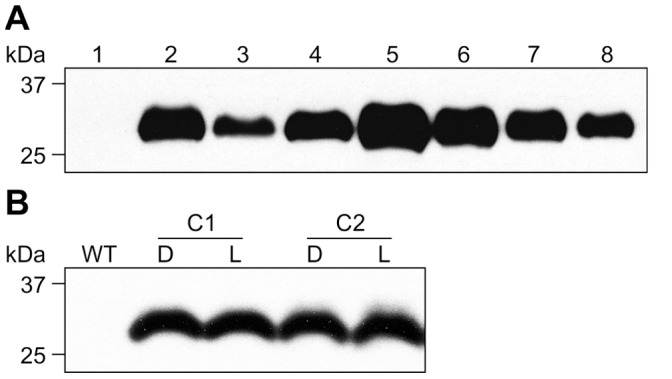
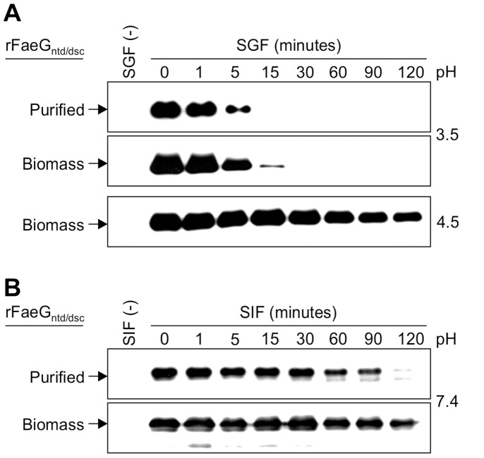
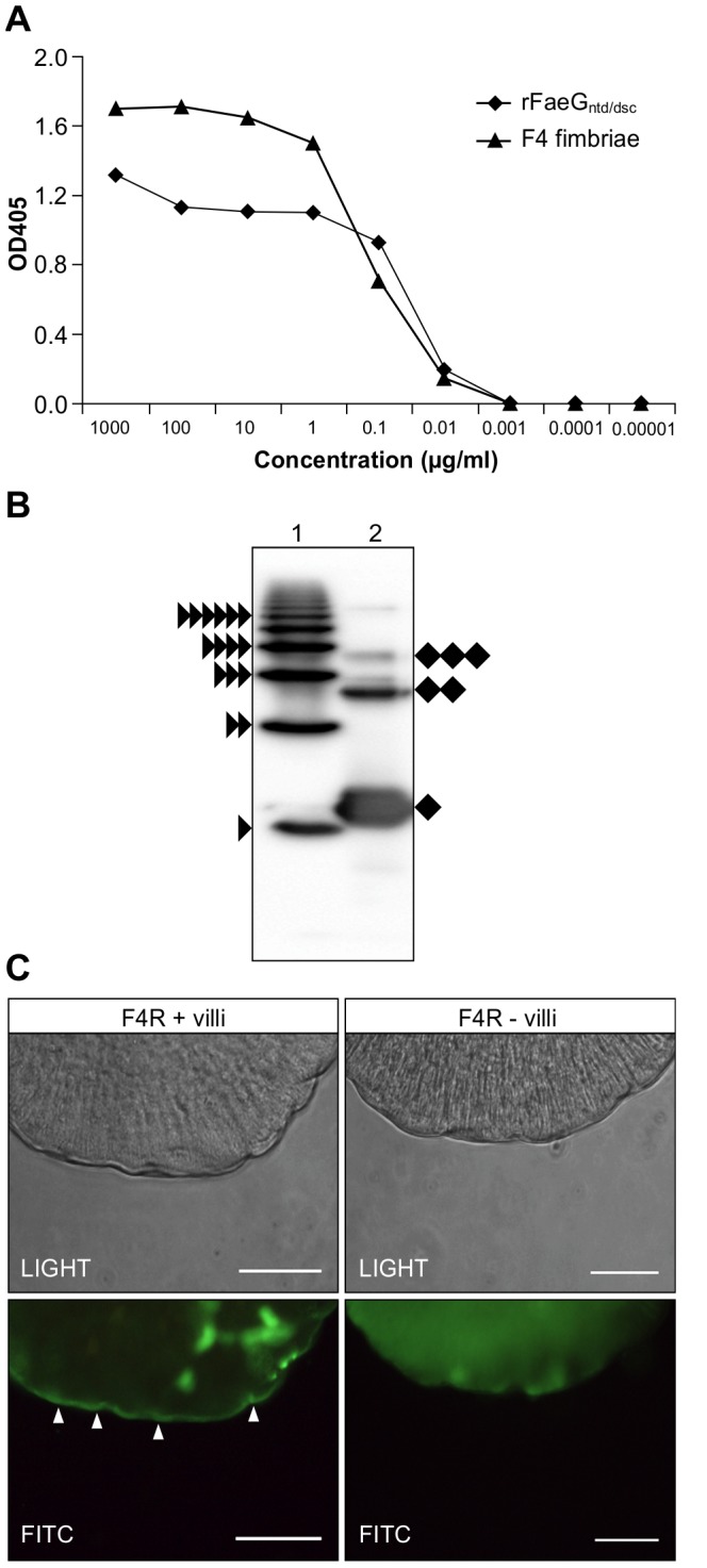
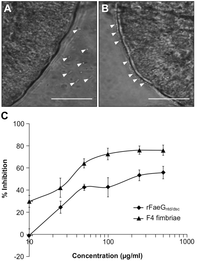
Post-weaning diarrhea (PWD) in piglets is a major problem in piggeries worldwide and results in severe economic losses. Infection with Enterotoxigenic Escherichia coli (ETEC) is the key culprit for the PWD disease. F4 fimbriae of ETEC are highly stable proteinaceous polymers, mainly composed of the major structural subunit FaeG, with a capacity to evoke mucosal immune responses, thus demonstrating a potential to act as an oral vaccine against ETEC-induced porcine PWD. In this study we used a transplastomic approach in tobacco to produce a recombinant variant of the FaeG protein, rFaeG(ntd/dsc), engineered for expression as a stable monomer by N-terminal deletion and donor strand-complementation (ntd/dsc). The generated transplastomic tobacco plants accumulated up to 2.0 g rFaeG(ntd/dsc) per 1 kg fresh leaf tissue (more than 1% of dry leaf tissue) and showed normal phenotype indistinguishable from wild type untransformed plants. We determined that chloroplast-produced rFaeG(ntd/dsc) protein retained the key properties of an oral vaccine, i.e. binding to porcine intestinal F4 receptors (F4R), and inhibition of the F4-possessing (F4+) ETEC attachment to F4R. Additionally, the plant biomass matrix was shown to delay degradation of the chloroplast-produced rFaeG(ntd/dsc) in gastrointestinal conditions, demonstrating a potential to function as a shelter-vehicle for vaccine delivery. These results suggest that transplastomic plants expressing the rFaeG(ntd/dsc) protein could be used for production and, possibly, delivery of an oral vaccine against porcine F4+ ETEC infections. Our findings therefore present a feasible approach for developing an oral vaccination strategy against porcine PWD.


Conflict of interest statement

Competing Interests: The authors have declared that no competing interests exist.

Figures

Figure 1
Figure 1. Accumulation of chloroplast-targeted, transiently-expressed rFaeGntd/dsc.
Transient expression of the rFaeGntd/dsc protein via agroinfiltration in Nicotiana benthamiana leaves was examined by SDS-PAGE and staining (a), and immunoblot analysis (b). Lanes 1 and 2−5.0 µg of protein extract of leaves co-infiltrated with Agrobacteria carrying chloroplast-targeted rFaeGntd/dsc and the p19 viral suppressor of post-transcriptional gene silencing (1), or p19 alone as negative control (2). rFaeGntd/dsc is indicated with a black rhomb, higher bands likely correspond to rFaeGntd/dsc with partially cleaved transit peptide; Lane 3−0.5 µg purified F4ad fimbriae as positive control, the F4 native FaeG is indicated with a black triangle; the ∼2 kDa difference in size of rFaeGntd/dsc (29 kDa) and the native FaeG (27 kDa) is due to the additional complementing fused domain.
Figure 2
Figure 2. Homoplastomic lines show normal phenotype.
(a) A schematic representation of the chloroplast transformation cassette (pCT-rFaeGntd/dsc). The cassette was designed to integrate between the trnI (tRNA-Ile) and trnA (tRNA-Ala) genes of the tobacco plastome. The wild type (WT) plastome trnI - trnA region is shown at the bottom. Expected sizes of Rsr II-digested fragments are indicated. Thick black lines represent hybridization sites for the probe used in Southern blot analyses. IEE  =  intercistronic expression element with the Shine-Dalgarno sequence from the 5′ UTR of bacteriophage T7 gene 10 fused to the 3′ end; aadA  =  gene encoding aminoglycoside 3′ adenylyltransferase for spectinomycin resistance; TpsbC  = 3′ UTR of psbC from white poplar plastome; PpsbA  = 5′ UTR and promoter of tobacco psbA gene. rfaeGntd/dsc  =  gene encoding the rFaeGntd/dsc protein variant. TrbcL  = 3′ UTR of rbcL from white poplar plastome. (b) Phenotypes of mature transplastomic tobacco cv. I 64 plants transformed with pCT-rFaeGntd/dsc (1 and 2) were indistinguishable from WT plants (3). A one-meter ruler was photographed to the left of each plant as size reference. (c) Confirmation of homoplastomy. Southern blot analysis of total plant DNA from 2 independent transformants and 1 untransformed plant displayed a single band of the expected size.
Figure 3
Figure 3. Spatial accumulation of rFaeGntd/dsc in transplastomic tobacco plants.
(a) Schematic showing the 10 leaves sampled to assess the spatial accumulation of rFaeGntd/dsc in transplastomic tobacco plants. (b) Samples examined on SDS-PAGE stained gel (upper panel) and western blot (lower panel). Each lane was loaded with an extract from either ∼2.3 mg of leaf tissue (stained gel), or ∼0.5 mg (immunoblotted gel). WT =  leaf 4 from an untransformed plant. A band of the predicted size (29 kDa, indicated with a black rhomb) corresponding to rFaeGntd/dsc was observed in all transplastomic leaf samples, but was absent in the WT. This band was immunoreactive with anti-FaeG serum on the Western blot. kDa - protein molecular weight marker.
Figure 4
Figure 4. Purification of rFaeGntd/dsc from crude plant extract and quantification.
(a) rFaeGntd/dsc was extracted from 5 g of mature transplastomic leaf tissue and purified. The initial volume of the extract was 50 ml; 3 µl of the extract from each step of the procedure were resolved by SDS-PAGE and stained. Lane 1 - Initial extract from leaf tissue, pH = 7.5; lane 2 - extract acidified to pH = 2 and centrifuged; lane 3 - clarified extract neutralized to pH = 7.4; Lane 4 - flowthrough from IMAC column; Lane 5 - wash with 20 mM imidazole; Lane 6 - elution of purified rFaeGntd/dsc; Lane 7 - 0.5 µg of BSA as loading control; kDa - protein molecular weight marker. (b) Purified rFaeGntd/dsc was quantified using densitometry. Dilutions of the purified rFaeGntd/dsc protein (lanes 1 through 7) were resolved in SDS-PAGE gel along with known amounts of BSA (lanes 8–14; 1.0, 0.8, 0.6, 0.4, 0.2, 0.1, 0.05 µg BSA, respectively) and stained. BSA bands were used for generation of a standard curve (R2 = 0.987; p = 0.01) and extrapolating rFaeGntd/dsc concentration. kDa - molecular weight marker.
Figure 5
Figure 5. Accumulation levels of rFaeGntd/dsc in transplastomic leaf tissue.
(a) Samples of equal volume (4 µl) were prepared from crude extract fractions. Lane 1 - WT extract (negative control); lanes 2, 3 and 4 represent crude extract of 0.4 mg of leaf tissue, re-extracted pellet, and clarified extract, respectively, where clarified extract contains 5 µg TSP. The rFaeGntd/dsc yield was estimated using a standard curve (R2 = 0.993) of known amounts of purified rFaeGntd/dsc (lanes 5 through 8∶2 µg, 1 µg, 0.5 µg and 0.25 µg, respectively). (b) No variation in rFaeGntd/dsc accumulation was observed in transplastomic clones (C1, C2) after dark (D) or after light (L) periods. Image is representative of sampling on three different days, 1 µg TSP was used per lane. WT =  untransformed control.
Figure 6
Figure 6. Stability of rFaeGntd/dsc under simulated gastrointestinal conditions.
Time course analysis of the stability of chloroplast-expressed rFaeGntd/dsc in simulated gastric fluid (SGF; a) and simulated intestinal fluid (SIF; b). rFaeGntd/dsc was present in similar amounts either as purified protein (“Purified”) or as lyophilized and powdered transplastomic leaf tissue (“Biomass”) and was visualized by western blotting. SGF digestion of leaf biomass was done at two different pH values: pH = 3.5 and pH = 4.5. SGF and SIF fluids with no substrate [SGF (−) and SIF (−), respectively] represent negative controls. The rFaeGntd/dsc band is indicated with an arrow.
Figure 7
Figure 7. Chloroplast-produced rFaeGntd/dsc protein is recognized in F4 fimbriae-specific ELISA, partially polymerizes and specifically binds to the brush border of F4R+ small intestinal villi.
(a) Both rFaeGntd/dsc and F4 fimbriae are recognized by a monoclonal anti-F4ad fimbriae antibody in ELISA. (b) Purified F4 fimbriae (lane 1) and purified rFaeGntd/dsc (lane 2) were resolved under non-reducing conditions to assess polymerization. The F4 fimbriae sample displayed the formation of native FaeG polymers, number of subunits is indicated by stacked black triangles next to each band. Most of the rFaeGntd/dsc is present as monomers (denoted by black rhomb); formation of rFaeGntd/dsc dimers and trimers was also observed (two and three stacked black rhombs). (c) Adhesion of the rFaeGntd/dsc protein to the brush border of F4R+ small intestinal villi. Binding to the F4-specific receptors present on the apical surface of the epithelial cells, which line the brush border of F4R+ small intestinal villi is shown as a bright line on the edge of the sample, the result of excited FITC fluorochrome (indicated with white arrows, lower panel). rFaeGntd/dsc fails to bind to brush border of F4R− small intestinal villi. Images are representative of rFaeGntd/dsc adhesion to isolated villi of three F4R+ and two F4R− piglets. Bar: 50 µm.
Figure 8
Figure 8. Chloroplast-produced rFaeGntd/dsc inhibits the adhesion of F4+ ETEC to porcine small intestinal villi.
Adhesion of F4+ ETEC to F4R− villi (a) and F4R+ villi (b), white arrows indicate bacterial cells. Bar: 50 µm. (c) Competitive inhibition of adhesion of F4+ ETEC to porcine small intestinal villi by the rFaeGntd/dsc protein or F4 fimbriae, determined at different protein concentrations. The data represent the mean ±SE (n = 4).

Similar articles

Cited by

References

    1. Fairbrother JM, Nadeau E, Gyles CL (2005) Escherichia coli in postweaning diarrhea in pigs: an update on bacterial types, pathogenesis, and prevention strategies. Anim Health Res Rev 6: 17–39. - PubMed
    1. Hampson DJ (1994) Postweaning Escherichia coli diarrhoea in pigs. In: Gyles CL, editor. Escherichia coli in domestic animals and humans. Wallingford: CAB International. 171–191.
    1. Bertschinger HU, Fairbrother JM (1999) Escherichia coli infections. In: Straw BE, D’Allaire S, Mengeling WL, Taylor DJ, editors. Diseases of Swine, 8th Edition. Iowa: Iowa State University Press. 431–454.
    1. Ciosek D, Truszczynski M, Jagodzinski M (1983) The effectiveness of inactivated vaccines applied parenterally to sows to control Escherichia coli diarrhea in piglets in an industrial fattening farm. Comp Immunol Microbiol Infect Dis 6: 313–319. - PubMed
    1. Van den Broeck W, Cox E, Goddeeris BM (1999) Receptor-dependent immune responses in pigs after oral immunization with F4 fimbriae. Infect Immun 67: 520–526. - PMC - PubMed

Publication types

MeSH terms