. 2012;7(8):e42432.
doi: 10.1371/journal.pone.0042432. Epub 2012 Aug 6.

Structure determination and functional analysis of a chromate reductase from Gluconacetobacter hansenii

Affiliations

Structure determination and functional analysis of a chromate reductase from Gluconacetobacter hansenii

Hongjun Jin et al. PLoS One. 2012.

Abstract

Environmental protection through biological mechanisms that aid in the reductive immobilization of toxic metals (e.g., chromate and uranyl) has been identified to involve specific NADH-dependent flavoproteins that promote cell viability. To understand the enzyme mechanisms responsible for metal reduction, the enzyme kinetics of a putative chromate reductase from Gluconacetobacter hansenii (Gh-ChrR) was measured and the crystal structure of the protein determined at 2.25 Å resolution. Gh-ChrR catalyzes the NADH-dependent reduction of chromate, ferricyanide, and uranyl anions under aerobic conditions. Kinetic measurements indicate that NADH acts as a substrate inhibitor; catalysis requires chromate binding prior to NADH association. The crystal structure of Gh-ChrR shows the protein is a homotetramer with one bound flavin mononucleotide (FMN) per subunit. A bound anion is visualized proximal to the FMN at the interface between adjacent subunits within a cationic pocket, which is positioned at an optimal distance for hydride transfer. Site-directed substitutions of residues proposed to involve in both NADH and metal anion binding (N85A or R101A) result in 90-95% reductions in enzyme efficiencies for NADH-dependent chromate reduction. In comparison site-directed substitution of a residue (S118A) participating in the coordination of FMN in the active site results in only modest (50%) reductions in catalytic efficiencies, consistent with the presence of a multitude of side chains that position the FMN in the active site. The proposed proximity relationships between metal anion binding site and enzyme cofactors is discussed in terms of rational design principles for the use of enzymes in chromate and uranyl bioremediation.


Conflict of interest statement

Competing Interests: The authors have declared that no competing interests exist.

Figures

Figure 1
Figure 1. Substrate inhibition by NADH in an ordered bireactant mechanism.
A. Double-reciprocal plots of initial velocities versus substrate concentrations assayed with fixed concentration of NADH: 9.5 µM (open diamond), 19 µM (closed diamond), 25 µM (open triangle), 37.5 µM (closed triangle), 50 µM (open square), 75 µM (closed square), 100 µM (open circle), and 200 µM (closed circle). The VMax is calculated based on the y-axis intercept on this plot. B. Relationship between the slopes (i.e., Slope 1/CrO4 2−) in Figure 1A at each of seven fixed NADH concentrations. C. Double-reciprocal plots of initial velocities versus substrate concentrations with fixed concentration of CrO4 2−: 31 µM (open triangle), 62 µM (closed triangle), 125 µM (open square), 250 µM (closed square), 500 µM (open circle), and 1000 µM (closed circle). At low NADH concentrations it is possible to fit the data with a straight line. However, at high NADH concentrations, individual curves bend upwards. Values for KmA, KmB, Kia and Ki were calculated from axes-intercepts and slopes in panels B and C (see Table S2) . D. Cleland notation depicting catalytic mechanism of Gh-ChrR, showing substrate inhibition by NADH binding to FMN-E to form a dead-end complex FMN-E-NADH that competes with metal complex formation, Mox-FMNH2-E-NADH.
Figure 2
Figure 2. Crystal structure of Gh-ChrR.
Monomeric (A) and tetrameric (B) depictions of the 2.25 Å structure of Gh-ChrR showing the backbone fold, a space-filling model of bound FMN (elements color: red  =  oxygen, blue  =  nitrogen, gray  =  carbon) and bound chloride anion (green sphere). Secondary structural elements including the 310 helices (η) are numbered sequentially from the N-terminus. C. Electrostatic potentials at the solvent-accessible surface of Gh-ChrR. A stick model of the FMN molecule and the associated chloride ion (gray sphere) is highlighted. The electrostatic potential are drawn (Pymol) at a level of −71.817 kT/e (red) to +71.817 kT/e (blue), where k is the Boltzman’s constant, T is the absolute temperature, and e is the magnitude of the electron charge.
Figure 3
Figure 3. Structure proximal to bound FMN.
A. Electron density surrounding FMN and chloride ion (gray sphere) contoured at 1.0 σ. B. Schematic representation of hydrophobic contacts (arc with radiating spokes) and potential hydrogen bonds (dashed lines) between FMN and two monomeric units (chain A and C) of the Gh-ChrR tetramer. Atoms are color-coded: black = carbon, red = oxygen, blue = nitrogen. This image was produced using the program LIGPLOT .
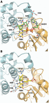
Figure 4
Figure 4. Putative Gh-ChrR NADH and substrate binding sites.
A. NADH was modeled into the Gh-ChrR structure by superimposing it with the NADH-containing structure of EmoB (PDB entry: 2VZJ, Figure S7). The nicotinamide ring of NADH (primarily green stick model) is stacked on top of the isoalloxazine ring of FMN (primarily yellow stick model), and the adenosine part of NADH points to ribtyl group of FMN. The black arrow indicates the distance from C4N of NADH to the si-face of the FMN isoalloxazine ring. Residues N53, D54, E57, S100, R101 and F137 from chain A (cyan) and residues N85, P119, and T154 from chain C (gold) interact with NADH. B. The putative active site of Gh-ChrR shown with bound FMN (primarily yellow stick model) and a chloride ion (green sphere). The black arrow indicates the distance from the Cl to the si-face of the FMN isoalloxazine ring. Key residue R101 holding chloride ion in place is shown in a stick model. Critical residues for hydride transfer, N85 and Y86 from chain A (cyan) and S118 from chain C (gold) are shown in a stick model. The green dash lines indicate the distance (∼3 Å) between N of amide group of N85/Y86 and O4, and the distance (∼3 Å) between OG of hydroxyl group of S118 and O2.

Similar articles

Cited by

References

    1. Wall JD, Krumholz LR (2006) Uranium reduction. Annu Rev Microbiol 60: 149–166. - PubMed
    1. Fredrickson JK, Zachara JM (2008) Electron transfer at the microbe-mineral interface: a grand challenge in biogeochemistry. Geobiology 6: 245–253. - PubMed
    1. Lovley DR, Phillips EJP (2002) Bioremediation of uranium contamination with enzymatic uranium reduction. Environmental Science & Technology 26: 2228–2234.
    1. Reguera G, Nevin KP, Nicoll JS, Covalla SF, Woodard TL, et al. (2006) Biofilm and nanowire production leads to increased current in Geobacter sulfurreducens fuel cells. Appl Environ Microbiol 72: 7345–7348. - PMC - PubMed
    1. Reguera G, McCarthy KD, Mehta T, Nicoll JS, Tuominen MT, et al. (2005) Extracellular electron transfer via microbial nanowires. Nature 435: 1098–1101. - PubMed

Publication types