. 2012;7(8):e42512.
doi: 10.1371/journal.pone.0042512. Epub 2012 Aug 6.

Waking action of ursodeoxycholic acid (UDCA) involves histamine and GABAA receptor block

Affiliations

Waking action of ursodeoxycholic acid (UDCA) involves histamine and GABAA receptor block

Yevgenij Yanovsky et al. PLoS One. 2012.

Abstract

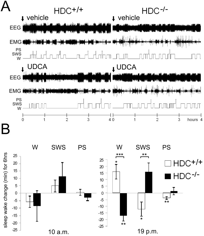
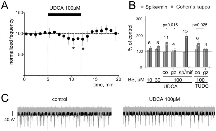
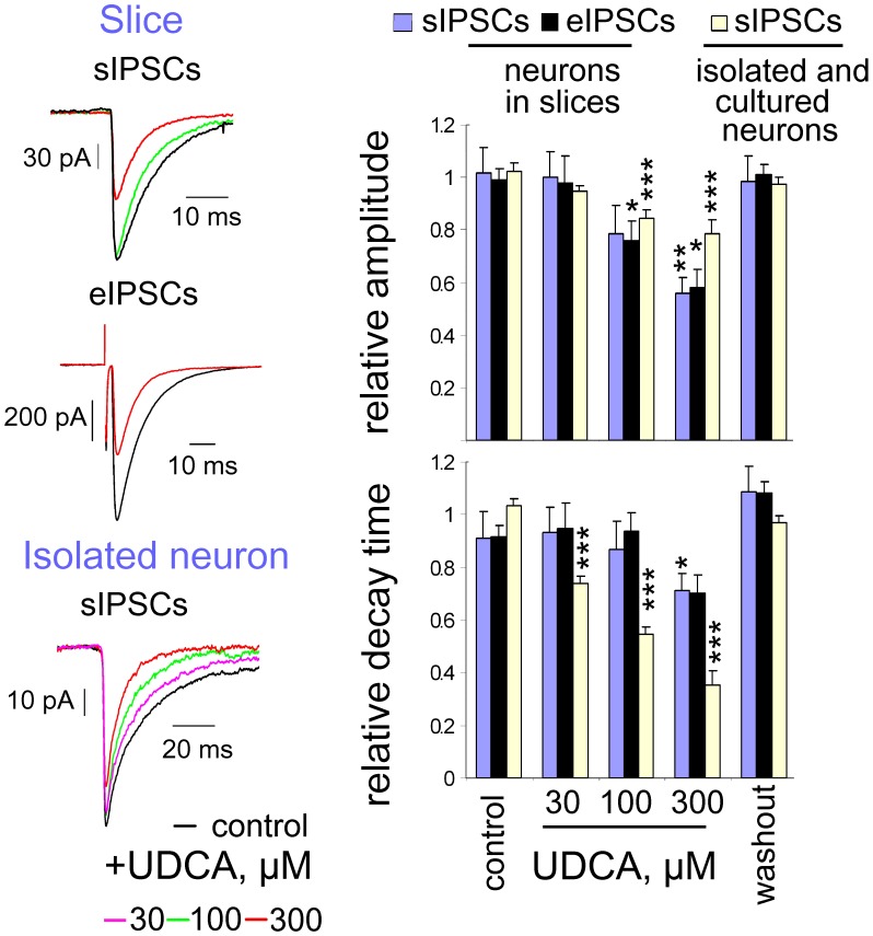
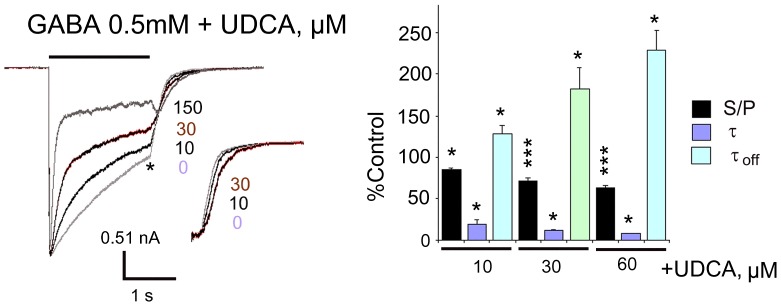
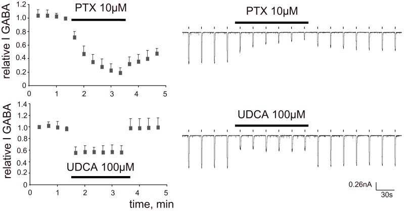
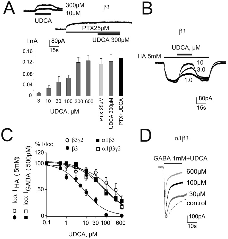
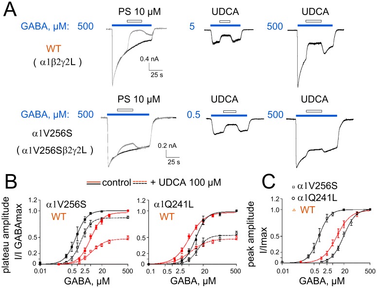
Since ancient times ursodeoxycholic acid (UDCA), a constituent of bile, is used against gallstone formation and cholestasis. A neuroprotective action of UDCA was demonstrated recently in models of Alzheimer's disease and retinal degeneration. The mechanisms of UDCA action in the nervous system are poorly understood. We show now that UDCA promotes wakefulness during the active period of the day, lacking this activity in histamine-deficient mice. In cultured hypothalamic neurons UDCA did not affect firing rate but synchronized the firing, an effect abolished by the GABA(A)R antagonist gabazine. In histaminergic neurons recorded in slices UDCA reduced amplitude and duration of spontaneous and evoked IPSCs. In acutely isolated histaminergic neurons UDCA inhibited GABA-evoked currents and sIPSCs starting at 10 µM (IC(50) = 70 µM) and did not affect NMDA- and AMPA-receptor mediated currents at 100 µM. Recombinant GABA(A) receptors composed of α1, β1-3 and γ2L subunits expressed in HEK293 cells displayed a sensitivity to UDCA similar to that of native GABA(A) receptors. The mutation α1V256S, known to reduce the inhibitory action of pregnenolone sulphate, reduced the potency of UDCA. The mutation α1Q241L, which abolishes GABA(A)R potentiation by several neurosteroids, had no effect on GABA(A)R inhibition by UDCA. In conclusion, UDCA enhances alertness through disinhibition, at least partially of the histaminergic system via GABA(A) receptors.


Conflict of interest statement

Competing Interests: The authors have declared that no competing interests exist.

Figures

Figure 1
Figure 1. UDCA given orally increases wakefulness.
A. Polygraphic recording (EEG and EMG) and corresponding hypnograms showing the effect of UDCA given at 7 p.m. B. Changes in sleep-wake duration after UDCA application relative to control (vehicle application) in wild type (HDC +/+) and histidine decarboxylase deficient (HDC−/−) mice. In both mouse genotypes UDCA given during the sleepy period of the day (10 a.m.) does not affect sleep-wake states, but does so during the active period of the day. Given at 7 p.m. UDCA increases waking (W) and decreases slow wave sleep (SWS) and paradoxical sleep (PS) in the HDC+/+ mice. In HDC−/− mice UDCA decreases waking. The significant difference between time spent in a given vigilance state in control and under UDCA is indicated with stars near the corresponding bars. Significant difference between genotypes is indicated with stars on top of brackets. * p<0.05; ** p<0.01; *** p<0.005.
Figure 2
Figure 2. UDCA synchronizes network activity like a GABAA receptor antagonist.
A. Firing rate of TMN neurons (n = 10) recorded in mouse hypothalamic slices is not significantly affected by UDCA during the first 5 min of UDCA perfusion. Each filled circle represents the average firing during 5 min. Significant difference from baseline is indicated with stars (* p<0.05, Wilcoxon test). B. Summary of MEA experiments illustrates the change in spikes/min (all spikes over all active electrodes) and Cohen's kappa (synchronization index). Note that gabazine blanks the effect of UDCA and tauroursodeoxycholate (TUDC) on synchronization. Mineralocorticoid- and glucocorticoid- receptor- antagonists (mifepristone and spironolactone, respectively) did not significantly change effects of UDCA (significance of modulation compared to the control indicated with stars within bars (*: p<0.05). C. Examples of neuronal firing patterns recorded from 2 electrodes in one hypothalamic culture (one electrode in black, another in grey color) during 1 second. Note more synchronous discharge of hypothalamic neurons in the presence of UDCA.
Figure 3
Figure 3. Spontaneous and evoked IPSCs in TMN neurons are inhibited by UDCA.
At the left: Examples of averaged (5 min periods) spontaneous and evoked IPSCs recorded from the same neuron in a slice in control (black) and in the presence of UDCA (100 µM: green trace; 300 µM: red trace) and of averaged sIPSCs recorded in an acutely isolated neuron below (each trace represents an average of 24 to 87 events). At the right: Comparison of the relative amplitudes and decay time constants of eIPSCs and sIPSCs from different neuronal preparations. Four to ten neurons were tested with each UDCA concentration. Data obtained from the 4 acutely isolated neurons were pooled with data from 6 cultured neurons. Data points were normalized on the averaged value from control and washout. Significance of difference from control is indicated above the bars (p<0.05; **:p<0.01; ***: p<0.005).
Figure 4
Figure 4. Kinetics of GABAA receptor inhibition by UDCA in TMN neurons.
UDCA affects apparent desensitisation of maximal GABA-mediated whole-cell currents. Termination of GABA+UDCA (150 µM) application displays tail current, not obvious at smaller UDCA concentrations, which prolong the decay of GABA-current (τoff) after agonist and drug removal (inset shows relaxation currents scaled to the control amplitude (*)). Note, that the peak current amplitude is not affected by UDCA. The bar graph at right shows the summary of UDCA effects on rate and degree of desensitisation obtained from 4–8 neurons. The ratio of steady-state versus peak current (S/P), fast decay time constant (τ) and current decay time constant after removal of drugs (τoff) relative to the control values are given. ** p<0.01; ***p<0.005, Wilcoxon test.
Figure 5
Figure 5. Time courses of GABA-response-block by UDCA and picrotoxin.
Picrotoxin (PTX) continuously present in the recording solution shows use-dependent block of GABA (10 µM, 1 s) - currents: block is not fully reversible after PTX withdrawal. In contrast, the UDCA-block is fully reversible and achieves maximum with the first GABA/UDCA co-application.
Figure 6
Figure 6. Recombinant α1β3γ2L GABAARs are blocked by UDCA in a way comparable to native receptors.
A. UDCA and picrotoxin (PTX) induce outward shift in baseline current in HEK293 cells transfected with β-plasmid (block of constitutively open channels). Plot below shows averaged amplitudes of UDCA- and PTX- responses (4–10 cells for each concentration). Note lack of additivity between maximal UDCA and PTX induced outward currents. B. Histamine (HA)-evoked inward currents and their reversible blockade by different concentrations of UDCA at homopentameric GABAA receptors. C. Concentration-response curves for UDCA-block of control (co) either GABA (0.5 mM)- or histamine (HA, 5 mM)- evoked currents obtained from 4 different receptor types fitted with the following IC50s(nH): α1β3γ2L: 73±16 µM (0.63); α1β: 81±16 µM (0.65); β3γ2L: 232±66 µM (0.62); β3: 7.4±2 µM (0.8). Four to ten cells were investigated with the whole concentration range for each receptor type. D. Examples of current recordings in α1β3-expressing HEK293 cell: GABA (dotted line)- or GABA+UDCA – responses are superimposed.
Figure 7
Figure 7. Impairment of GABAAR-block by UDCA in the mutant α1V256S- but not α1Q241L-containing receptors.
A. GABA-evoked currents, in response to maximal concentrations and concentrations around EC25, and their inhibition by pregnenolone sulphate (PS) or UDCA 100 µM. B. GABA dose-response curves constructed for the plateau amplitudes measured for control: immediately before UDCA application, which started 15–25 s after the beginning of the GABA application; and for the amplitude of blocked current: at the beginning of UDCA application (first point at a steady-state level). The inhibition of maximal GABA-responses by UDCA is significantly smaller in WT (47±3% of control, n = 7) and mutant α1Q241L receptors (54±2% of control, n = 5, no difference with WT) than in the mutant α1V256S receptors (87±1% of control, n = 5; p = 0.0047 vs WT). UDCA does not significantly modify the EC50 and nH values for GABA in the WT (6±1 µM; 1.2±0.2 versus 5.1±0.5 µM; 1.4±0.1 in control), in the mutant α1Q241L (11.5±1.2 µM; 2.0±0.4 versus 9.8±0.7 µM; 1.9±0.2 in control) and α1V256S (1.5±0.07 µM; 1.6±0.1 versus 0.83±0.03 µM; 1.5±0.06 in control) receptors. C. GABA dose-response curves constructed from peak current amplitude values normalized on maximal GABA-evoked current for the WT (EC50 = 8±0.5 µM; nH = 1.2±0.1; n = 8), α1V256S (EC50 = 0.9±0.03 µM; nH = 1.5±0.06; n = 5) and α1Q241L (EC50 = 20±1 µM; nH = 1.4±0.1; n = 5) receptors of α1β2γ2L-composition expressed in HEK293 cells.

References

    1. Festi D, Montagnani M, Azzaroli F, Lodato F, Mazzella G, et al. (2007) Clinical efficacy and effectiveness of ursodeoxycholic acid in cholestatic liver diseases. Curr Clin Pharmacol 2: 155–177. - PubMed
    1. Woo SJ, Kim JH, Yu HG (2010) Ursodeoxycholic acid and tauroursodeoxycholic acid suppress choroidal neovascularization in a laser-treated rat model. J Ocul Pharmacol Ther 26: 223–229. - PubMed
    1. Boatright JH, Moring AG, McElroy C, Phillips MJ, Do VT, et al. (2006) Tool from ancient pharmacopoeia prevents vision loss. Mol Vis 12: 1706–14.: 1706–1714. - PubMed
    1. Keene CD, Rodrigues CM, Eich T, Chhabra MS, Steer CJ, et al. (2002) Tauroursodeoxycholic acid, a bile acid, is neuroprotective in a transgenic animal model of Huntington's disease. Proc Natl Acad Sci U S A 99: 10671–10676. - PMC - PubMed
    1. Sola S, Amaral JD, Borralho PM, Ramalho RM, Castro RE, et al. (2006) Rodrigues,C.M., Functional modulation of nuclear steroid receptors by tauroursodeoxycholic acid reduces amyloid beta-peptide-induced apoptosis. Mol Endocrinol 20: 2292–2303. - PubMed

Publication types

MeSH terms