. 2012;7(8):e42652.
doi: 10.1371/journal.pone.0042652. Epub 2012 Aug 6.

The role of the subthalamic nucleus in L-DOPA induced dyskinesia in 6-hydroxydopamine lesioned rats

Affiliations

The role of the subthalamic nucleus in L-DOPA induced dyskinesia in 6-hydroxydopamine lesioned rats

Asier Aristieta et al. PLoS One. 2012.

Abstract

L-DOPA is the most effective treatment for Parkinson's disease (PD), but prolonged use leads to disabling motor complications including dyskinesia. Strong evidence supports a role of the subthalamic nucleus (STN) in the pathophysiology of PD whereas its role in dyskinesia is a matter of controversy. Here, we investigated the involvement of STN in dyskinesia, using single-unit extracellular recording, behavioural and molecular approaches in hemi-parkinsonian rats rendered dyskinetic by chronic L-DOPA administration. Our results show that chronic L-DOPA treatment does not modify the abnormal STN activity induced by the 6-hydroxydopamine lesion of the nigrostriatal pathway in this model. Likewise, we observed a loss of STN responsiveness to a single L-DOPA dose both in lesioned and sham animals that received daily L-DOPA treatment. We did not find any correlation between the abnormal involuntary movement (AIM) scores and the electrophysiological parameters of STN neurons recorded 24 h or 20-120 min after the last L-DOPA injection, except for the axial subscores. Nonetheless, unilateral chemical ablation of the STN with ibotenic acid resulted in a reduction in global AIM scores and peak-severity of dyskinesia. In addition, STN lesion decreased the anti-dyskinetogenic effect of buspirone in a reciprocal manner. Striatal protein expression was altered in dyskinetic animals with increases in ΔFosB, phosphoDARPP-32, dopamine receptor (DR) D3 and DRD2/DRD1 ratio. The STN lesion attenuated the striatal molecular changes and normalized the DRD2/DRD1 ratio. Taken together, our results show that the STN plays a role, if modest, in the physiopathology of dyskinesias.


Conflict of interest statement

Competing Interests: The authors have declared that no competing interests exist.

Figures

Figure 1
Figure 1. Schematic representation of experimental design and groups.
At the beginning of the study, animals received a vehicle or 6-OHDA injection into the right medial forebrain bundle and were screened by amphetamine-induced rotations 2 weeks later. Rats were treated daily with saline or L-DOPA injections (6 mg/kg plus 12 mg/kg benserazide) for 21 days. AIMs were rated 2–3 days per week (testing sessions are marked in black). Experiment 1: After the chronic treatment was completed, L-DOPA was administered twice per week, and electrophysiological experiments were performed. All animals were perfused transcardially and processed for histology. Experiment 2: After the last testing session (day 21), the animals received a vehicle (in white) or ibotenic acid (in red) injection into the right STN. After a second period of L-DOPA treatment and AIMs rating (in black) animals were killed and the striata were dissected and frozen for western blot, and STN was postfixed for histology. Experiment 3: After chronic treatment and AIMs rating (in black), animals were killed and striata removed and frozen for western blot analysis.
Figure 2
Figure 2. Characterization of hemiparkinsonian rats with L-DOPA induced dyskinesia.
Evolution of dykinesia scores showing ( A ) the sum of AIM scores for axial, limb and orolingual ratings and ( B ) locomotive score, during L-DOPA chronic treatment. Time course of ( C ) axial, limb and orolingual AIMs score and ( D ) locomotive scores evaluated after a single injection of L-DOPA on the last testing session (day 21st). Groups: sham saline (n = 9), sham L-DOPA (n = 12), 6-OHDA saline (n = 12) and 6-OHDA L-DOPA (n = 13). Note that animals in the sham saline, sham L-DOPA and 6-OHDA saline groups did not develop any AIMs. ( E ) Photomicrographs showing the extensive loss of TH positive fibers in the striatum (upper right panel) and TH positive cells in the substantia nigra (lower right panel) ipsilateral to 6-OHDA lesion. All 6-OHDA lesioned animals included in this experiment showed >95% reduction in TH-fiber optical density in the lesioned striatum. Groups: sham saline (n = 9), sham L-DOPA (n = 12), 6-OHDA saline (n = 12) and 6-OHDA L-DOPA (n = 13). ( F ) Western blot results showing a significant reduction of TH expression in all the 6-OHDA lesioned groups. Groups: sham L-DOPA (n = 7), 6-OHDA saline (n = 9), 6-OHDA L-DOPA (n = 10) and 6-OHDA L-DOPA+STN lesion (n = 8). R = right, ipsilateral to sham or 6-OHDA injection; L = left, contralateral to sham or 6-OHDA injection. Data are expressed as mean ± S.E.M. *** p<0.001 (one-way ANOVA, followed by Bonferroni post-hoc).
Figure 3
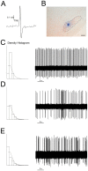
Figure 3. Electrophysiological characterization of neurons in the subthalamic nucleus.
( A ) A single spike from an STN neuron recorded in vivo. ( B ) Histological verification of the recording site in the STN, counterstained with neutral red. Scale bar: 200 µm. Examples of action potential traces, showing the three characteristic firing patterns of STN neurons. ( C ) Tonic firing pattern, in which the density histogram follows a Gaussian distribution. ( D ) Random firing pattern, in which the density histogram represents a Poisson distribution. ( E ) Bursting firing pattern, in which the density histogram represents a distribution significantly different from a Poisson distribution, with a significantly positive skewness of the density discharge distribution histogram.
Figure 4
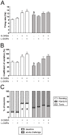
Figure 4. Electrophysiological characterization of the subthalamic nucleus.
( A ) Firing rate of STN glutamatergic neurons. Baseline firing rate (white bars) was significantly different between the groups (F(4,307) = 15.25, p<0.0001, one-way ANOVA). The acute L-DOPA challenge (grey bars), induced an increase in firing rate in sham saline animals only. ( B ) Coefficient of variation (%). Baseline activity recording (white bars) showed significant differences between groups (F(4,307) = 11.85, p<0.0001, one-way ANOVA) and acute L-DOPA treatment produced a significant reduction in the sham saline group. ( C ) Percentage of bursting, random and tonic neurons. Baseline burst firing pattern (white bars) was significantly increased in 6-OHDA lesioned groups (p<0.05, Fisher's test). Both sham groups showed a trend to increase the number of bursting neurons after an acute L-DOPA dose (sham saline, p = 0.08, and sham L-DOPA, p = 0.06 vs its baseline). Data are expressed as mean ± S.E.M. All data included in the acute L-DOPA challenge were obtained between 20 and 120 min after L-DOPA injection (time period corresponding to high dyskinetic behaviour). * p<0.05 vs sham saline and sham L-DOPA (one-way ANOVA, followed by Bonferroni post-hoc). & p<0.05 vs before L-DOPA administration (unpaired t test). # p<0.05 vs sham saline and sham L-DOPA (Fisher's exact test for firing pattern).
Figure 5
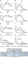
Figure 5. Effect of subthalamic nucleus lesion on the severity of L-DOPA-induced dyskinesia.
Evolution of ( A ) AIM scores/session, showing significant reduction after STN lesion in the sum of axial, limb and orolingual AIMs scored per session, whereas ( B ) locomotive AIMs scored per session were similar in both groups. The arrow marks the time of STN injection at day 22. Graphical representation of AIM scores/time point (AUC) comparing the day before STN injection (day 21st, black triangle) and the last testing session after STN injection (day 48th, black square). Time course of axial, limb and orolingual AIMs scored in ( C ) STN-lesion and ( D ) STN-sham animal groups. The results show a significant decrease in AIMs scores after STN lesion. Groups: 6-OHDA L-DOPA+STN-lesion (n = 14) and 6-OHDA L-DOPA+STN-sham (n = 5). Data are expressed as mean ± S.E.M. & p<0.05 vs day 21st (one-way RM ANOVA, followed by Bonferroni post-hoc). * p<0.05 vs STN-sham (two-way RM ANOVA, followed by Bonferroni post-hoc). # p<0.05 (two-way RM ANOVA, followed by Bonferroni post-hoc).
Figure 6
Figure 6. Effect of subthalamic nucleus lesion on the severity of the different subtypes of L-DOPA-induced dyskinesia.
Evolution of AIM scores/session of the ( A ) axial, ( C ) limb and ( E ) orolingual AIMs pre- and post-STN injection, comparing STN-lesion and STN-sham groups. Time course of score for each subtype of AIM pre- and post-STN injection; there were significant differences between pre- and post- injection in the STN-lesion in ( B ) axial, ( D ) limb and ( F ) oral subtypes. Groups: 6-OHDA L-DOPA+STN-lesion (n = 14) and 6-OHDA L-DOPA+STN-sham (n = 5). ( G ) The response to buspirone was maintained after the STN lesion, but the effect was significantly smaller than in the ( H ) STN-sham group. Groups: 6-OHDA L-DOPA+STN-lesion (n = 6) and 6-OHDA L-DOPA+STN-sham (n = 5). ( I ) Thionine-staining of the STN (upper panel). The bottom panels correspond to higher magnification of the ipsilateral (right) and contralateral (left) sides. Scale bar: 400 µm (upper panel) and 200 µm (lower panel). Data are expressed as mean ± S.E.M. & p<0.05 vs day 21st (one-way RM ANOVA, followed by Bonferroni). * p<0.05 vs STN-sham (two-way RM ANOVA, followed by Bonferroni post-hoc). # p<0.05 (two-way RM ANOVA, followed by Bonferroni post-hoc).
Figure 7
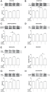
Figure 7. Effect of subthalamic nucleus lesion in protein expression in the striatum.
( A ) Western blot representative images (upper panel) and quantification (lower panel) of cFos protein showing no significant differences between the groups. ( B ) Quantification of ΔFosB/FosB western blot showed significant increased in 6-OHDA L-DOPA group versus 6-OHDA saline group ratio, indicating that L-DOPA treatment increase ΔFosB expression in the lesioned striatum. ( C ) pDARPP32/DARPP ratio was significantly higher in lesioned animal treated chronically with L-DOPA. ( D ) Results of the analysis of DRD1 and ( E ) DRD2 expression showing no significant differences between the groups. ( F ) Analysis of DRD2/DRD1 ratio showed a significant increase in 6-OHDA L-DOPA group. ( G ) DRD3 was significantly higher in 6-OHDA treated with L-DOPA compared to the animals that received saline. ( H ) mGluR5 protein expression quantification revealed a significant reduction in 6-OHDA L-DOPA group versus 6-OHDA saline group. Groups: sham L-DOPA (n = 7), 6-OHDA saline (n = 9), 6-OHDA L-DOPA (n = 10) and 6-OHDA L-DOPA+STN-lesion (n = 8). R = right, ipsilateral to sham or 6-OHDA injection; L = left, contralateral to sham or 6-OHDA injection. Data are expressed as mean ± S.E.M. * p<0.05 and ** p<0.01 (one-way ANOVA, followed by Bonferroni post-hoc).

Similar articles

Cited by

References

    1. Kish SJ, Shannak K, Hornykiewicz O (1988) Uneven pattern of dopamine loss in the striatum of patients with idiopathic Parkinson's disease. Pathophysiologic and clinical implications. N Engl J Med 318: 876–880. - PubMed
    1. Ahlskog JE, Muenter MD (2001) Frequency of levodopa-related dyskinesias and motor fluctuations as estimated from the cumulative literature. Mov Disord 16: 448–458. - PubMed
    1. Wichmann T, Dostrovsky JO (2011) Pathological basal ganglia activity in movement disorders. Neuroscience 198: 232–244. - PMC - PubMed
    1. Obeso JA, Olanow CW, Nutt JG (2000) Levodopa motor complications in Parkinson's disease. Trends Neurosci 23: S2–S7. - PubMed
    1. Cesaro P, Damier P, Nguyen JP, Palfi S, Ollat H (2003) La maladie de Parkinson.

Publication types

MeSH terms