. 2012;7(8):e42668.
doi: 10.1371/journal.pone.0042668. Epub 2012 Aug 6.

The influence of therapeutic radiation on the patterns of bone marrow in ovary-intact and ovariectomized mice

Affiliations

The influence of therapeutic radiation on the patterns of bone marrow in ovary-intact and ovariectomized mice

Susanta K Hui et al. PLoS One. 2012.

Abstract

Background: The functional components of bone marrow (i.e., the hematopoietic and stromal populations) and the adjacent bone have traditionally been evaluated incompletely as distinct entities rather than the integrated system. We perturbed this system in vivo using a medically relevant radiation model in the presence or absence of ovarian function to understand integrated tissue interaction.

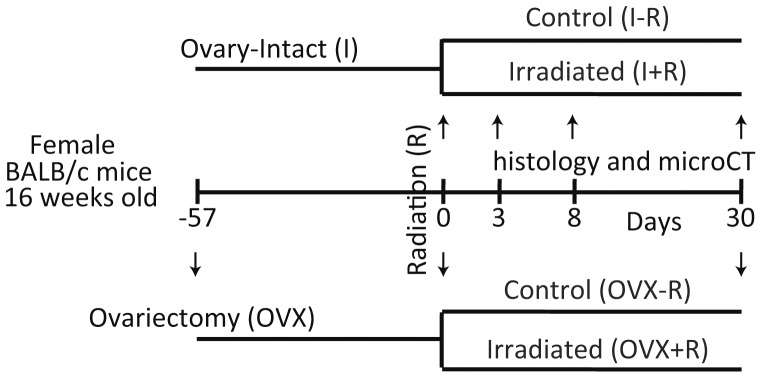
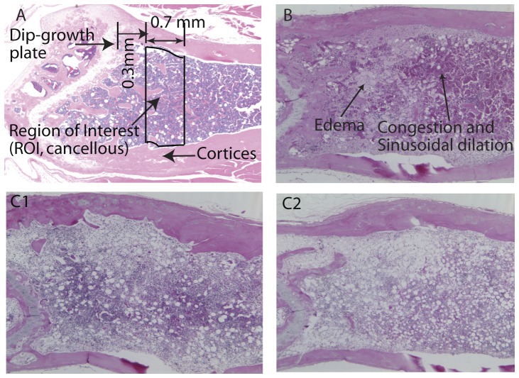
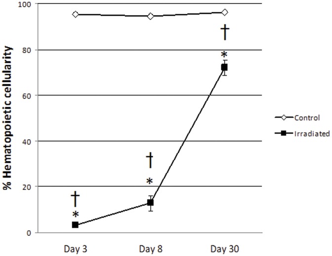
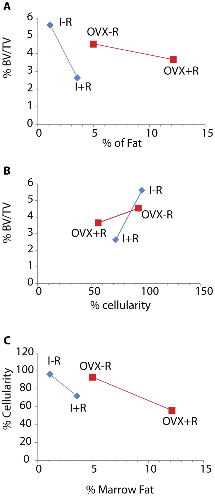
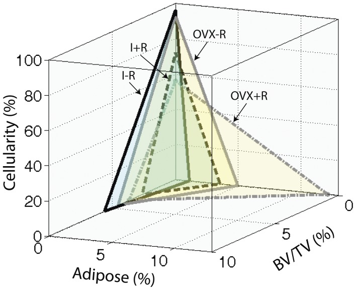
Methodology/principal findings: Ovary-intact and ovariectomized mice underwent either no radiation or single fractional 16 Gy radiation to the caudal skeleton (I ± R, OVX ± R). Marrow fat, hematopoietic cellularity, and cancellous bone volume fraction (BV/TV %) were assessed. Ovariectomy alone did not significantly reduce marrow cellularity in non-irradiated mice (OVX-R vs. I-R, p = 0.8445) after 30 days; however it impaired the hematopoietic recovery of marrow following radiation exposure (OVX+R vs. I+R, p = 0.0092). The combination of radiation and OVX dramatically increases marrow fat compared to either factor alone (p = 0.0062). The synergistic effect was also apparent in the reduction of hematopoietic marrow cellularity (p = 0.0661); however it was absent in BV/TV% changes (p = 0.2520). The expected inverse relationship between marrow adiposity vs. hematopoietic cellularity and bone volume was observed. Interestingly compared with OVX mice, intact mice demonstrated double the reduction in hematopoietic cellularity and a tenfold greater degree of bone loss for a given unit of expansion in marrow fat.

Conclusions/significance: Ovariectomy prior to delivery of a clinically-relevant focal radiation exposure in mice, exacerbated post-radiation adipose accumulation in the marrow space but blunted bone loss and hematopoietic suppression. In the normally coupled homeostatic relationship between the bone and marrow domains, OVX appears to alter feedback mechanisms. Confirmation of this non-linear phenomenon (presumably due to differential radiosensitivity) and demonstration of the mechanism of action is needed to provide strategies to diminish the effect of radiation on exposed tissues.


Conflict of interest statement

Competing Interests: The authors have declared that no competing interests exist.

Figures

Figure 1
Figure 1. Experimental Plan Schematic.
Sixteen week old BALB/c mice were ovariectomized (OVX) and maintained in the vivarium for 57 days in order to attain skeletal hemostasis. Both intact (I) and OVX mice were then irradiated with 16Gy delivered to the caudal skeleton or maintained as controls. Groups of animals were euthanized at 3, 8, and 30 days post irradiation in order to perform histological evaluations of the distal femur; microCT measurements were conducted at the 30 day time point only.
Figure 2
Figure 2. Schematic representation of bone and marrow.
A. region of interest in sagittal view of a normal control mouse. B. Sagittal section of bone marrow from an intact mouse 8 days post-irradiation. Note the areas of congestion and sinusoidal dilation characterized by increased density of red blood cell within the expanded vascular spaces; also present are areas of edema characterized by increased volume of pale pink fluid in the interstitial spaces. C. Sagittal sections of bone marrow 30 days after irradiation in intact (C1) and OVX (C2) mice. Note the more intensely purple areas indicative of higher hematopoietic activity in C1 compared with C2, as well as expanded adipose in C2 compared with C1.
Figure 3
Figure 3. Percent of the marrow space (mean ±SEM) occupied by hematopoietic precursors in intact control (I−R) and irradiated (I+R) mice estimated by visual inspection by a board certified veterinary clinical pathologist (LS) employing standard protocols.
Radiation caused marked hypocellularity of the hematopoietic components of the marrow by Day 3, with significant increases but still very low hematopoietic activity on Day 8 and marked but still incomplete recovery of hematopoietic activity with normal precursor maturation at Day 30. There were no changes over time in non-irradiated animals. Two-way ANOVA with Tukey's post-tests: * Irradiated mice had significantly lower cellularity than control mice of the same day, p<0.0001, † Cellularity of irradiated mice increased significantly from Day 3 through Day 30, p: 0.0292 – <0.0001.
Figure 4
Figure 4. Ovariectomy influences the relationships between bone volume (BV/TV%), adipogenesis (% marrow fat), and hematopoiesis (% cellularity) at Day 30 with or without 16 Gy radiation to the hind limb in mice.
Data of every two measurements are represented as least squares mean values for the group. Panel A: OVX results in a 10 fold reduction in the rate of bone volume loss per unit increase in marrow fat after radiation treatment compared with intact mice. Panel B: OVX blunts the rate of bone volume loss relative to reduced hematopoietic cellularity after radiation compared with intact mice by a factor of 6. Panel C: OVX halves the reduction in hematopoietic cellularity compared with increased marrow fat after radiation treatment compared with intact mice.
Figure 5
Figure 5. 3-D illustration of interrelationships among the three tissue components of bone and marrow: hemopoietic component measured by cellularity, stromal damage component measured by marrow fat or adipose content, and osseous component measured by the cancellous bone BV/TV%.
Cumulative increases in marrow fat after irradiation, in the absence of ovarian function (10 fold) was not reflected by equivalent losses of either cancellous bone or hematopoietic cellularity. The proportionality of changes in these tissue components were maintained among irradiated intact mice.

References

    1. Hui Susanta K, Khalil Ali, Zhang Yan, Coghill Kathleen, Le Chap, et al. (2010) Longitudinal assessment of bone loss from diagnostic CT scans in gynecologic cancer patients treated with chemotherapy and radiation. American Journal of Obstetrics & Gynecology 203: 353.e351–353.e357. - PMC - PubMed
    1. Chen Z, Maricic M, Bassford T, Pettinger M, Ritenbaugh C, et al. (2005) Fracture risk among breast cancer survivors: Results from the Women's Health Initiative Observational Study. Archives of Internal Medicine 165: 552. - PubMed
    1. Baxter N, Habermann E, Tepper J, Durham S, Virnig B (2005) Risk of pelvic fractures in older women following pelvic irradiation. JAMA 294: 2587–2593. - PubMed
    1. Hui SK, Fairchild GR, Kidder LS, Sharma M, Bhattacharya M, et al. (2012) Skeletal Remodeling Following Clinically Relevant Radiation-Induced Bone Damage Treated with Zoledronic Acid. Calcified Tissue International 90: 40–49. - PubMed
    1. Despars G, St-Pierre Y (2011) Bidirectional interactions between bone metabolism and hematopoiesis. Experimental hematology 39: 809–816. - PubMed

Publication types

MeSH terms