. 2012;7(8):e42692.
doi: 10.1371/journal.pone.0042692. Epub 2012 Aug 6.

The "CPC clip motif": a conserved structural signature for heparin-binding proteins

Affiliations

The "CPC clip motif": a conserved structural signature for heparin-binding proteins

Marc Torrent et al. PLoS One. 2012.

Abstract

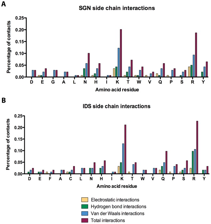
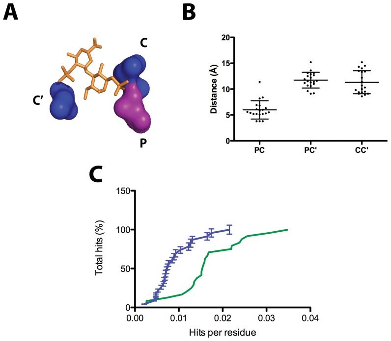
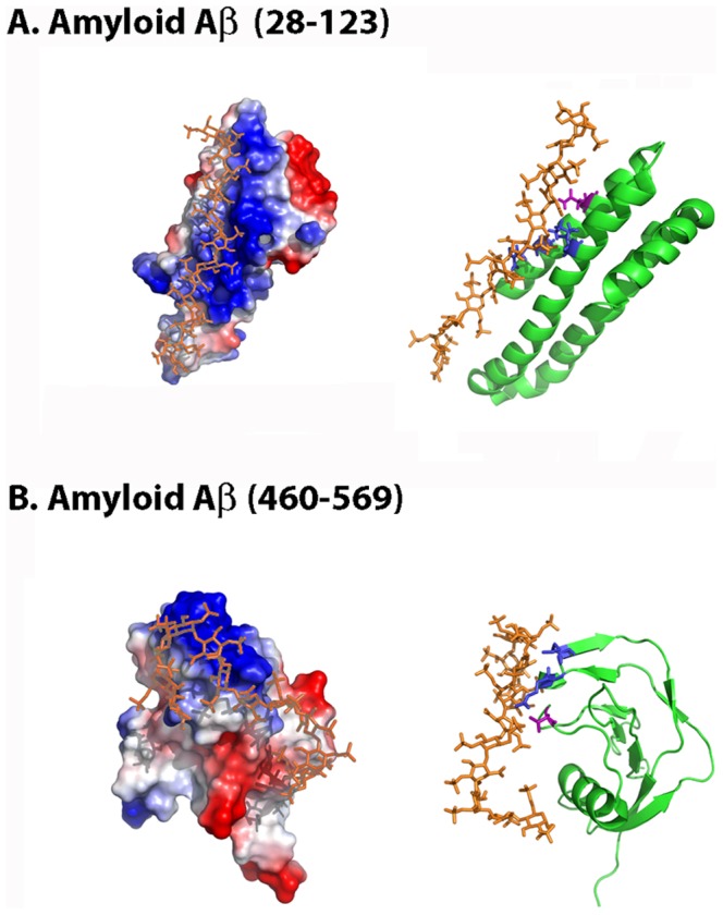
Glycosaminoglycans (GAGs) are essential molecules that regulate diverse biological processes including cell adhesion, differentiation, signaling and growth, by interaction with a wide variety of proteins. However, despite the efforts committed to understand the molecular nature of the interactions in protein-GAG complexes, the answer to this question remains elusive.In the present study the interphases of 20 heparin-binding proteins have been analyzed searching for a conserved structural pattern. We have found that a structural motif encompassing one polar and two cationic residues (which has been named the CPC clip motif) is conserved among all the proteins deposited in the PDB. The distances between the α carbons and the side chain center of gravity of the residues composing this motif are also conserved. Furthermore, this pattern can be found in other proteins suggested to bind heparin for which no structural information is available. Hence we propose that the CPC clip motif, working like a staple, is a primary contributor to the attachment of heparin and other sulfated GAGs to heparin-binding proteins.


Conflict of interest statement

Competing Interests: The authors have declared that no competing interests exist.

Figures

Figure 1
Figure 1. Summary of side-chain amino acid interactions for protein-heparin complexes deposited in the PDB.
Molecular contacts were inspected for (A) SGN and (B) IDS, the two major components of heparin. The fraction of contacts is represented for each amino acid.
Figure 2
Figure 2. Molecular representation of the CPC clip motif for the 20 reference protein-heparin complexes.
For each complex, the ligand is colored in orange, the amino acids belonging to the CPC clip motif in green and suggested polar interactions are depicted as blue dashed lines. Images were generated with Pymol.
Figure 3
Figure 3. Statistical analysis of the Cα and side chain center of gravity distances between the amino acids conforming the CPC clip motif.
(A) Schematic representation of the CPC clip motif, composed of one polar (P) and two cationic residues (namely C and C′, being C the closest to the polar residue). Image was generated with Pymol. (B) Measured PC, PC′ and CC′ distances for the 20 reference proteins described. (C) Enrichment of CPC clip motif in heparin-binding proteins. The negative and positive testing databases were analyzed by SPASM and a cumulative frequency histogram plotting the number of hits per residue is depicted. The positive testing dataset is colored in green and the negative dataset in blue. Each point in the negative dataset represents the average of five independent tests and errors bars are depicted. See the Materials and Methods section for further information.
Figure 4
Figure 4. Molecular docking simulation of lymphotactin and fractalkine heparin-binding sites.
The figure displays the protein electrostatic potential (left) and the protein cartoon highlighting in red the CPC clip motif (right) of lymphotactin Ltn10 (A) and Ltn40 (B) and CDF fractalkine domain (C). CPC residues are colored in blue (cationic) and magenta (polar). Heparin dodecasaccharide ligand used in docking simulations is colored in orange. PDB codes: 1J8I (Ltn10), 2JP1 (Ltn40), 1B2T (fractalkine) and 1HPN (heparin ligand).
Figure 5
Figure 5. Molecular docking simulation of Aβ28–123 and Aβ460–569 heparin-binding sites.
The figure displays the protein electrostatic potential (left) and the protein cartoon highlighting in red the CPC clip motif (right) of (A) Aβ28–123 and (B) Aβ460–569. CPC residues are colored in blue (cationic) and magenta (polar). Heparin dodecasaccharide ligand used in docking simulations is colored in orange. PDB codes: 1MWR (Aβ28–123), 1TKN (Aβ460–569) and 1HPN (heparin ligand).

References

    1. Gandhi NS, Mancera RL (2008) The structure of glycosaminoglycans and their interactions with proteins. Chem Biol Drug Des 72: 455–482. - PubMed
    1. Peplow PV (2005) Glycosaminoglycan: a candidate to stimulate the repair of chronic wounds. Thromb Haemost 94: 4–16. - PubMed
    1. Casu B, Guerrini M, Torri G (2004) Structural and conformational aspects of the anticoagulant and anti-thrombotic activity of heparin and dermatan sulfate. Curr Pharm Des 10: 939–949. - PubMed
    1. Kovensky J (2009) Sulfated oligosaccharides: new targets for drug development? Curr Med Chem 16: 2338–2344. - PubMed
    1. Liu D, Shriver Z, Qi Y, Venkataraman G, Sasisekharan R (2002) Dynamic regulation of tumor growth and metastasis by heparan sulfate glycosaminoglycans. Semin Thromb Hemost 28: 67–78. - PubMed

Publication types

Associated data