. 2013 Mar 1;18(7):784-94.
doi: 10.1089/ars.2011.4440. Epub 2012 Sep 24.

Naturally occurring variation in the Glutathione-S-Transferase 4 gene determines neurodegeneration after traumatic brain injury

Affiliations

Naturally occurring variation in the Glutathione-S-Transferase 4 gene determines neurodegeneration after traumatic brain injury

Faiez Al Nimer et al. Antioxid Redox Signal. .

Abstract

Aim: Genetic factors are important for outcome after traumatic brain injury (TBI), although exact knowledge of relevant genes/pathways is still lacking. We here used an unbiased approach to define differentially activated pathways between the inbred DA and PVG rat strains. The results prompted us to study further if a naturally occurring genetic variation in glutathione-S-transferase alpha 4 (Gsta4) affects the outcome after TBI.

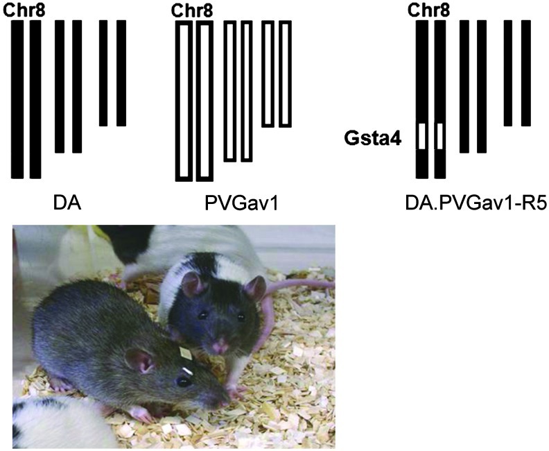
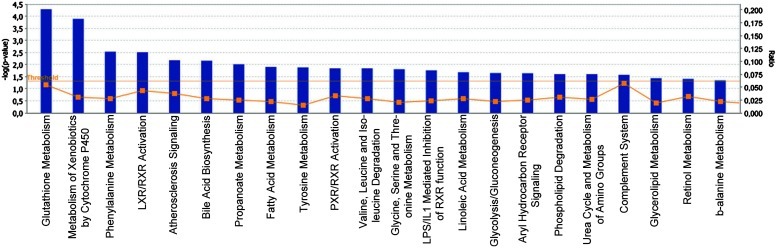
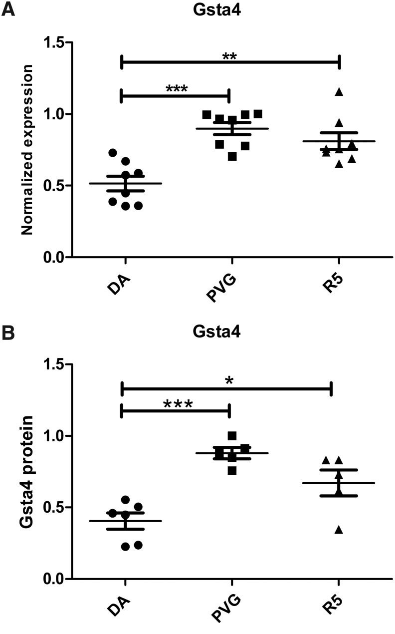
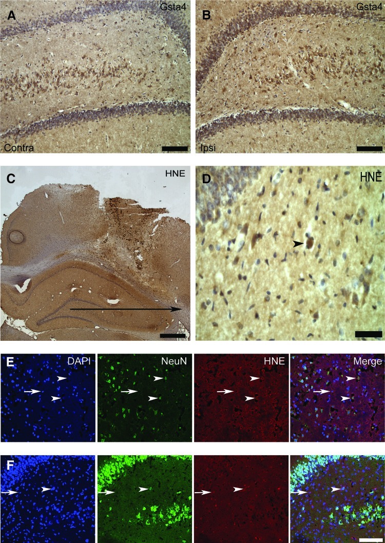
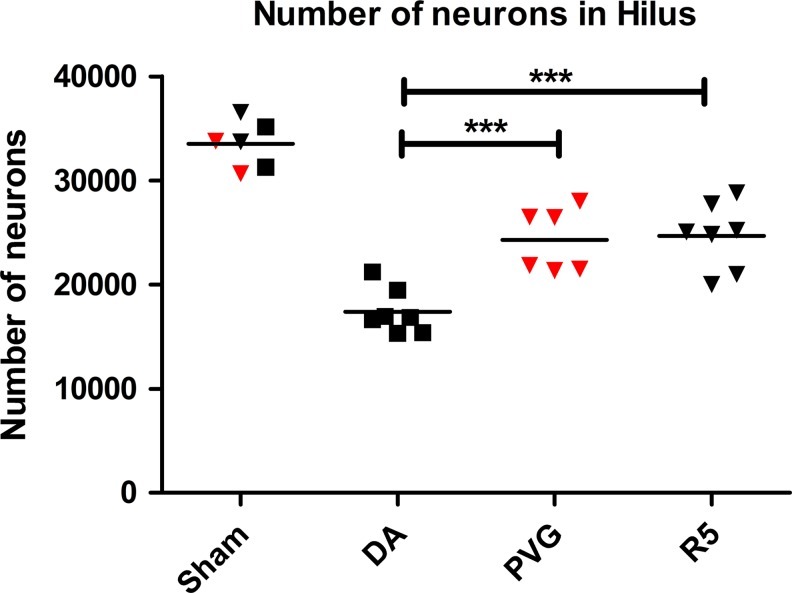
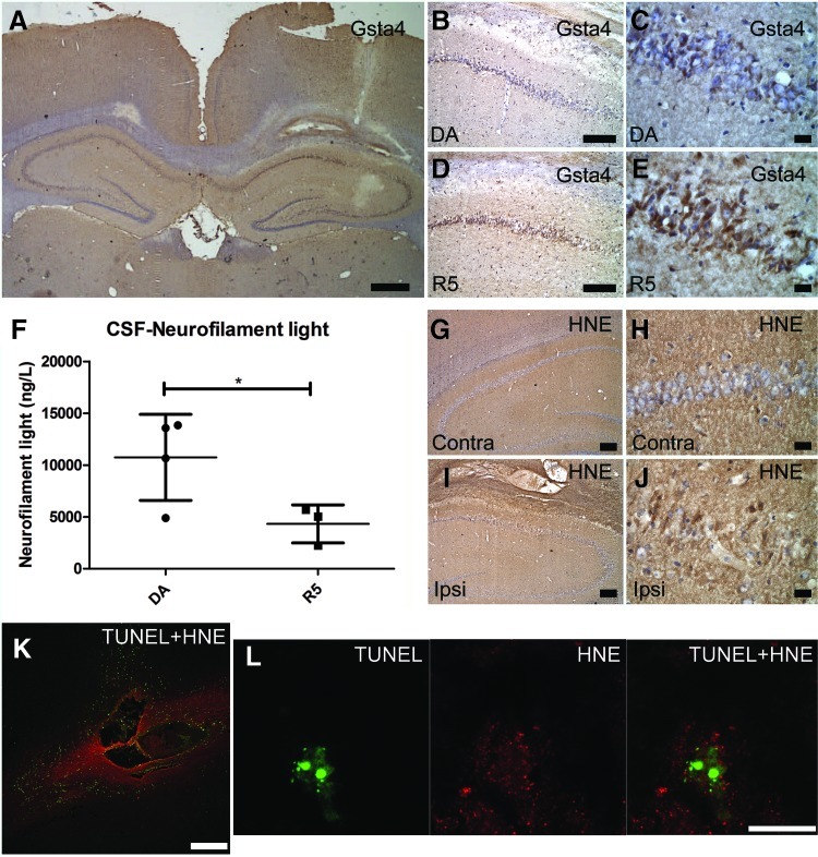
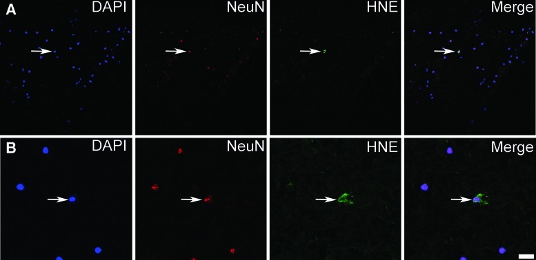
Results: Survival of neurons after experimental TBI is increased in PVG compared to the DA strain. Global expression profiling analysis shows the glutathione metabolism pathway to be the most regulated between the strains, with increased Gsta4 in PVG among top regulated transcripts. A congenic strain (R5) with a PVG genomic insert containing the Gsta4 gene on DA background displays a reversal of the strain pattern for Gsta4 expression and increased survival of neurons compared to DA. Gsta4 is known to effectively reduce 4-hydroxynonenal (4-HNE), a noxious by-product of lipid peroxidation. Immunostaining of 4-HNE was evident in both rat and human TBI. Intracerebral injection of 4-HNE resulted in neurodegeneration with increased levels of a marker for nerve injury in cerebrospinal fluid of DA compared to R5.

Innovation: These findings provide strong support for the notion that the inherent capability of coping with increased 4-HNE after TBI affects outcome in terms of nerve cell loss.

Conclusion: A naturally occurring variation in Gsta4 expression in rats affects neurodegeneration after TBI. Further studies are needed to explore if genetic variability in Gsta4 can be associated to outcome also in human TBI.


Figures

FIG. 1.
FIG. 1.
Experimental set up used to dissect the effect of genetic variability in the Gsta4 gene. Experimental traumatic brain injury was performed in the two inbred rat strains DA and PVGav1 and their genome is shown in black or white color, respectively. The genome of the R5 congenic rat has all genes from the DA strain illustrated in black color except for a fragment with 35 genes, including Gsta4 (illustrated as a small white segment at chromosome 8) from the PVGav1 strain. (To see this illustration in color, the reader is referred to the web version of this article at www.liebertpub.com/ars.)
FIG. 2.
FIG. 2.
Differentially expressed genes between DA and PVGav1 after TBI. Partek Express (Partek, St Louis, MO) was used to carry out statistical analyses of microarray data files. A t-test and a fold change value were used to examine the significance and degree of the differences between DA and PVGav1 for every expressed target. Transcripts with a p<0.005 and a ≥1.4-fold change were uploaded for analysis with the Ingenuity Pathway Analysis software in order to identify top canonical pathways affected by strain. IPA identified the canonical pathways that were most significant in the data set in 2 different ways: 1) By using Fisher's exact test to calculate the probability (p value) that the association between the genes in the data set and the canonical pathway is explained by chance alone; 2) By calculating the ratio of the number of genes from the data set that map to the pathway divided by the total number of genes that map to the canonical pathway. The bars represent −log p value and the percentage of genes present in the data set compared to the total number of genes present in each selected pathway in the IPA data base. The yellow line represents the ratio of affected genes to the total number of genes in a pathway. Canonical pathways above the threshold (horizontal line) are shown. (To see this illustration in color, the reader is referred to the web version of this article at www.liebertpub.com/ars.)
FIG. 3.
FIG. 3.
Genetic differences in Gsta4 expression after TBI. Gsta4 transcriptional levels were analyzed by quantitative PCR in the pericontusional area one day after TBI. The DA strain had lower expression of Gsta4 compared to the PVGav1 strain. The congenic R5 strain with the DA genomic background but with the Gsta4 haplotype from PVGav1 had similar levels to the PVGav1 (A). The protein level of Gsta4 was measured with Western blot using beta-actin as reference protein. Data show that the difference in expression is reflected also at the protein level. (B). Statistical analyses were made using one-way ANOVA with a Bonferroni post-hoc test (*p<0.05, **p<0.01, ***p<0.0001).
FIG. 4.
FIG. 4.
Gsta4 and 4-HNE-protein adduct immunolocalization. (A, B) Immunohistochemical labeling for Gsta4 in the hilus of a R5 rat at one day after traumatic brain injury on the contralateral and ipsilateral hilus, dentate gyrus, and part of CA3. (C, D) Immunohistochemical labeling of 4-HNE-protein adducts in the lesion and the surrounding cortex and hippocampus of a DA rat at one day after TBI with (D) showing a picture of hilus in higher magnification. (E, F) Confocal micrograph in the same DA rat of fluorescent triple immunolabeling with DAPI, the neuronal marker NeuN, and 4-HNE-protein adducts in the cortex (E) and hilus (F). Neuronal cells both at the contralateral (A) and ipsilateral (B) site of the injury express Gsta4 with a stronger labeling on the ipsilateral site. In addition, a strong labeling for 4-HNE-protein adducts is present in the blood derived infiltrating cells in the injury, as well as a diffuse labeling in the surrounding cortex and the hippocampus (C). Positive staining of a neuron in the hilus is indicated (D, arrow head). Cytoplasmic staining for 4-HNE-protein adducts is present in both neurons (arrow heads) and glia (arrows) in the cortex (E) and the hilus (F). Scale bars are 100 μm in A, B, E and F; 1 cm in C, and 15 μm in D. (To see this illustration in color, the reader is referred to the web version of this article at www.liebertpub.com/ars.)
FIG. 5.
FIG. 5.
Surviving neuronal cells in the hilus 30 days after TBI. Design-based stereology was used to count the neurons in the hilus that had survived TBI 30 days after the injury. The estimated total number of neurons is shown for the DA (black boxes), PVGav1 (red triangles), and R5 (black triangles) rat. Sham operated rats were used as controls. Comparison between the groups were evaluated by one-way ANOVA with a Bonferroni post-hoc test (***p<0.001). (To see this illustration in color, the reader is referred to the web version of this article at www.liebertpub.com/ars.)
FIG. 6.
FIG. 6.
Gsta4 expression, 4-HNE detoxification, and apoptotic cell death following intracerebral 4-HNE injection. (A–E) Gsta4 immunohistochemical staining, 24 h after intraparenchymal injection of 4-HNE showing expression of Gsta4 in the site ipsilateral to the injection versus the contralateral site and differences between the DA and R5 strain particularly in the CA1 area. (F) CSF levels of neurofilament light as assessed with ELISA, and (G–J) imunohistochemical detection of 4-HNE-protein adducts in the ipsilateral versus the contralateral site. (K, L) Double immunofluorescent labeling for apoptotic cell death as assessed with TUNEL (green) and 4-HNE-protein adducts (red). Note the upregulation of Gsta4 around the injection site (A) and that neurons of the CA1 area display more Gsta4 staining for the R5 strain compared to the DA (B–E). Also, note in (F) that CSF neurofilament light levels are elevated in the DA strain compared to the R5. Furthermore, in (G–I) note that 4-HNE has diffused in the tissue surrounding the injection to form adducts which are present both in glia and neurons. Finally, (K) shows that injection of 4-HNE results in apoptotic cell death around the injection site and (L) shows a neuron of the CA1 area with an apoptotic fragmented nucleus and the presence of 4-HNE-Michael adducts in the cytoplasm. Scale bars are 1 mm in (A); 250 μm in (K), 100 μm in (B), (D), (G), and (I); 15 μm in (C), (E), (H), (J), and (L). Comparison between the two groups were evaluated by the Student's t test (*p<0.05). (To see this illustration in color, the reader is referred to the web version of this article at www.liebertpub.com/ars.)
FIG. 7.
FIG. 7.
Presence of 4-HNE-protein adducts in neurons in human TBI. (A, B) Triple immunolabeling of human pericontusional tissue, 15 h after trauma, with DAPI, the neuronal marker NeuN, and 4-HNE-protein adducts. Note the presence of 4-HNE-protein adducts in the cytoplasm of neurons in human pericontusional tissue. Two areas (A, B) in the tissue are shown with the second (B) in higher magnification. Scale bars are 50 μm in (A) and 12.5 μm in (B). (To see this illustration in color, the reader is referred to the web version of this article at www.liebertpub.com/ars.)

References

    1. Abel EL. Angel JM. Riggs PK. Langfield L. Lo HH. Person MD. Awasthi YC. Wang LE. Strom SS. Wei Q. DiGiovanni J. Evidence that Gsta4 modifies susceptibility to skin tumor development in mice and humans. J Natl Cancer Inst. 2010;102:1663–1675. - PMC - PubMed
    1. Al Nimer F. Beyeen AD. Lindblom R. Strom M. Aeinehband S. Lidman O. Piehl F. Both MHC and non-MHC genes regulate inflammation and T-cell response after traumatic brain injury. Brain Behav Immun. 2011;25:981–990. - PubMed
    1. Arakawa M. Ishimura A. Arai Y. Kawabe K. Suzuki S. Ishige K. Ito Y. N-Acetylcysteine and ebselen but not nifedipine protected cerebellar granule neurons against 4-hydroxynonenal-induced neuronal death. Neurosci Res. 2007;57:220–229. - PubMed
    1. Balogh LM. Atkins WM. Interactions of glutathione transferases with 4-hydroxynonenal. Drug Metab Rev. 2011;43:165–178. - PMC - PubMed
    1. Beyeen AD. Adzemovic MZ. Ockinger J. Stridh P. Becanovic K. Laaksonen H. Lassmann H. Harris RA. Hillert J. Alfredsson L. Celius EG. Harbo HF. Kockum I. Jagodic M. Olsson T. IL-22RA2 associates with multiple sclerosis and macrophage effector mechanisms in experimental neuroinflammation. J Immunol. 2010;185:6883–6890. - PubMed

Publication types