. 2012 Oct;67(10):1215-22.
doi: 10.1111/j.1398-9995.2012.02875.x.. Epub 2012 Aug 1.

Mesenchymal stem cells derived from human induced pluripotent stem cells modulate T-cell phenotypes in allergic rhinitis

Affiliations
Free PMC article

Mesenchymal stem cells derived from human induced pluripotent stem cells modulate T-cell phenotypes in allergic rhinitis

Q L Fu et al. Allergy. 2012 Oct.
Free PMC article

Abstract

Background: Human induced pluripotent stem cells (iPSCs) possess remarkable self-renewal capacity and the potential to differentiate into novel cell types, such as mesenchymal stem cells (MSCs). iPSC-MSCs have been shown to enhance tissue regeneration and attenuate tissue ischaemia; however, their contribution to the immune regulation of Th2-skewed allergic rhinitis (AR) and asthma remains unclear.

Objective: This study compared the immunomodulatory effects of iPSC-MSCs and bone marrow-derived MSCs (BM-MSCs) on lymphocyte proliferation, T-cell phenotypes and cytokine production in peripheral blood mononuclear cells (PBMCs) in patients with AR, and investigated the possible molecular mechanisms underlying the immunomodulatory properties of iPSC-MSCs.

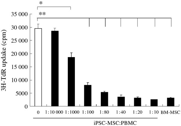
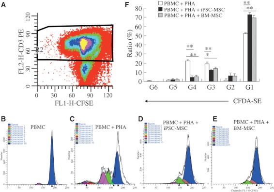
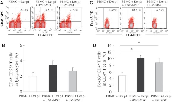
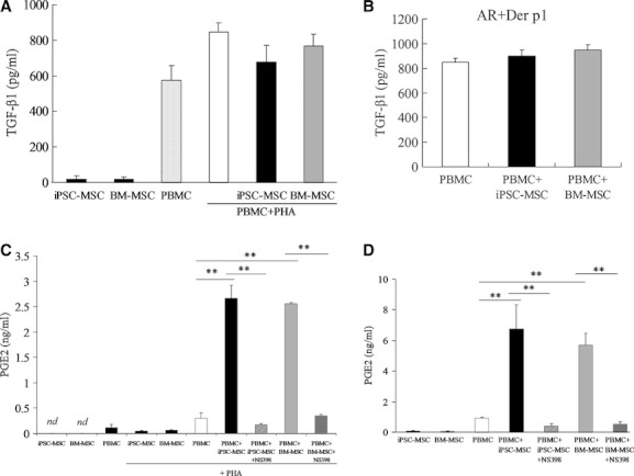
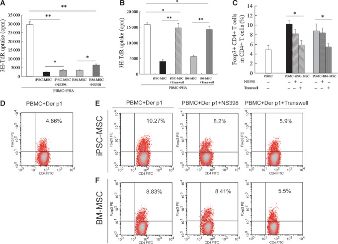
Methods: In co-cultures of PBMCs with iPSC-MSCs or BM-MSCs, lymphocyte proliferation was evaluated using 3H-thymidine (3H-TdR) uptake, carboxyfluorescein diacetate, succinimidyl ester (CFDA-SE) assays; the regulatory T-cell (Treg) phenotype was determined by flow cytometry, and cytokine levels were measured using an enzyme-linked immunosorbent assay. The immunomodulatory properties of both MSCs were further evaluated using NS398 and transwell experiments.

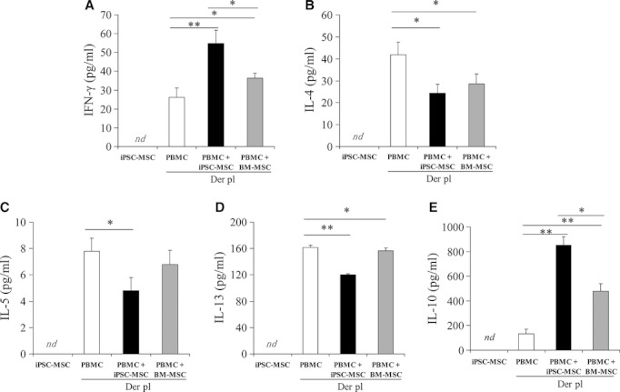
Results: Similar to BM-MSCs, we determined that iPSC-MSCs significantly inhibit lymphocyte proliferation and promote Treg response in PBMCs (P < 0.05). Accordingly, the cytokine milieu (IFN-γ, IL-4, IL-5, IL-10 and IL-13) in the supernatants of PBMCs changed significantly (P < 0.05). The immunomodulatory properties of iPSC-MSCs and BM-MSCs were associated with prostaglandin E2 (PGE2) production and cell-cell contact.

Conclusions: These data demonstrate that iPSC-MSCs are capable of modulating T-cell phenotypes towards Th2 suppression through inducing Treg expansion, suggesting that iPSC-MSCs can be used as an alternative candidate to adult MSCs to treat allergic airway diseases.


Figures

Figure 1
Figure 1
The immunosuppressive effect of iPSC-MSCs on PHA-stimulated lymphocyte proliferation. iPSC-MSCs significantly inhibited PHA-stimulated lymphocyte proliferation in a number-dependent manner, as determined by 3H-TdR uptake. BM-MSCs were used as a control. PBMCs from healthy volunteers (1 × 10⁵/well) were stimulated by PHA (5 μg/ml) in a 96-well plate for 3 days in the presence or absence of different numbers of iPSC-MSCs or BM-MSCs (1 × 10⁴/well) (n = 10). The data are expressed as the means ± SEM. *P < 0.05; **P < 0.01.
Figure 2
Figure 2
The immunosuppressive effect of iPSC-MSCs on PHA-stimulated T-cell proliferation. iPSC-MSCs suppressed T-cell proliferation in PHA-stimulated PBMCs, as determined by multicolour flow cytometric analysis. CFDA-SE-labelled PBMCs from healthy volunteers (5 × 10⁵ cells/well) were cultured in a 24-well plate for 3 days, and PBMCs were gated for flow cytometric analysis based on CD3+ staining (A). Representative results of T-cell proliferation without stimulation (B), PHA (5 μg/ml) stimulation (C), PHA stimulation in the presence of iPSC-MSCs (5 × 10⁴/well) (D) or BM-MSCs (E) are shown. Both iPSC-MSCs and BM-MSCs significantly increased the ratio of G1 T cells but decreased the ratio of G3 and G4 T cells in PHA-stimulated PBMCs (n = 10) (F). The data are expressed as the means ± SEM. G (generation) indicates the number of cell divisions. *P < 0.05; **P < 0.01.
Figure 3
Figure 3
Induced pluripotent stem cells-MSCs enhanced the Treg frequency in Der p1-stimulated PBMCs, as determined by flow cytometry. PBMCs from allergic rhinitis (AR) patients (1 × 10⁶ cells/well) were cultured in a 24-well plate following Der p1 (10 μg/ml) stimulation in the presence or absence of iPSC-MSCs or BM-MSCs (5 × 10⁴/well) for 3 days. Representative flow cytometric results of CD4+CD25+ cells (A) and Foxp3+CD4+ cells (C) are shown. Both iPSC-MSCs and BM-MSCs significantly increased the frequency of Foxp3+CD4+ cells (D), but not CD4+CD25+ T cells (B), in the CD4+ subpopulation after Der p1 stimulation (n = 5 for each group). The data are expressed as the means ± SEM. *P < 0.05.
Figure 4
Figure 4
Induced pluripotent stem cells-MSCs-modulated cytokine levels in the supernatants of Der p1-stimulated PBMCs. PBMCs (1 × 10⁶ cells/well) collected from patients with AR were cultured in a 24-well plate following Der p1 (10 μg/ml) stimulation in the presence or absence of iPSC-MSCs or BM-MSCs (5 × 10⁴/well) for 3 days. The levels of IFN-γ, IL-4, IL-5, IL-13 and IL-10 in the supernatants of Der p1-stimulated PBMCs were determined using ELISA. iPSC-MSCs significantly increased the levels of IFN-γ (A) and IL-10 (B) but decreased the levels of IL-4 (C), IL-5 (D) and IL-13 (E) in the supernatants of Der p1-stimulated PBMCs. For each group, n = 11. The data are expressed as the means ± SEM. *P < 0.05; **P < 0.01. nd: no detectable.
Figure 5
Figure 5
Induced pluripotent stem cells-MSCs or BM-MSCs modulated the levels of TGF-β1 and PGE2 in the supernatants of PHA or Der p1-stimulated PBMCs. (A, C) PBMCs from healthy donors (1 × 10⁵ cells/well) were cultured in a 96-well plate in the presence of iPSC-MSCs or BM-MSCs (1 × 10⁴ cells/well) for 3 days under the indicated conditions. (B, D) PBMCs from patients with AR (1 × 10⁶ cells/well) were cultured in a 24-well plate with 10 μg/ml Der p1 in the presence of iPSC-MSCs or BM-MSCs (5 × 10⁴ cells/well) for 3 days. Serum-free medium was used for the TGF-β1 experiments. The levels of TGF-β1 and PGE2 in the supernatants were determined using ELISA. A significant effect of both iPSC-MSCs and BM-MSCs on TGF-β1 level was not observed in the supernatants of PHA- or Der p1-stimulated PBMCs (A, B). However, both iPSC-MSCs and BM-MSCs significantly increased the PGE2 level in the supernatants of PHA- or Derp1-stimulated PBMCs, which was significantly inhibited by NS398 (C, D). For each group, n = 6. The data are expressed as the means ± SEM. **P < 0.01. nd: no detectable.
Figure 6
Figure 6
The immunomodulatory effects of iPSC-MSCs or BM-MSCs on lymphocyte proliferation and T regulatory cells' association with PGE2 production and cell–cell contact suppression. PBMCs from healthy volunteers were stimulated by PHA (5 μg/ml) for 3 days in the presence of iPSC-MSCs or BM-MSCs with NS398 (5 μM) or with transwell separation from MSCs. NS398 (A) and transwell separation (B) significantly reversed the immunomodulatory effect of iPSC-MSCs and BM-MSCs on PHA-stimulated lymphocyte proliferation, as suggested by 3H-TdR uptake (n = 10). PBMCs (1 × 10⁶ cells/well) collected from patients with AR were cultured in a 24-well plate with Der p1 (10 μg/ml) stimulation in the presence of iPSC-MSCs or BM-MSCs (5 × 10⁴ cells/well) with NS398 or without transwell separation from MSCs for 3 days. The transwell experiment significantly reversed the immunomodulatory effects of iPSC-MSC and BM-MSCs on the frequency of Foxp3+CD4+ cells in the CD4+ subpopulation of cultured PBMCs (C). Representative flow cytometric results in groups (D, Der p1-stimulated PBMCs; E, Der p1-stimulated PBMCs plus iPSC-MSCs; F, Der p1-stimulated PBMCs plus BM-MSCs) in PBMCs are shown. For each group, n = 5. The data are expressed as the means ± SEM. *P < 0.05, **P < 0.01. nd: no detectable.

References

    1. Bousquet J, Khaltaev N, Cruz AA, Denburg J, Fokkens WJ, Togias A, et al. Allergic Rhinitis and its Impact on Asthma (ARIA) 2008 update (in collaboration with the World Health Organization, GA(2)LEN and AllerGen) Allergy. 2008;63(Suppl. 86):8–160. - PubMed
    1. Galli SJ, Tsai M, Piliponsky AM. The development of allergic inflammation. Nature. 2008;454:445–454. - PMC - PubMed
    1. Shi HZ, Qin XJ. CD4CD25 regulatory T lymphocytes in allergy and asthma. Allergy. 2005;60:986–995. - PubMed
    1. Kearley J, Barker JE, Robinson DS, Lloyd CM. Resolution of airway inflammation and hyperreactivity after in vivo transfer of CD4+ CD25+ regulatory T cells is interleukin 10 dependent. J Exp Med. 2005;202:1539–1547. - PMC - PubMed
    1. Kearley J, Robinson DS, Lloyd CM. CD4+ CD25+ regulatory T cells reverse established allergic airway inflammation and prevent airway remodeling. J Allergy Clin Immunol. 2008;122:617–624. - PMC - PubMed

Publication types

MeSH terms