

P2X7 receptor regulation of non-classical secretion from immune effector cells
- PMID: 22882764
- PMCID: PMC3473166
- DOI: 10.1111/cmi.12001
P2X7 receptor regulation of non-classical secretion from immune effector cells
Abstract
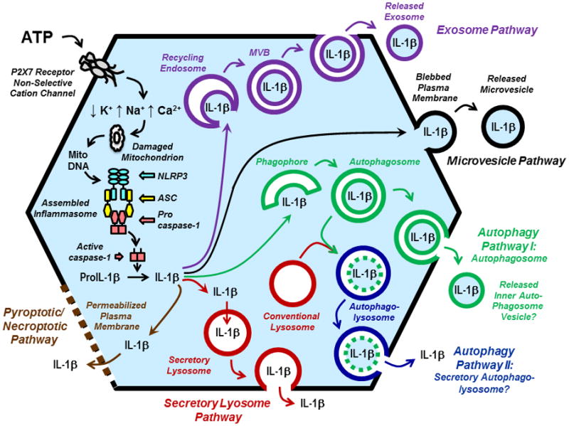
P2X7 receptors (P2X7R) are extracellular ATP-gated ion channels expressed in the immune effector cells that carry out critical protective responses during the early phases of microbial infection or acute tissue trauma. P2X7R-positive cells include monocytes, macrophages, dendritic cells and T cells. Given its presence in all host and pathogen cell types, ATP can be readily released into extracellular compartments at local sites of tissue damage and microbial invasion. Thus, extracellular ATP and its target receptors on host effector cells can be considered as additional elements of the innate immune system. In this regard, stimulation of P2X7R rapidly triggers a key step of the inflammatory response: induction of NLRP3/caspase-1 inflammasome signalling complexes that drive the proteolytic maturation and secretion of the proinflammatory cytokines interleukin-1β (IL-1β) and interleukin-18 (IL-18). IL-1β (and IL-18) lacks a signal sequence for compartmentation within the Golgi and classical secretory vesicles and the proIL-1β precursor accumulates within the cytosol following translation on free ribosomes. Thus, ATP-induced accumulation of the mature IL-1β cytokine within extracellular compartments requires non-classical mechanisms of export from the cytosolic compartment. Five proposed mechanisms include: (i) exocytosis of secretory lysosomes that accumulate cytosolic IL-1β via undefined protein transporters; (ii) release of membrane-delimited microvesicles derived from plasma membrane blebs formed by evaginationsof the surface membrane that entrap cytosolic IL-β; (iii) release of membrane-delimited exosomes secondary to the exocytosis of multivesicular bodies formed by invaginations of recycling endosomes that entrap cytosolic IL-β; (iv) exocytosis of autophagosomes or autophagolysosomes that accumulate cytosolic IL-1β via entrapment during formation of the initial autophagic isolation membrane or omegasome and (v) direct release of cytosolic IL-1β secondary to regulated cell death by pyroptosis or necroptosis. These mechanisms are not mutually exclusive and may represent engagement of parallel or intersecting membrane trafficking responses to P2X7R activation.
© 2012 Blackwell Publishing Ltd.
Figures
References
-
- Andrews NW. Regulated secretion of conventional lysosomes. Trends Cell Biol. 2000;10:316–321. - PubMed
Publication types
MeSH terms
Substances
Grants and funding
LinkOut - more resources
Full Text Sources
Miscellaneous
