Comparative Study
. 2012 Nov;12(11):2920-9.
doi: 10.1111/j.1600-6143.2012.04203.x. Epub 2012 Aug 6.

Intragraft CD11b(+) IDO(+) cells mediate cardiac allograft tolerance by ECDI-fixed donor splenocyte infusions

Affiliations
Comparative Study

Intragraft CD11b(+) IDO(+) cells mediate cardiac allograft tolerance by ECDI-fixed donor splenocyte infusions

G Chen et al. Am J Transplant. 2012 Nov.

Abstract

We have previously shown that pre- and post-transplant infusions of donor splenocytes treated with 1-ethyl-3-(3'-dimethylaminopropyl)-carbodiimide (ECDI-SPs) provide permanent donor-specific protection of islet allografts. The efficacy of donor ECDI-SPs in protecting vascularized cardiac allografts and mechanism(s) of protection are unknown. In this study, we show that infusions of ECDI-SPs significantly prolong cardiac allograft survival concomitant with an impressive accumulation of CD11b(+) IDO(+) cells in the cardiac allograft, and that the presence of this population is dependent on Gr1(+) cells. Consequently, depletion of Gr1(+) cells or inhibition of indoleamine 2,3 dioxygenase (IDO) activity abrogates graft protection by ECDI-SPs infusions. In addition, T cells from ECDI-SPs treated recipients secrete high levels of interleukin 10 and interleukin 13 upon in vitro restimulation, which are also dampened in recipients treated with the IDO inhibitor. Furthermore, combination of donor ECDI-SPs with a short course of rapamycin provides indefinite cardiac allograft survival in 100% of the recipients. These findings reveal a novel mechanism of donor ECDI-SPs in inducing cardiac transplant tolerance and provide several targets that are amenable to therapeutic manipulations for tolerance induction for cardiac transplantation.


Conflict of interest statement

CONFLICT OF INTEREST DISCLOSURE

The authors of this manuscript have no conflicts of interest to disclose as described by the American Journal of Transplantation.

Figures

FIG. 1
FIG. 1. Donor ECDI-SPs significantly prolong cardiac allograft survival, and in combination with rapamycin promote permanent cardiac allograft function
Heterotopic heart transplant was performed using BALB/c as donors and B6 as recipients. SJL mice were used as donor for third party cardiac grafts. A: Timeline of treatment for different experimental groups. SPs: untreated donor splenocytes; ECDI-SPs: ECDI treated donor splenocytes; Rapa: rapamycin. B: Donor ECDI-SPs treatment alone prolongs cardiac allograft survival in a dose-dependent and donor-specific fashion. C: Donor ECDI-SPs treatment in combination with rapamycin allows permanent protection of cardiac allografts.
FIG. 2
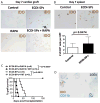
FIG. 2. Graft protection by ECDI-SPs treatment is concomitant with intra-graft IDO-expressing cells
A: Graft histology showing immunohistochemical staining of IDO+ cells. Magnification: x10. Data shown is representative of at least three cardiac grafts obtained and sectioned from each treatment group on post transplant day 7. B: Spleen histology showing immunohistochemical staining of IDO+ cells. Magnification: x20. Data shown is representative of two spleens obtained and sectioned from each treatment group on post transplant day 7. Bar graphs show average cell numbers per high power field counted from 10–12 different sections of two different spleens from each group. C: Effect of the IDO inhibitor 1-MT on cardiac graft protection by ECDI-SPs treatment alone or ECDI-SPs treatment in combination with rapamycin. 1-MT was given at 10 mg/day i.p. from day −7 to day +7 as described in Materials and Methods. D: Dual staining of cardiac grafts with CD11b (blue) and IDO (brown). Magnification: x100. Data shown is from a cardiac graft from the ECDI-SPs alone treatment group, but is representative of 3 cardiac grafts each obtained and sectioned from the ECDI-SPs alone or ECDI-SPs + rapamycin treatment groups.
FIG. 3
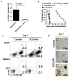
FIG. 3. Depletion of Gr1+ cells diminishes intra-graft IDO-expressing cells and abrogates the graft protection provided by ECDI-SPs treatment
200 μg of anti-Gr1 mAb (anti-mLy-6G, clone RB6-8C5) was given i.p. on day −8, and additionally 100 μg on days −7, −3, −1, and +1. ECDI-SPs were given on day −7 and day +1. Cardiac transplant was performed on day 0. A: Heart grafts were retrieved on day 7, sectioned and stained for IDO as in FIG. 2A. IDO+ cells were enumerated from 8 sections under low power view by 2 different individuals. Results were averaged and presented as the bar graph shown. B: Effect of anti-Gr1 mAb on cardiac graft protection by ECDI-SPs treatment. Graft survival in groups treated with anti-Gr1 mAb alone or ECDI-SPs alone was shown for comparison. C: FACS analysis of cells from cardiac grafts (top panels) or the spleen (lower panels) from control vs. ECDI-SPs treated recipients (day 7 post transplantation). Plots were gated on live cells. Data presented is representative of 4 mice examined from each group in two different experiments. D: Dual staining of day 7 cardiac grafts with Gr1 (dark blue) and IDO (brown). Top panel: control graft; middle and lower panels: ECDI-SPs graft. Magnifications are shown on the micrographs.
FIG. 4
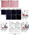
FIG. 4. Protected cardiac grafts show intact histology, diminished graft infiltrating CD8+ cells, but increased graft infiltrating CD4+Foxp3+ cells
A: A representative day 7 and day 90 cardiac graft retrieved from ECDI-SPs + rapamycin treated recipients. Grafts were stained by hematoxylin and eosin. For BD, cardiac grafts from different treatment groups and control were retrieved on the indicated days post transplant, sectioned and stained for CD8 in B, CD4 in C and Foxp3 in D. Bar graphs show average cell numbers per low power field counted by two different individuals from 12 – 18 different sections from 3–4 different cardiac grafts from each group. Magnifications: A and B: x10; D: x40.
FIG. 5
FIG. 5. Cardiac allograft protection is associated with altered anti-donor cellular and humoral responses
For A and B, in vitro re-stimulation by MLRs were set up using BALB/c splenocytes as stimulators and splenic T cells from different treatment groups and control (on day 7 and/or day 90 post transplant) as responders. A: Proliferation was measured by 3H-thymidine uptake. B: Cytokine production after in vitro re-stimulation was measured by multiplex cytokine assay kit as detailed in Materials and Methods. C: Serum total anti-donor IgG was measured on day 14 and day 90 from recipients treated with ECDI-SPs + rapamycin or control untreated recipients. Results shown in A and B are representative of 3 independent experiments. Results shown in C are representative of serum samples tested from 6 mice in each group.
FIG. 6
FIG. 6. Inhibition of IDO abolishes suppression of anti-donor proliferation and antibody responses
For A and B, in vitro re-stimulation by MLRs were set up using BALB/c splenocytes as stimulators and splenic T cells from recipients treated with ECDI-SPs + rapamycin with or without 1-MT treatment (all on day 7 post transplant) as responders. A: Proliferation was measured by 3H-thumidine uptake. B: Cytokine production after in vitro re-stimulation was measured by multiplex cytokine assay kit as detailed in Materials and Methods. C: Serum total anti-donor IgG was measured on day 14 from recipients treated with ECDI-SPs + rapamycin with or without 1-MT treatment, and compared with that from control untreated recipients. Results shown in A and B are representative of 3 independent experiments. Results shown in C are representative of serum samples tested from 5–6 mice in each group.

Similar articles

Cited by

References

    1. Lechler RI, Sykes M, Thomson AW, Turka LA. Organ transplantation--how much of the promise has been realized? Nat Med. 2005 Jun;11(6):605–13. - PubMed
    1. Dugast AS, Haudebourg T, Coulon F, Heslan M, Haspot F, Poirier N, et al. Myeloid-derived suppressor cells accumulate in kidney allograft tolerance and specifically suppress effector T cell expansion. J Immunol. 2008 Jun 15;180(12):7898–906. - PubMed
    1. Garcia MR, Ledgerwood L, Yang Y, Xu J, Lal G, Burrell B, et al. Monocytic suppressive cells mediate cardiovascular transplantation tolerance in mice. J Clin Invest. 2010 Jul 1;120(7):2486–96. - PMC - PubMed
    1. Jia W, Jackson-Cook C, Graf MR. Tumor-infiltrating, myeloid-derived suppressor cells inhibit T cell activity by nitric oxide production in an intracranial rat glioma + vaccination model. J Neuroimmunol. 2010 Jun;223(1–2):20–30. - PMC - PubMed
    1. Obermajer N, Muthuswamy R, Lesnock J, Edwards RP, Kalinski P. Positive feedback between PGE2 and COX2 redirects the differentiation of human dendritic cells toward stable myeloid-derived suppressor cells. Blood. 2011 Nov 17;118(20):5498–505. - PMC - PubMed

Publication types

MeSH terms