. 2012 Aug 8;16(2):250-64.
doi: 10.1016/j.cmet.2012.07.007.

IRE1α induces thioredoxin-interacting protein to activate the NLRP3 inflammasome and promote programmed cell death under irremediable ER stress

Affiliations

IRE1α induces thioredoxin-interacting protein to activate the NLRP3 inflammasome and promote programmed cell death under irremediable ER stress

Alana G Lerner et al. Cell Metab. .

Abstract

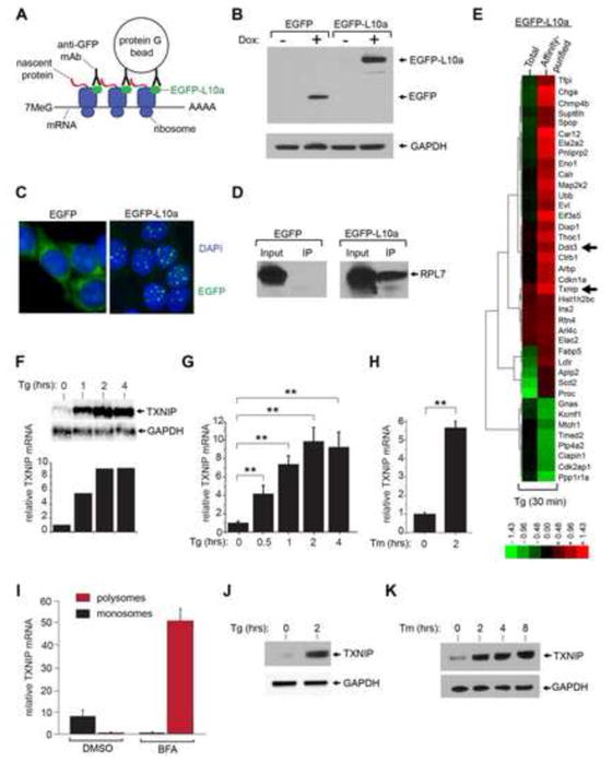
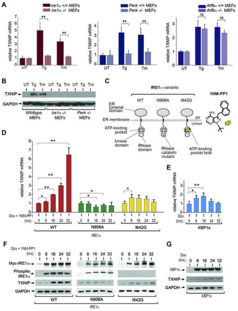
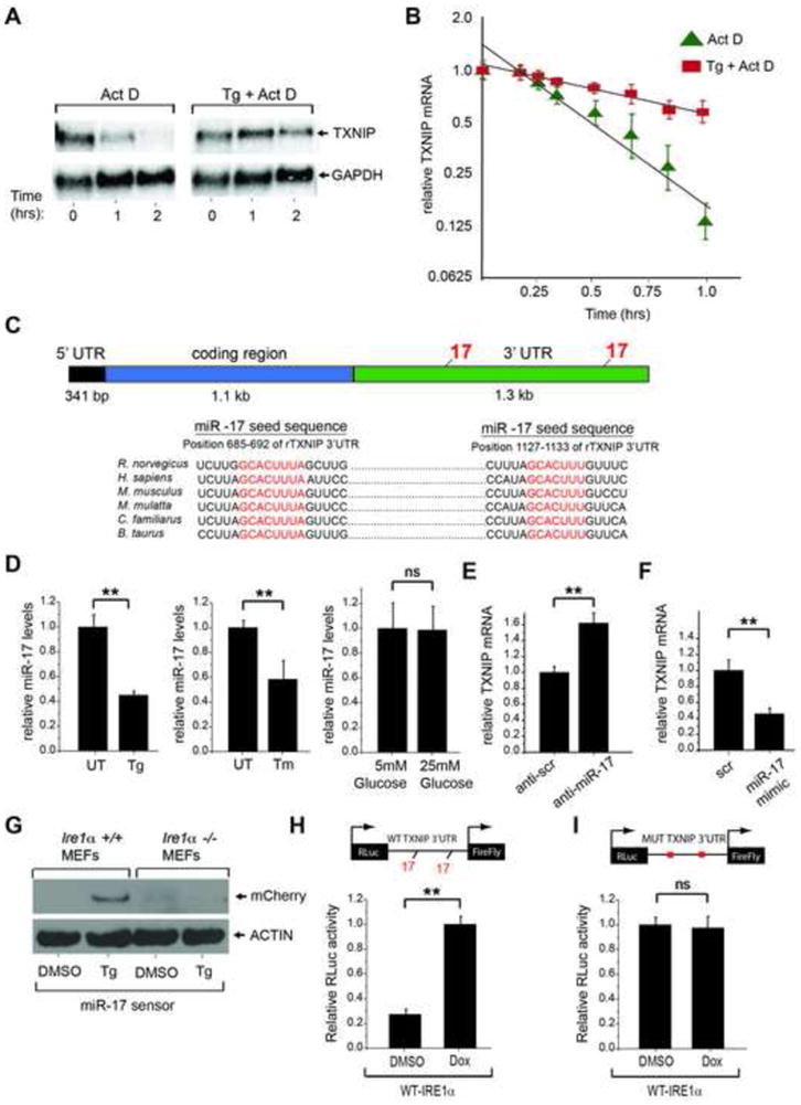
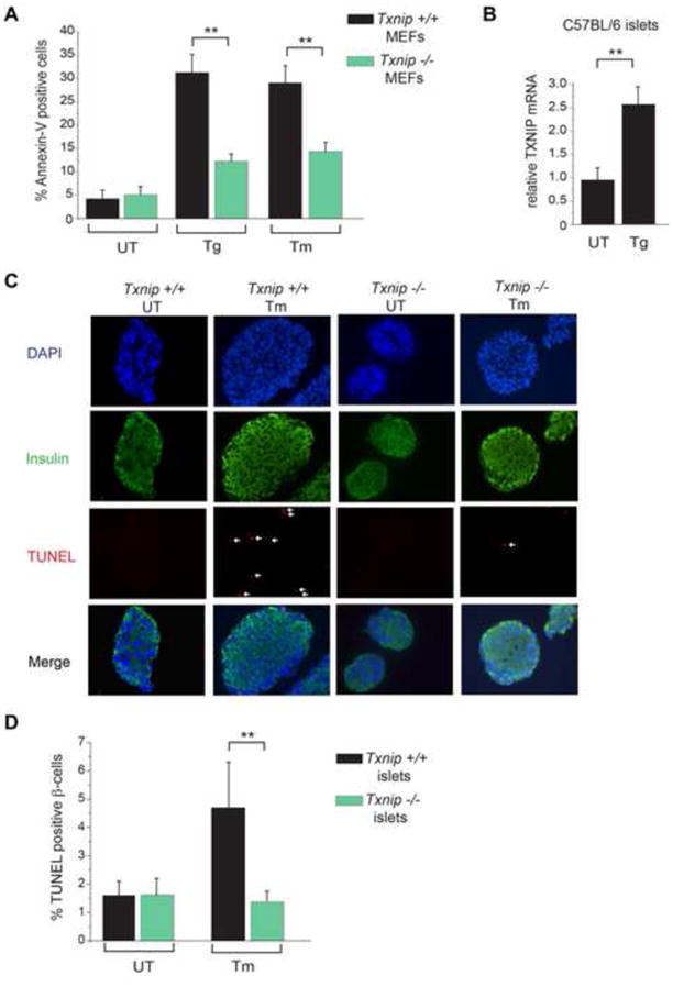
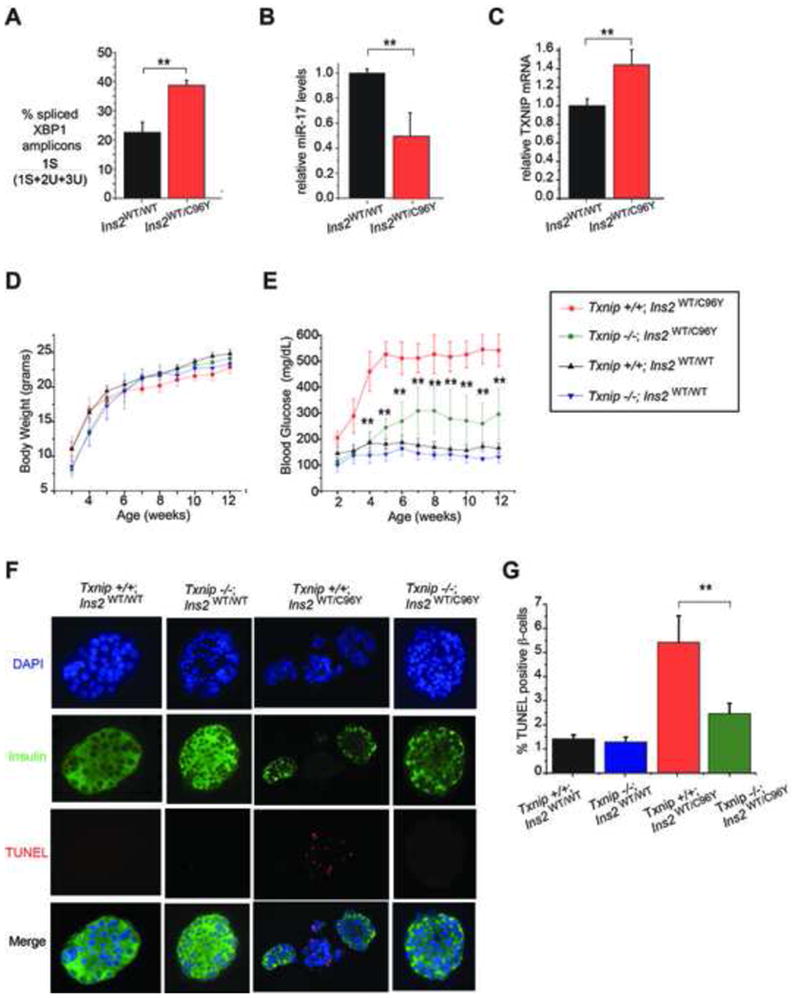
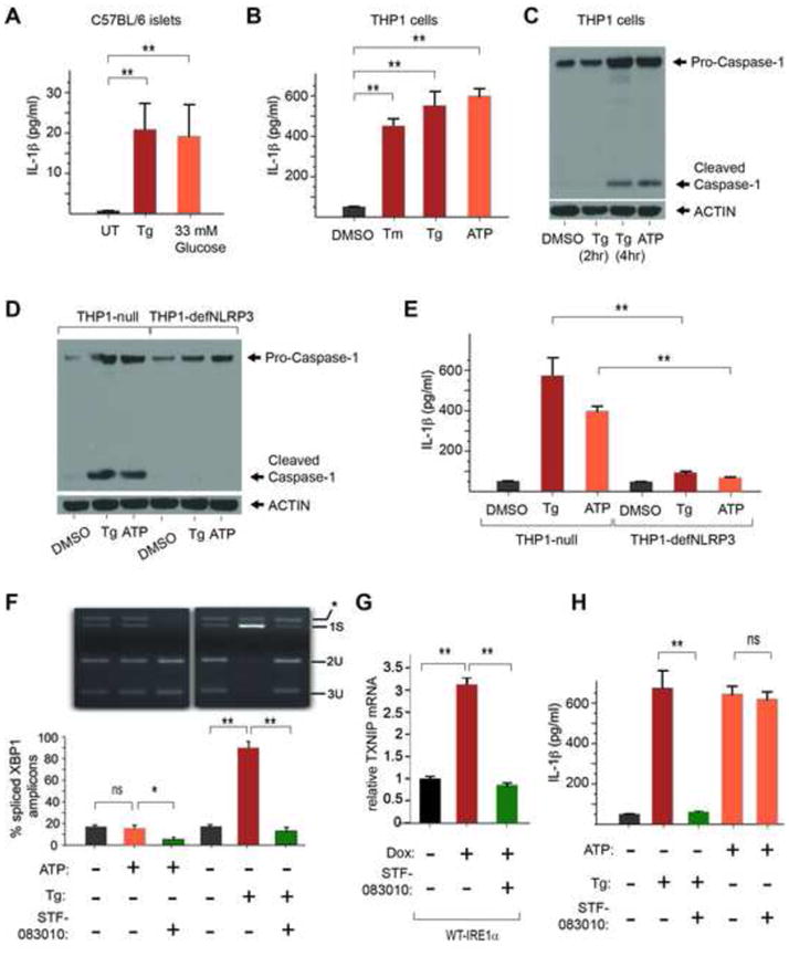
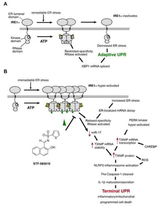
When unfolded proteins accumulate to irremediably high levels within the endoplasmic reticulum (ER), intracellular signaling pathways called the unfolded protein response (UPR) become hyperactivated to cause programmed cell death. We discovered that thioredoxin-interacting protein (TXNIP) is a critical node in this "terminal UPR." TXNIP becomes rapidly induced by IRE1α, an ER bifunctional kinase/endoribonuclease (RNase). Hyperactivated IRE1α increases TXNIP mRNA stability by reducing levels of a TXNIP destabilizing microRNA, miR-17. In turn, elevated TXNIP protein activates the NLRP3 inflammasome, causing procaspase-1 cleavage and interleukin 1β (IL-1β) secretion. Txnip gene deletion reduces pancreatic β cell death during ER stress and suppresses diabetes caused by proinsulin misfolding in the Akita mouse. Finally, small molecule IRE1α RNase inhibitors suppress TXNIP production to block IL-1β secretion. In summary, the IRE1α-TXNIP pathway is used in the terminal UPR to promote sterile inflammation and programmed cell death and may be targeted to develop effective treatments for cell degenerative diseases.


Figures

Figure 1
Figure 1. TXNIP mRNA and protein are rapidly induced in cells undergoing endoplasmic reticulum stress
(A) Schematic of affinity purification of poly-ribosomes using a translational fusion of enhanced green fluorescent protein (EGFP) to the large ribosomal subunit protein L10a (EGFP-L10a). (B) Immunoblot analysis of whole cell extracts from 24 hour untreated and 1μg/ml doxycycline (Dox)- treated insulinoma (INS-1) cell lines expressing EGFP, or a EGFP translational fusion to the large ribosomal subunit protein L10a (EGFP-L10a) under a Dox inducible promoter. (C) Confocal images of INS-1 cells expressing EGFP or EGFP-L10a. Prior to imaging, cells were induced with 1μg/ml Dox for 24 hours, fixed with paraformaldehyde, and stained with 4′,6-diamidino-2-phenylindo (DAPI). (D) Immunoblot analysis of ribosomal protein L7 (RPL7) after anti-EGFP immunoprecipitation (i.p) confirms co-immunoprecipitation of ribosomes in INS-1 cells expressing EGFP-L10a (but not in cells expressing EGFP) following 24 hours treatment with 1μg/ml Dox. (E) Hierarchical clustering analysis of gene expression changes in INS-1 EGFP-L10a-expressing cells (Dox 1μg/ml for 24 hours) under ER stress through the use of DNA microarrays. cDNAs for hybridization were generated from total cellular mRNAs, or from mRNAs collected from anti-EGFP-L10a affinity-purified ribosomes. Indicated genes are those whose expression increased (red) or decreased (green) at least 2 fold under 1 μM thapsigargin (Tg) for 30 minutes (compared to no treatment). See Table S1 for gene identities, log2 expression changes, and statistics. (F & G) Time course analysis of TXNIP mRNA expression (normalized to GAPDH) during ER stress (1 μM Tg) in INS-1 cells by Northern blot, (F), or quantitative real-time PCR (Q-PCR), (G). (H) Analysis of TXNIP mRNA expression (normalized to GAPDH) during ER stress with 5 μg/ml tunicamycin (Tm) or 1 μM Tg in INS-1 cells by Q-PCR. (I) Poly-ribosome profiling demonstrates recruitment of TXNIP mRNA from monosomes into poly-ribosomes under treatment with 2.5 μg/ml brefeldin A (BFA) at 30 minutes. (J) Immunoblot detection of TXNIP protein in INS-1 cells during ER stress (1 μM Tg). (K) Immunoblot detection of TXNIP protein in INS-1 cells during ER stress (5 μg/ml Tm). Three independent biological samples were used for Q-PCR experiments. Data are shown as mean ± SD. **p < 0.005.
Figure 2
Figure 2. Robust induction of TXNIP requires activation of IRE1α’s bifunctional kinase and RNase domains
(A) Analysis of TXNIP mRNA expression (normalized to GAPDH) by Q-PCR during ER stress treatment in UPR sensor signaling mutants. Atf6α −/−, Perk−/−, and Ire1α−/− MEFs (and wild-type counterparts) were treated with 1 μM Tg, or 5 μg/ml Tm, for 6 hours. (B) Immunoblot for TXNIP protein from whole cell lysates of wild-type, Ire1α−/− and Perk−/− MEFs untreated or treated with 1 μM Tg or 5 μg/ml Tm for 3 hrs. (C) Schematic representation of IRE1α variants used in this study, and chemical structure of 1NM-PP1. (D) Time course analysis of TXNIP mRNA expression (normalized to GAPDH) by Q-PCR through ER stress-independent forcible activation of IRE1α and mutants, and forced expression of XBP1s, in INS-1 cells with 1μg/ml Dox and 5μM 1NM-PP1. (E) Time course analysis of TXNIP proteins (by immunoblot) following forced activation of IRE1α and mutants, and forced expression of XBP1s, in INS-1 cells untreated or treated with 1μg/ml Dox and 5μM 1NM-PP1. Three independent biological samples were used for Q-PCR experiments. Data are shown as mean ± SD. **p < 0.005, *p < 0.01.
Figure 3
Figure 3. IRE1α Increases TXNIP mRNA Stability Through Decreasing miR-17
(A and B) Analysis by Northern blotting and Q-PCR shows that TXNIP mRNA is short lived, but becomes stabilized under ER stress. Total RNA extracts from INS-1 cells treated with 5 μg/ml Actinomycin D plus/minus 1 μM Tg were probed for TXNIP mRNA (or GAPDH). B) Early time course (1st hour) Q-PCR of TXNIP mRNA levels (relative to GAPDH) in INS-1 cells treated with 5 μg/ml Actinomycin D plus/minus 1 μM Tg with best fit line. (C) Schematic showing miR-17 binding sites within the 3′-UTR of TXNIP mRNA across multiple species. (D) Q-PCR of miR-17 levels from HEK293 cells untreated or treated with 1 μM Tg, or 5 μg/ml Tm, for 6 hours. (E) TXNIP mRNA levels as analyzed by Q-PCR from HEK293 cells 24 hrs post transfection with scrambled or miR-17 anti-miR. (F) TXNIP mRNA levels as analyzed by Q-PCR from HEK293 cells 24 hrs post transfection with scrambled or miR-17 mimic. (G) Immunoblot analysis of miR-17 mCherry sensor in wild-type and Ire1α−/− MEFs (36 post-transfection) following treatment with DMSO control or 1 μM Tg for 12 hours. (H) IRE1α induction of TXNIP luciferase reporter is dependent on miR-17 binding sites. Dox-inducible WT-IRE1α HEK293 cells were transfected (24 hours) with a luciferase reporter construct containing wild-type or miR-17 binding mutant TXNIP 3′-UTR. The cells were treated with DMSO control or 1μg/ml Dox for 24 hours, lysed and then analyzed for luciferase activity. Three independent biological samples were used for Q-PCR and luciferase experiments. Data are shown as mean ± SD. **p < 0.005, ns = not significant.
Figure 4
Figure 4. Loss of Txnip protects MEFs and pancreatic islets against ER stress-induced apoptosis
(A) Wild-type and Txnip−/− MEFs were challenged with 1 μM Tg or 5 μg/ml Tm for 24hrs and assessed for apoptosis by flow cytometry for Annexin-V binding. (B) Pancreatic islets were isolated from 6 week old C57BL/6 mice and left untreated or treated with 1 μM Tg for 6hrs. TXNIP mRNA (relative to GAPDH) was measured by Q-PCR. (C) Pancreatic islets were isolated from 6 week old Txnip+/+ and Txnip−/− mice, cultured in the absence or presence of 5 μg/ml Tm for 12hrs, and then subjected to DAPI, anti-insulin and TUNEL staining. (D) Quantification of TUNEL positive β-cells from C. Bar graphs represent three independent biological samples. All mice were on C57BL/6 genetic background. Data are shown as mean ± SD. **p < 0.005.
Figure 5
Figure 5. Txnip deficiency protects against β-cell loss and diabetes in the Ins2WT/C96Y mouse
(A–C) Pancreatic islets from 3 week old Ins2WT/C96Y mice show evidence of ER stress at baseline, including increased XBP-1 splicing, decreased miR-17, and elevated TXNIP mRNA as assessed by Q-PCR. (D) Indicated genotypes showed no significant differences in body weight up to 12 weeks of age. For the 12 week timecourse, N=9 for Txnip+/+ Ins2WT/C96Y mice, N=10 for Txnip+/+Ins2WT/WT, and N=8 for both Txnip−/−Ins2WT/WT and Txnip−/−Ins2WT/C96Ymice. (E) Body glucose levels for indicated genotypes up to 12 weeks of age. Note that Txnip−/− Ins2WT/C96Ymice have significantly lower blood glucose levels compared to Txnip+/+ Ins2WT/C96Ymice at all time points. (F) Pancreatic islets were isolated from mice of indicated genotypes at 5 weeks of age and assessed by DAPI, anti-insulin and TUNEL staining. (G) Quantification of TUNEL positive β-cells from experiments in F. Bar graphs represent three independent biological samples. All mice were on C57BL/6 genetic background. Data are shown as mean ± SD. **p < 0.005.
Figure 6
Figure 6. ER stress leads to IRE1 α-dependent TXNIP upregulation, NLRP3 inflammasome activation, Caspase-1 cleavage, and IL-1β secretion
(A) IL-1β secretion from C57BL/6 murine islets exposed to 1μM Tg or 33 mM glucose. (B) IL-1β secretion from human THP-1 cells after 4hr treatment with DMSO control, 10μg/ml Tm, 1μM Tg, or 5 mM ATP as assessed by ELISA. (C) Caspase-1 cleavage from Pro-caspase-1 in THP_1 cells (detected by immunoblot) in response to ER stress 1μM Tg (at 2 hours and 4 hours), or 5 mM ATP at 4 hours. (D) Caspase-1 cleavage in response to ER stress (1μM Tg) is abrogated in THP-1 cells lacking the NLRP3 inflammasome (THP1-defNLRP3); compare to THP1-null positive control cells. Control DAMP, ATP, is at 5 mM. (E) (E) IL-1β secretion in response to ER stress (1μM Tg) is abrogated in THP-1 cells lacking the NLRP3 inflammasome (THP1-defNLRP3); compare to THP1-null positive control cells. Control DAMP ATP is at 5 mM. (F) STF-083010 blocks IRE1α RNase. Shown is an EtBr-stained agarose gel of XBP1 cDNA amplicons after induction of ER stress for 4 hrs in THP-1 cells using 1 μM Tg, with or without pre-treatment with STF-083010 at 50μM for 2 hours. The cDNA amplicon of unspliced XBP1 mRNA is cleaved by a PstI site within a 26 nucleotide intron to give 2U and 3U. IRE1α-mediated cleavage of the intron and re-ligation in vivo removes the PstI site to give the 1S (spliced) amplicon. *is a spliced/unspliced XBP1 hybrid amplicon. The ratio of spliced over (spliced + unspliced) amplicons—1S/(1S+2U+3U)—is reported as % spliced XBP1 amplicons. STF-083010 blocks: TXNIP mRNA upregulation in WT- IRE1α-overexpressing INS-1 cells (G), and IL-1β secretion from THP-1 cells (H). IL-1β secretion in response to 5mM ATP is unaffected by STF-083010. (E–F) Bar graphs represent three independent biological samples. Data are shown as mean ± SD. **p < 0.005, ns = not significant.
Figure 7
Figure 7. Illustrative models of adaptive (A) and terminal (B) UPR signaling
A. Under remediable levels of ER stress, adaptive UPR outputs through XBP1 mRNA splicing reduces ER stress, in turn closing negative feedback loops to shut down low-level IRE1α signaling. B. Alternatively, under irremediable levels of ER stress, hyperactivated IRE1α induces TXNIP as a potentiating step in the Terminal UPR, in part through stabilizing TXNIP mRNA by reducing levels of a repressive miR that targets TXNIP mRNA. This event combines with de novo transcription of TXNIP, through PERK kinase and ChREBP, to result in rapid elevation of TXNIP mRNA to new steady-state levels. TXNIP protein activates the NLRP3 inflammasome, which cleaves Pro-Caspase-1 to its active form, in turn causing maturation and secretion of Interleukin 1-β (IL-1β), thus promoting sterile inflammation and programmed cell death. Moreoever, ER-localized mRNA decay by hyperactivated IRE1α (requiring both a functional kinase and RNase activity) furthers—rather than corrects—ER stress, thus promoting vicious cycles of cell destruction. Also shown is the RNAse inhibitor—STF-083010—which reduces terminal UPR endpoints by inhibiting IRE1α RNAse activity.

Comment in

Similar articles

Cited by

References

    1. Aragon T, van Anken E, Pincus D, Serafimova IM, Korennykh AV, Rubio CA, Walter P. Messenger RNA targeting to endoplasmic reticulum stress signalling sites. Nature. 2009;457:736–740. - PMC - PubMed
    1. Calfon M, Zeng H, Urano F, Till JH, Hubbard SR, Harding HP, Clark SG, Ron D. IRE1 couples endoplasmic reticulum load to secretory capacity by processing the XBP-1 mRNA. Nature. 2002;415:92–96. - PubMed
    1. Cha-Molstad H, Saxena G, Chen J, Shalev A. Glucose-stimulated expression of Txnip is mediated by carbohydrate response element-binding protein, p300, and histone H4 acetylation in pancreatic beta cells. J Biol Chem. 2009;284:16898–16905. - PMC - PubMed
    1. Chen J, Saxena G, Mungrue IN, Lusis AJ, Shalev A. Thioredoxin-interacting protein: a critical link between glucose toxicity and beta-cell apoptosis. Diabetes. 2008;57:938–944. - PMC - PubMed
    1. Credle JJ, Finer-Moore JS, Papa FR, Stroud RM, Walter P. Inaugural Article: On the mechanism of sensing unfolded protein in the endoplasmic reticulum. Proc Natl Acad Sci U S A. 2005;102:18773–18784. - PMC - PubMed

Publication types

MeSH terms

Associated data