

Volume transmission of beta-endorphin via the cerebrospinal fluid; a review
- PMID: 22883598
- PMCID: PMC3439317
- DOI: 10.1186/2045-8118-9-16
Volume transmission of beta-endorphin via the cerebrospinal fluid; a review
Abstract
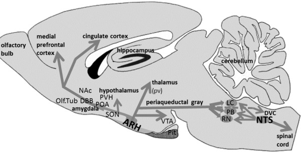
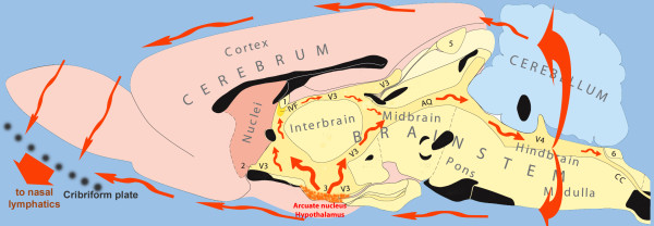
There is increasing evidence that non-synaptic communication by volume transmission in the flowing CSF plays an important role in neural mechanisms, especially for extending the duration of behavioral effects. In the present review, we explore the mechanisms involved in the behavioral and physiological effects of β-endorphin (β-END), especially those involving the cerebrospinal fluid (CSF), as a message transport system to reach distant brain areas. The major source of β-END are the pro-opio-melano-cortin (POMC) neurons, located in the arcuate hypothalamic nucleus (ARH), bordering the 3rd ventricle. In addition, numerous varicose β-END-immunoreactive fibers are situated close to the ventricular surfaces. In the present paper we surveyed the evidence that volume transmission via the CSF can be considered as an option for messages to reach remote brain areas. Some of the points discussed in the present review are: release mechanisms of β-END, independence of peripheral versus central levels, central β-END migration over considerable distances, behavioral effects of β-END depend on location of ventricular administration, and abundance of mu and delta opioid receptors in the periventricular regions of the brain.
Figures
References
-
- Agnati LF, Cortelli P, Biagini G, Bjelke B, Fuxe K. Different classes of volume transmission signals exist in the central nervous system and are affected by metabolic signals, temperature gradients and pressure waves. Neuroreport. 1994;6:9–12. - PubMed
-
- Agnati LF, Guidolin D, Guescini M, Genedani S, Fuxe K. Understanding wiring and volume transmission. Brain Res Rev. 2010;64:137–159. - PubMed
-
- Fuxe K, Dahlstrom AB, Jonsson G, Marcellino D, Guescini M, Dam M, Manger P, Agnati L. The discovery of central monoamine neurons gave volume transmission to the wired brain. Prog Neurobiol. 2010;90:82–100. - PubMed
-
- Veening JG, de Jong T, Barendregt HP. Oxytocin-messages via the cerebrospinal fluid: behavioral effects; a review. Physiol Behav. 2010;101:193–210. - PubMed
-
- Frederickson RC, Geary LE. Endogenous opioid peptides: review of physiological, pharmacological and clinical aspects. Prog Neurobiol. 1982;19:19–69. - PubMed
LinkOut - more resources
Full Text Sources
Other Literature Sources
Research Materials
Miscellaneous
