

Removal of adhesive wound dressing and its effects on the stratum corneum of the skin: comparison of eight different adhesive wound dressings
- PMID: 22883604
- PMCID: PMC7950515
- DOI: 10.1111/j.1742-481X.2012.01061.x
Removal of adhesive wound dressing and its effects on the stratum corneum of the skin: comparison of eight different adhesive wound dressings
Abstract
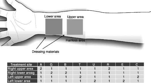
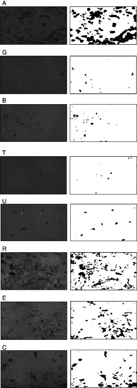
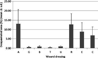
In recent years, adhesive wound dressings have been increasingly applied postoperatively because of their ease of use as they can be kept in place without having to cut and apply surgical tapes and they can cover a wound securely. However, if a wound dressing strongly adheres to the wound, a large amount of stratum corneum is removed from the newly formed epithelium or healthy periwound skin. Various types of adhesives are used on adhesive wound dressings and the extent of skin damage depends on how much an adhesive sticks to the wound or skin surface. We quantitatively determined and compared the amount of stratum corneum removed by eight different wound dressings including polyurethane foam using acrylic adhesive, silicone-based adhesive dressing, composite hydrocolloid and self-adhesive polyurethane foam in healthy volunteers. The results showed that wound dressings with silicone adhesive and self-adhesive polyurethane foam removed less stratum corneum, whereas composite hydrocolloid and polyurethane foam using acrylic adhesive removed more stratum corneum.
Keywords: Adhesive wound dressings; Dressing removal; Periwound skin; Stratum corneum; Wound dressing.
© 2012 The Authors. International Wound Journal © 2012 Medicalhelplines.com Inc and John Wiley & Sons Ltd.
Figures
Similar articles
-
A model for quantitative evaluation of skin damage at adhesive wound dressing removal.Int Wound J. 2013 Jun;10(3):291-4. doi: 10.1111/j.1742-481X.2012.00975.x. Epub 2012 Apr 26. Int Wound J. 2013. PMID: 22533468 Free PMC article.
-
Investigation of adhesion of modern wound dressings: a comparative analysis of 56 different wound dressings.J Eur Acad Dermatol Venereol. 2011 Aug;25(8):933-9. doi: 10.1111/j.1468-3083.2010.03886.x. Epub 2010 Nov 10. J Eur Acad Dermatol Venereol. 2011. PMID: 21062365
-
Effects of adhesive dressings on the stratum corneum of the skin.J Wound Care. 2001 Feb;10(2):7-10. doi: 10.12968/jowc.2001.10.2.26054. J Wound Care. 2001. PMID: 12964220 Clinical Trial.
-
Impact of adhesive surgical tape and wound dressings on the skin, with reference to skin stripping.J Wound Care. 2008 Apr;17(4):157-8, 160-2. doi: 10.12968/jowc.2008.17.4.28836. J Wound Care. 2008. PMID: 18494433 Review.
-
Fluid handling by foam wound dressings: From engineering theory to advanced laboratory performance evaluations.Int Wound J. 2024 Feb;21(2):e14674. doi: 10.1111/iwj.14674. Int Wound J. 2024. PMID: 38353372 Free PMC article. Review.
Cited by
-
Thermoregulation, incubator humidity, and skincare practices in appropriate for gestational age ultra-low birth weight infants: need for more evidence.World J Pediatr. 2024 Jul;20(7):643-652. doi: 10.1007/s12519-024-00818-x. Epub 2024 Jun 12. World J Pediatr. 2024. PMID: 38864998 Free PMC article. Review.
-
How Should Clinical Wound Care and Management Translate to Effective Engineering Standard Testing Requirements from Foam Dressings? Mapping the Existing Gaps and Needs.Adv Wound Care (New Rochelle). 2024 Jan;13(1):34-52. doi: 10.1089/wound.2021.0173. Epub 2022 Apr 5. Adv Wound Care (New Rochelle). 2024. PMID: 35216532 Free PMC article. Review.
-
Repeated Application and Removal of Polyisocyanopeptide Hydrogel Wound Dressings in a Splinted Full-Thickness Wound Model.Int J Mol Sci. 2023 Mar 7;24(6):5127. doi: 10.3390/ijms24065127. Int J Mol Sci. 2023. PMID: 36982201 Free PMC article.
-
Development and Characterization of Biocompatible Chitosan-Aloe Vera Films Functionalized with Gluconolactone and Sorbitol for Advanced Wound Healing Applications.ACS Appl Mater Interfaces. 2025 Mar 12;17(10):15196-15207. doi: 10.1021/acsami.5c00715. Epub 2025 Feb 25. ACS Appl Mater Interfaces. 2025. PMID: 39999379 Free PMC article.
-
Hydrogel Foams Containing Superabsorbent Particles for Wound Care Applications.ACS Appl Bio Mater. 2025 Jun 16;8(6):4633-4646. doi: 10.1021/acsabm.4c01719. Epub 2025 May 23. ACS Appl Bio Mater. 2025. PMID: 40407609
References
-
- Hollinworth H, White R. The clinical significance of wound pain. In: White R, Harding K, editors. Trauma and pain in wound care. Wounds UK, 2006:3–16.
-
- Waring M, Bielfeldt S, Matzold K, Wilhelm KP, Butcher M. An evaluation of the skin stripping of wound dressing adhesives. J Wound Care 2011;20:412, <414, 416–22. - PubMed
-
- van der Valk PG, Maibach HI. A functional study of the skin barrier to evaporative water loss by means of repeated cellophane‐tape stripping. Clin Exp Dermatol 1990;15:180–2. - PubMed
-
- Saap L, Fahim S, Arsenault E, Pratt M, Pierscianowski T, Falanga V, Pedvis‐Leftick A. Contact sensitivity in patients with leg ulcerations: a North American study. Arch Dermatol 2004;140:1241–6. - PubMed
Publication types
MeSH terms
Substances
LinkOut - more resources
Full Text Sources
