

Diagnosing preperimetric glaucoma with spectral domain optical coherence tomography
- PMID: 22883689
- PMCID: PMC3787835
- DOI: 10.1016/j.ophtha.2012.06.009
Diagnosing preperimetric glaucoma with spectral domain optical coherence tomography
Abstract
Purpose: To evaluate the diagnostic accuracy of spectral domain optical coherence tomography (SD-OCT) for detection of preperimetric glaucoma and compare it with the performance of confocal scanning laser ophthalmoscopy (CSLO).
Design: Cohort study.
Participants: A cohort of 134 eyes of 88 glaucoma suspects based on the appearance of the optic disc.
Methods: Patients were recruited from the Diagnostic Innovations in Glaucoma Study (DIGS). All eyes underwent retinal nerve fiber layer (RNFL) imaging with Spectralis SD-OCT (Heidelberg Engineering, Carlsbad, CA) and topographic imaging with Heidelberg Retinal Tomograph III (HRT-III) (Heidelberg Engineering) CSLO within 6 months of each other. All patients had normal visual fields at the time of imaging and were classified on the basis of history of documented stereophotographic evidence of progressive glaucomatous change in the appearance of the optic nerve occurring before the imaging sessions.
Main outcome measures: Areas under the receiver operating characteristic curves (AUCs) were calculated to summarize diagnostic accuracies of the SD-OCT and CSLO. Likelihood ratios (LRs) were reported using the diagnostic categorization provided by each instrument after comparison to its normative database.
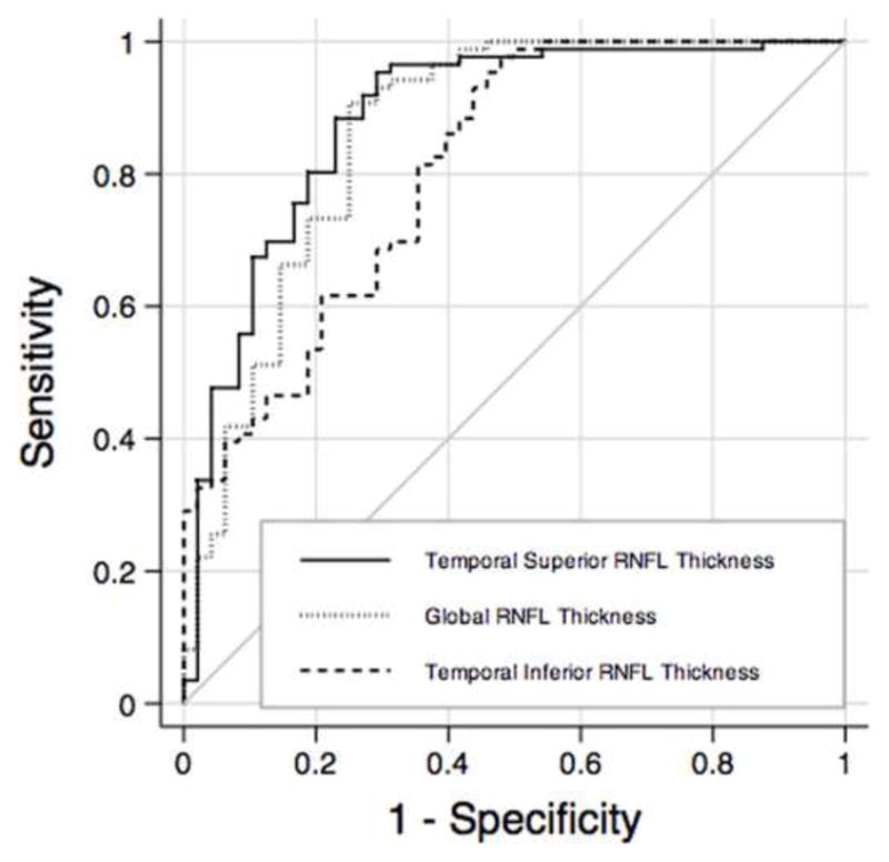
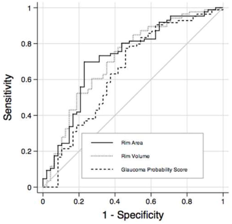
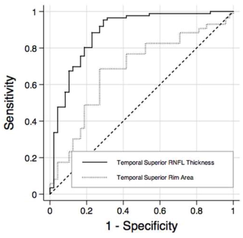
Results: Forty-eight eyes of 42 patients had evidence of progressive glaucomatous change and were included in the preperimetric glaucoma group. Eighty-six eyes of 46 patients without any evidence of progressive glaucomatous change followed untreated for an average of 14.0 ± 3.6 years were included in the control group. The parameter with the largest AUC obtained with the SD-OCT was the temporal superior RNFL thickness (0.88 ± 0.03), followed by global RNFL thickness (0.86 ± 0.03) and temporal inferior RNFL thickness (0.81 ± 0.04). The parameter with the largest AUC obtained with the CSLO was rim area (0.72 ± 0.05), followed by rim volume (0.71 ± 0.05) and linear cup-to-disk ratio (0.66 ± 0.05). Temporal superior RNFL average thickness measured by SD-OCT performed significantly better than rim area measurements from CSLO (0.88 vs. 0.72; P=0.008). Outside normal limits results for SD-OCT parameters were associated with strongly positive LRs.
Conclusions: The RNFL assessment with SD-OCT performed well in detecting preperimetric glaucomatous damage in a cohort of glaucoma suspects and had a better performance than CSLO.
Copyright © 2012 American Academy of Ophthalmology. Published by Elsevier Inc. All rights reserved.
Figures
References
-
- Nassif N, Cense B, Park B, et al. In vivo high-resolution video-rate spectral-domain optical coherence tomography of the human retina and optic nerve. [Accessed May 21, 2012];Opt Express [serial online] 2004 12:367–76. Available at: http://www.opticsinfobase.org/oe/abstract.cfm?uri=oe-12-3-367. - PubMed
-
- Wojtkowski M, Srinivasan V, Ko T, et al. Ultrahigh-resolution, high-speed, Fourier domain optical coherence tomography and methods for dispersion compensation. [Accessed May 21, 2012];Opt Express [serial online] 2004 12:2404–22. Available at: http://www.opticsinfobase.org/oe/abstract.cfm?uri=oe-12-11-2404. - PubMed
-
- Mistlberger A, Liebmann JM, Greenfield DS, et al. Heidelberg retina tomography and optical coherence tomography in normal, ocular-hypertensive, and glaucomatous eyes. Ophthalmology. 1999;106:2027–32. - PubMed
-
- Zangwill LM, Bowd C, Berry CC, et al. Discriminating between normal and glaucomatous eyes using the Heidelberg Retina Tomograph, GDx Nerve Fiber Analyzer, and Optical Coherence Tomograph. Arch Ophthalmol. 2001;119:985–93. - PubMed
-
- Greaney MJ, Hoffman DC, Garway-Heath DF, et al. Comparison of optic nerve imaging methods to distinguish normal eyes from those with glaucoma. Invest Ophthalmol Vis Sci. 2002;43:140–5. - PubMed
Publication types
MeSH terms
Grants and funding
LinkOut - more resources
Full Text Sources
Medical
