. 2012 Aug 9;75(3):402-9.
doi: 10.1016/j.neuron.2012.06.039.

Functional significance of isoform diversification in the protocadherin gamma gene cluster

Affiliations

Functional significance of isoform diversification in the protocadherin gamma gene cluster

Weisheng V Chen et al. Neuron. .

Erratum in

  • Neuron. 2012 Sep 6;75(5):928-9

Abstract

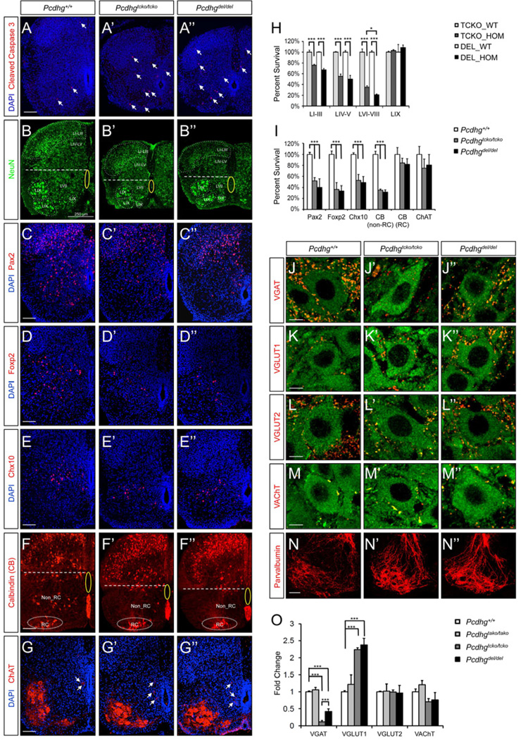
The mammalian Protocadherin (Pcdh) alpha, beta, and gamma gene clusters encode a large family of cadherin-like transmembrane proteins that are differentially expressed in individual neurons. The 22 isoforms of the Pcdhg gene cluster are diversified into A-, B-, and C-types, and the C-type isoforms differ from all other clustered Pcdhs in sequence and expression. Here, we show that mice lacking the three C-type isoforms are phenotypically indistinguishable from the Pcdhg null mutants, displaying virtually identical cellular and synaptic alterations resulting from neuronal apoptosis. By contrast, mice lacking three A-type isoforms exhibit no detectable phenotypes. Remarkably, however, genetically blocking apoptosis rescues the neonatal lethality of the C-type isoform knockouts, but not that of the Pcdhg null mutants. We conclude that the role of the Pcdhg gene cluster in neuronal survival is primarily, if not specifically, mediated by its C-type isoforms, whereas a separate role essential for postnatal development, likely in neuronal wiring, requires isoform diversity.


Figures

Figure 1
Figure 1. Pcdhg mutant alleles and phenotypes
(A) Schematic representations of Pcdhg wild type and mutant alleles. (B) Validation of deletions in homozygous animals by genomic PCR using exon-specific primers. (C–E) Gross phenotypes of Pcdhgtcko/tcko (P0), Pcdhgdel/del (P0) and Pcdhgtako/tako (P60) mutants as compared to littermate controls. Homozygous mutants are indicated with a red arrow. (F–G) Striking phenotype similarities between Pcdhgtcko/tcko and Pcdhgdel/del mutants at P0. Both have hunched posture and lack of voluntary movements and reflexes, exhibiting limb tremor, intense muscle stiffness and umbilical hernia (arrow), and die within hours after birth.
Figure 2
Figure 2. Similar levels and patterns of neuronal loss and synaptic changes in the spinal cord of Pcdhgtcko/tcko and Pcdhgdel/del mutants
(A–A”) Both mutants exhibit increased levels of apoptosis as indicated by the increased numbers of cleaved caspase 3+ profiles (arrows). Scale bar: 100 µm. (B–B”) Quantitative analyses of surviving neuronal populations in different lamina. NeuN+ neurons of distinct laminar regions are indicated. The central canal area is highlighted with a yellow oval, and the spinal cord is horizontally bisected with a dashed line to indicate the dorsal horn and the ventral horn areas. Scale bar: 250 µm. (C–G) Interneuron subpopulations are differentially affected to similar extents in both mutants. Shown are representative images of Pax2 (C-C”), Foxp2 (D-D”), Chx10 (E-E”), CB (F-F”) and ChAT (G-G”) immunolabeled P0 spinal cords of Pcdhg+/+, Pcdhgtcko/tcko, Pcdhgdel/del mice. Clusters of Renshaw cells (RC, F–F”) are encircled, and partition cells (G-G”) are indicated with arrows. Note the motor neurons are prominently labeled with anti-ChAT (G-G”). Scale bars: 100 µm. (H) Percent survival of NeuN+ neuronal populations according to their laminar locations (LI-LIX) in homozygous mutants (TCKO_HOM or DEL_HOM), calculated by normalizing neuronal counts to wild type littermate controls (TCKO_WT and DEL_WT). Details of the quantitative analyses can be found in Figure S2B. *p<0.05, ***p<0.001. (I) Percent survival of interneuron subpopulations in the ventral horn. ***p<0.001. (J–M) Specific types of synaptic inputs onto motor neurons of wild type and mutant spinal cords at P0. Motor neurons are labeled with anti-ChAT in green, and synaptic inputs with antibodies against different vesicular transporters in red. Scale bars: 5 µm. (N-N”) Terminal arborization of Parvalbumin+ Ia primary afferents surrounding motor pools is altered in both lines of mutants. Scale bar: 50 µm. (O) Fold changes in synaptic density for each type of synapse in the mutants as calculated by normalizing to wild type controls. Details of the quantitative analyses can be found in Figure S2C. ***p<0.001.
Figure 3
Figure 3. Similar levels and patterns of neuronal loss in the retina of Pcdhgtcko/tcko and Pcdhgdel/del mutants
(A-A”’) Nuclear and synaptic layers of retina sections from P18 control and mutants are labeled with Po-Pro1 or anti-Bassoon, respectively. Retinal lamination is normal, and the ONL and OPL thickness are unaffected. INL and IPL thickness are similarly reduced in Pcdhgfcon3/tcko and Pcdhgfcon3/fcon3 mutants but are unaffected in Pcdhgfcon3/tako mutants. (B–C) Numbers of Chx10+ bipolar (red), Pax6+ GABAergic amacrine interneurons (green), as well as Brn3a+ projection retinal ganglion cells (magenta) are similarly reduced in Pcdhgfcon3/tcko and Pcdhgfcon3/fcon3 mutants. (D) Quantification of INL and IPL thickness in control and mutant retinas. (E, F) Quantification of bipolar interneurons (Chx10+ BP) and projection retinal ganglion cells (Brn3a+ RGC). (F-F”) Increased number of apoptotic cells (cleaved caspase 3+) is detected in the GCL and INL of the mutant retina (P4), as seen in Pcdhg null retinas. (G-G”) Syt2+ OFF bipolar processes (red) and Gγ13+ ON bipolar processes (green) ramify in the upper and lower regions of the IPL, respectively (P18). Laminar specificity is retained in mutants deficient in C-type Pcdhg isoforms, but marker-laminae are reduced and disrupted, identical as shown for Pcdhg null mutants. ONL, outer nuclear layer; OPL, outer plexiform layer; INL, inner nuclear layer; IPL, inner plexiform layer; GCL, ganglion cell layer. *p < 0.05, *** p < 0.001. Scale bars: 50 µm.
Figure 4
Figure 4. Expression and function of A- and B-type Pcdhg isoforms are not appreciably affected in the absence of C-type genes
(A) Schematic representation of Pcdhg genes, transcripts and proteins. Positions of specific primers (red arrows) are indicated. ECD, extracellular domain; ICD, intracellular domain. (B) RT-PCR of individual Pcdhg transcripts in wild type and both lines of mutants. (C) Western blot indicates that the expression level of Pcdhg proteins in the Pcdhgtcko/tcko mutant brain is similar to the wild type level, but higher than that of Pcdhg+/del. Alpha tubulin is used as loading control. (D–E) A-type and B-type Pcdhg isoforms in Pcdhgtcko/tcko mutant brains still form complexes with Pcdha proteins, remain tyrosine phosphorylated, and mediate signaling. Western blots of P0 brain lysate from wild type and both mutants are shown in D, and blots of pan-Pcdhg immunoprecipitates in E. (F) Schematic representation of Pcdh clusters. Three identified cluster-wide enhancers (HS5-1, HS7 and HS16-20) and their respectively regulated regions are indicated. (G) Relative expression levels of individual exons in the Pcdh cluster region in E13.5 wild type and mutant spinal cords based on RNA-Seq analysis. Several genes with extremely low expression levels are excluded but can be found in Table S1. Significance levels are not indicated but can be found in Table S1. The red arrow in F and G indicates the EST gene AK149307.

References

    1. Asada H, Kawamura Y, Maruyama K, Kume H, Ding RG, Kanbara N, Kuzume H, Sanbo M, Yagi T, Obata K. Cleft palate and decreased brain gamma-aminobutyric acid in mice lacking the 67-kDa isoform of glutamic acid decarboxylase. Proc Natl Acad Sci U S A. 1997;94:6496–6499. - PMC - PubMed
    1. Buss RR, Sun W, Oppenheim RW. Adaptive roles of programmed cell death during nervous system development. Annu Rev Neurosci. 2006;29:1–35. - PubMed
    1. Chen HH, Hippenmeyer S, Arber S, Frank E. Development of the monosynaptic stretch reflex circuit. Curr Opin Neurobiol. 2003;13:96–102. - PubMed
    1. Esumi S, Kakazu N, Taguchi Y, Hirayama T, Sasaki A, Hirabayashi T, Koide T, Kitsukawa T, Hamada S, Yagi T. Monoallelic yet combinatorial expression of variable exons of the protocadherin-alpha gene cluster in single neurons. Nat Genet. 2005;37:171–176. - PubMed
    1. Feng G, Tintrup H, Kirsch J, Nichol MC, Kuhse J, Betz H, Sanes JR. Dual requirement for gephyrin in glycine receptor clustering and molybdoenzyme activity. Science. 1998;282:1321–1324. - PubMed

Publication types