

Mitoxantrone repression of astrocyte activation: relevance to multiple sclerosis
- PMID: 22884503
- PMCID: PMC3460523
- DOI: 10.1016/j.brainres.2012.07.054
Mitoxantrone repression of astrocyte activation: relevance to multiple sclerosis
Abstract
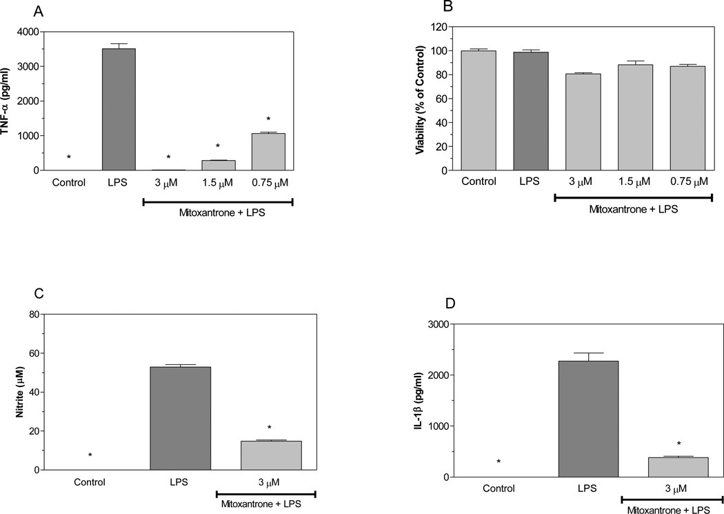
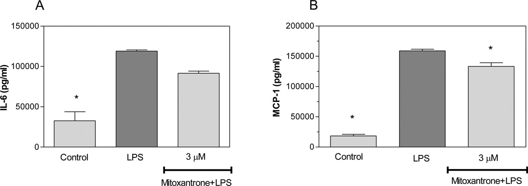
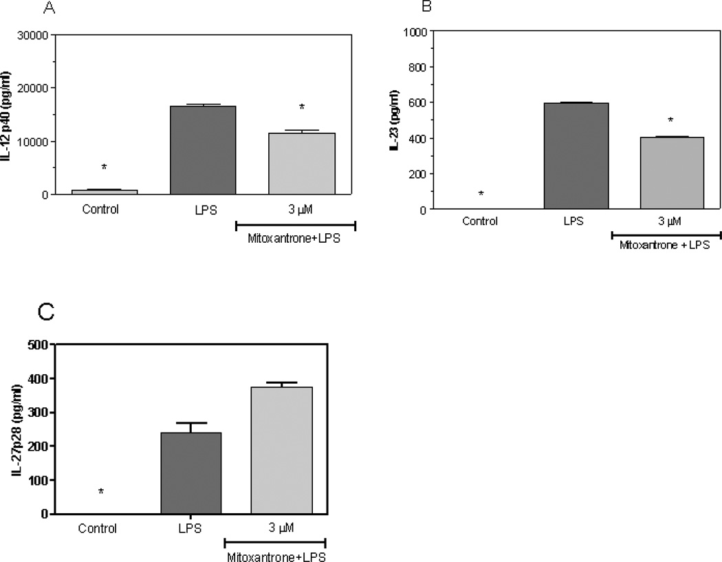
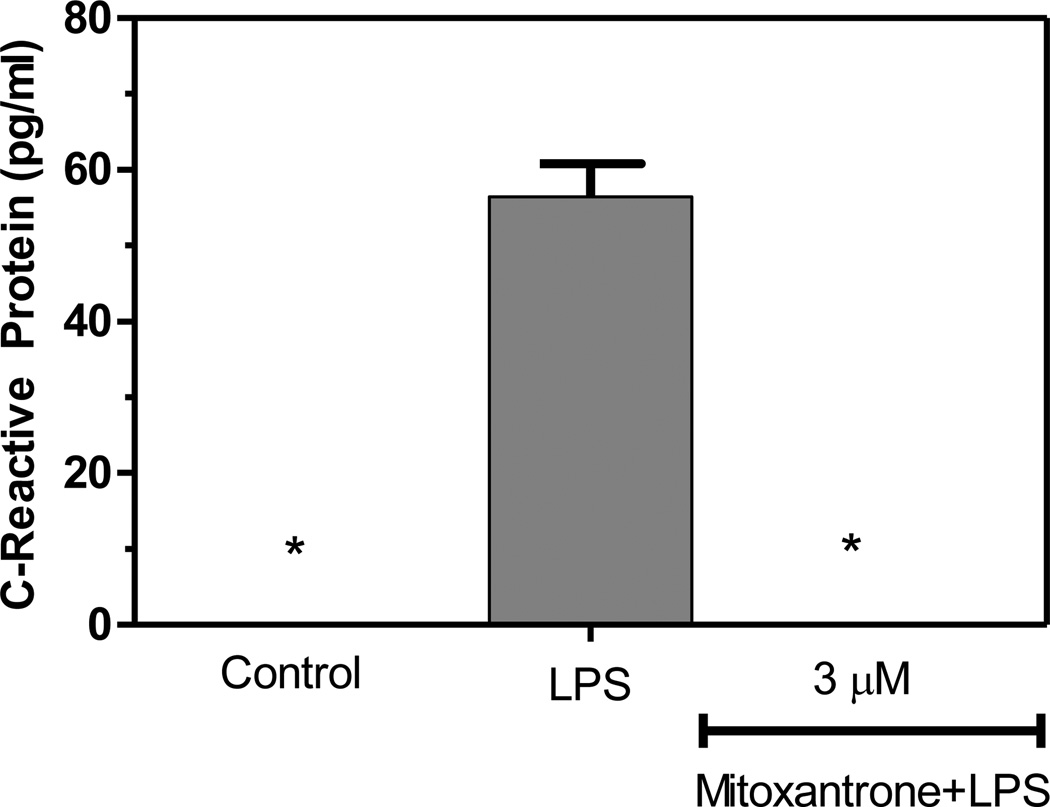
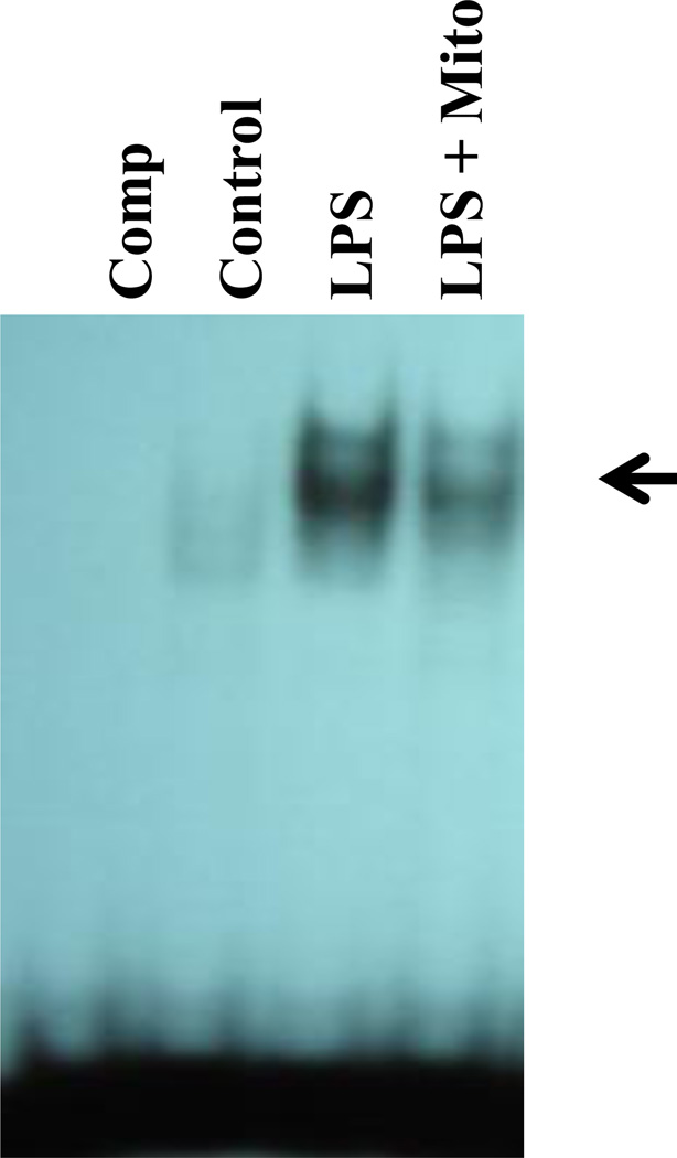
Mitoxantrone has been approved by the FDA for the treatment of multiple sclerosis (MS). However, the mechanisms by which mitoxantrone modulates MS are largely unknown. Activated astrocytes produce nitric oxide (NO), TNF-α, and IL-1β, molecules which can be toxic to central nervous system (CNS) cells including oligodendrocytes, thus potentially contributing to the pathology associated with MS. MCP-1 is a chemokine believed to modulate the migration of monocytes to inflammatory lesions present in the CNS of MS patients. IL-12 and IL-23 have been demonstrated to play critical roles in the pathogenesis of experimental autoimmune encephalomyelitis (EAE), an animal model of MS, by contributing to the development of CD4(+) T cell lineages termed Th1 and Th17, respectively. The current study demonstrates that mitoxantrone inhibits lipopolysachharide (LPS) induction of NO, TNF-α, IL-1β, and MCP-1 production by primary astrocytes. Mitoxantrone also inhibited IL-12 and IL-23 production by these cells. Furthermore, mitoxantrone suppressed the expression of C-reactive protein (CRP). Finally, we demonstrate that mitoxantrone suppressed LPS induction of NF-κB DNA-binding activity, suggesting a novel mechanism by which mitoxantrone suppresses the expression of proinflammatory molecules. Collectively, these studies demonstrate that mitoxantrone represses astrocyte production of potentially cytotoxic molecules, as well as molecules capable of altering T-cell phenotype. These in vitro studies suggest mechanisms by which mitoxantrone may modulate inflammatory diseases including MS.
Copyright © 2012 Elsevier B.V. All rights reserved.
Figures
Similar articles
-
Huperzine A inhibits CCL2 production in experimental autoimmune encephalomyelitis mice and in cultured astrocyte.Int J Immunopathol Pharmacol. 2013 Jul-Sep;26(3):757-64. doi: 10.1177/039463201302600320. Int J Immunopathol Pharmacol. 2013. PMID: 24067473
-
Resveratrol differentially modulates inflammatory responses of microglia and astrocytes.J Neuroinflammation. 2010 Aug 17;7:46. doi: 10.1186/1742-2094-7-46. J Neuroinflammation. 2010. PMID: 20712904 Free PMC article.
-
MiR-409-3p and MiR-1896 co-operatively participate in IL-17-induced inflammatory cytokine production in astrocytes and pathogenesis of EAE mice via targeting SOCS3/STAT3 signaling.Glia. 2019 Jan;67(1):101-112. doi: 10.1002/glia.23530. Epub 2018 Oct 7. Glia. 2019. PMID: 30294880
-
Mitoxantrone: a review of its use in multiple sclerosis.CNS Drugs. 2004;18(6):379-96. doi: 10.2165/00023210-200418060-00010. CNS Drugs. 2004. PMID: 15089110 Review.
-
Astrocytes in multiple sclerosis and experimental autoimmune encephalomyelitis: Star-shaped cells illuminating the darkness of CNS autoimmunity.Brain Behav Immun. 2019 Aug;80:10-24. doi: 10.1016/j.bbi.2019.05.029. Epub 2019 May 21. Brain Behav Immun. 2019. PMID: 31125711 Review.
Cited by
-
Mitoxantrone in NMO Spectrum Disorder in a Large Multicenter Cohort in French Caribbean.Neurol Neuroimmunol Neuroinflamm. 2023 Nov 10;11(1):e200175. doi: 10.1212/NXI.0000000000200175. Print 2024 Jan. Neurol Neuroimmunol Neuroinflamm. 2023. PMID: 37949668 Free PMC article.
-
Breast cancer tumor microenvironment affects Treg/IL-17-producing Treg/Th17 cell axis: Molecular and therapeutic perspectives.Mol Ther Oncolytics. 2023 Jan 11;28:132-157. doi: 10.1016/j.omto.2023.01.001. eCollection 2023 Mar 16. Mol Ther Oncolytics. 2023. PMID: 36816749 Free PMC article. Review.
-
A Fluorescence-Polarization-Based Lipopolysaccharide-Caspase-4 Interaction Assay for the Development of Inhibitors.Molecules. 2022 Apr 11;27(8):2458. doi: 10.3390/molecules27082458. Molecules. 2022. PMID: 35458656 Free PMC article.
-
Progress in the Application of Drugs for the Treatment of Multiple Sclerosis.Front Pharmacol. 2021 Jul 13;12:724718. doi: 10.3389/fphar.2021.724718. eCollection 2021. Front Pharmacol. 2021. PMID: 34326775 Free PMC article. Review.
-
Teriflunomide and its mechanism of action in multiple sclerosis.Drugs. 2014 Apr;74(6):659-74. doi: 10.1007/s40265-014-0212-x. Drugs. 2014. PMID: 24740824 Free PMC article. Review.
References
-
- Barger SW, Chavis JA, Drew PD. Dehydroepiandrosterone inhibits microglial nitric oxide production in a stimulus-specific manner. J Neurosci Res. 2000;62:503–509. - PubMed
-
- Bettelli E, et al. Reciprocal developmental pathways for the generation of pathogenic effector TH17 and regulatory T cells. Nature. 2006;441:235–238. - PubMed
-
- De Keyser J, Zeinstra E, Frohman E. Are astrocytes central players in the pathophysiology of multiple sclerosis? Arch Neurol. 2003;60:132–136. - PubMed
-
- Dong Y, Benveniste EN. Immune function of astrocytes. Glia. 2001;36:180–190. - PubMed
-
- Farina C, Aloisi F, Meinl E. Astrocytes are active players in cerebral innate immunity. Trends Immunol. 2007;28:138–145. - PubMed
Publication types
MeSH terms
Substances
Grants and funding
LinkOut - more resources
Full Text Sources
Other Literature Sources
Medical
Research Materials
Miscellaneous
