. 2012 Sep 30;210(2):119-24.
doi: 10.1016/j.jneumeth.2012.07.019. Epub 2012 Jul 31.

Optogenetic field potential recording in cortical slices

Affiliations

Optogenetic field potential recording in cortical slices

Wenhui Xiong et al. J Neurosci Methods. .

Abstract

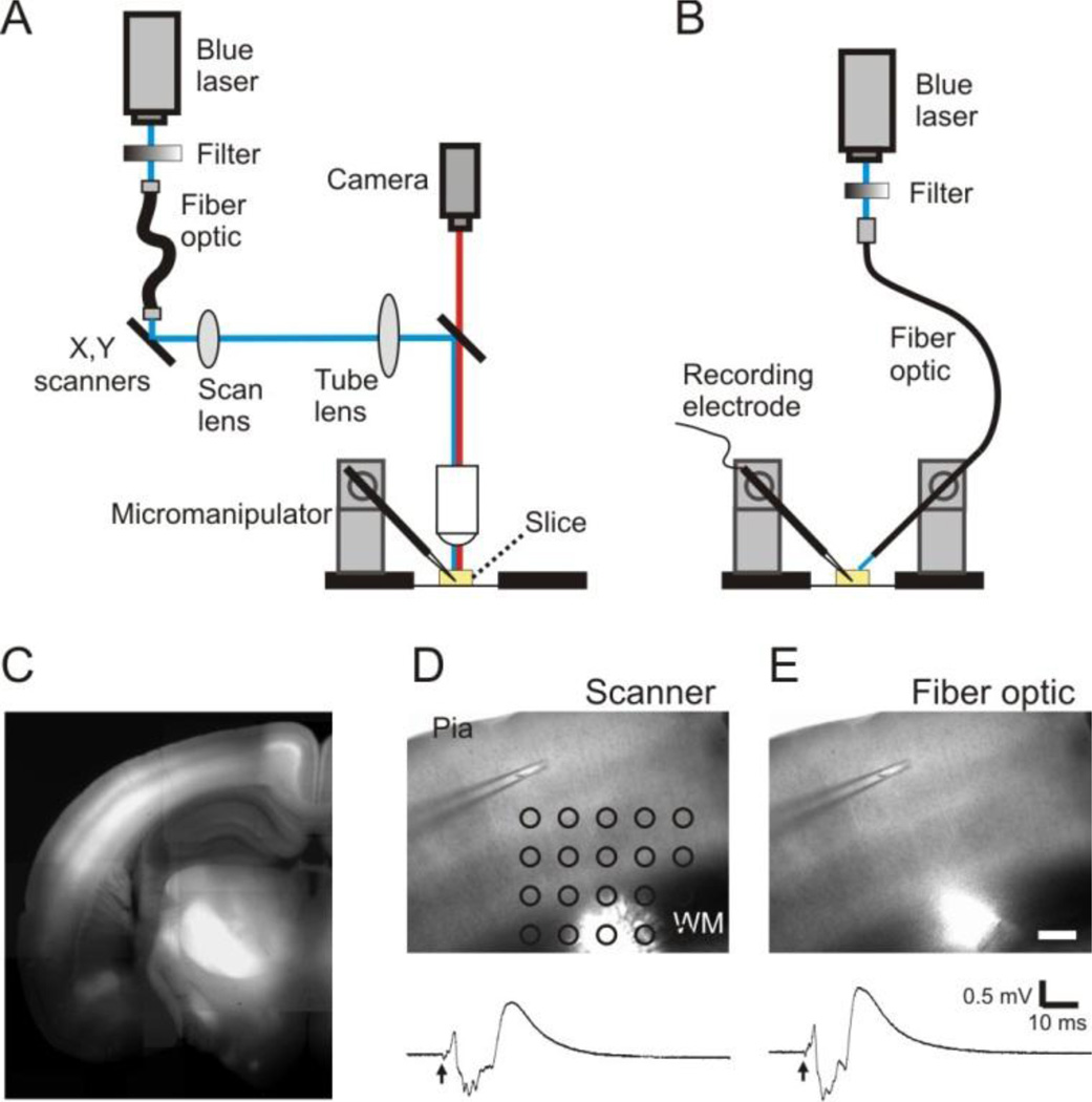
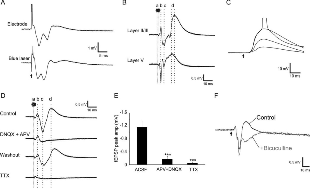
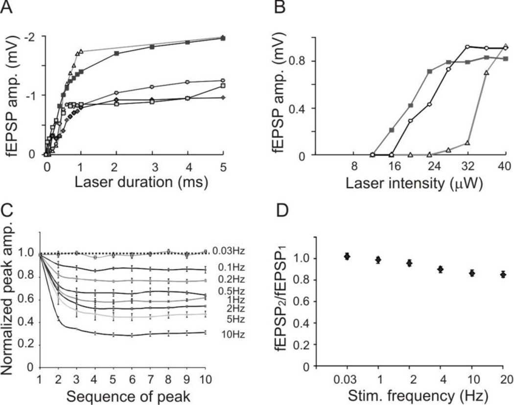
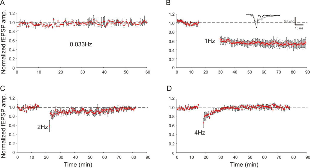
We introduce a method that uses optogenetic stimulation to evoke field potentials in brain slices prepared from transgenic mice expressing channelrhodopsin-2-YFP. Cortical slices in a recording chamber were stimulated with a 473 nm blue laser via either a laser scanning photostimulation setup or by direct guidance of a fiber optic. Field potentials evoked by either of the two optogenetic stimulation methods had stable amplitude, consistent waveform, and similar components as events evoked with a conventional stimulating electrode. The amplitude of evoked excitatory postsynaptic potentials increased with increasing laser intensity or pulse duration. We further demonstrated that optogenetic stimulation can be used for the induction and monitoring of long-term depression. We conclude that this technique allows for efficient and reliable activation of field potentials in brain slice preparation, and will be useful for studying short and long term synaptic plasticity.


Figures

Figure 1
Figure 1
A method of oFPR. A and B: Schematic representation showing that a laser scanning photostimulation system (A) or a fiber optic attached to a micromanipulator (B) was used for delivering pulses of blue laser (473 nm) onto brain slices. C. A fluorescence image of a coronal cortical slice prepared from a ChR2-YFP expressing transgenic mouse. ChR2-YFP expression is clearly visible in the cortex, particularly in layer V and layer II/III. D and E: Optogenetic stimulation delivered with the methods illustrated in either A (1-ms laser pulse at 0.04 mW) or B (1-ms laser pulse at 0.07 mW) evoked similar responses. Arrows indicate the time of laser flashes. Scale bar in E: 200 µm.
Figure 2
Figure 2
Field potentials evoked by optogenetic stimulation. A. Extracellular field potentials evoked in cortical layer II/III by electrical (0.2-ms pulse of square current at 0.5 mA, top trace) or optogenetic (2-ms pulse of blue laser at 0.04 mW, bottom trace) stimulation of the white matter. B. The components of optogenetically evoked responses: Following a laser flash on the white matter (a, 1 ms at 0.04 mW), the first negative (layer V) or positive (Layer II/III) peak appeared at 3.3 ms (b), which resulted from direction activation of ChR2 expressing axons. The second peak(s) of fEPSP occurred at ~7.1ms (c), which was followed by a slower large positive peak of fIPSP (d). C. Whole cell current clamp recording was made from a layer II/III pyramidal neuron when laser pulses at increasing intensities (1 ms, 0.01–0.04 mW) were applied to layer V. The constant onset latency of the evoked EPSPs indicates the monosynaptic nature of the responses. The action potential trace is truncated for better demonstration of the EPSPs. D–F. Pharmacology of optogenetically evoked responses. D. Addition of AMPA and NMDA receptor antagonists DNQX (20 µM) and APV (100 µM) blocked fEPSP (b) and fIPSP (c) but not the direct activation (a). This blockade was reversible by washing with normal ACSF. Application of 1 µM TTX abolished almost all components of the evoked response (bottom trace). E. The effects of those drugs on the fEPSP peak amplitudes (−1.16 ± 0.19 mV in control; −0.16 ± 0.09 mV in APV + DNQX, and −0.05 ± 0.03 mV in TTX, ***: P < 0.001). F. Addition of 10 µM GABAA receptor antagonist bicuculline in the ACSF blocked the third outward peak (gray trace), confirming that this component was an IPSP. The arrow indicates the time of laser flash.
Figure 3
Figure 3
Relationships between parameters of optogenetic stimulation and the evoked responses. A. Relationship between laser duration at maximum intensity (0.04 mW) and peak amplitude of evoked fEPSPs. B. Relationship between laser intensity of 1 ms pulses and peak amplitude of evoked fEPSPs. In both A and B, each line represents recordings from a slice. C. Significant desensitization of ChR2 activation at higher stimulating frequencies: The peak amplitudes of direct activation (peaks b in Fig. 2B) recorded in layer II/III rapidly depressed with increasing stimulating frequency. D. Relationship between fEPSP2/fEPSP1 ratio and stimulating frequency. Optogenetically evoked fEPSPs only slightly depressed at higher stimulating frequencies.
Figure 4
Figure 4
Stability of oFPR and optogenetic induction of LTD. Field potentials were elicited in layer II/III by applying laser flashes in layer V (1 ms at 0.04 mW). A. Normalized fEPSP amplitudes evoked by pulses of blue laser at 0.033 Hz were stable throughout the 60-min recording period (n=3). B–D. LTD induction by LFS: The graphs show normalized fEPSP peak amplitudes recorded before and after conditioning stimulation (900 pulses) at 1 Hz (B, n=5), 2 Hz (C, n=4), or 4 Hz (D, n=4). Only optogenetic stimulation at 1 Hz resulted in long-term depression of fEPSP amplitude (B), suggesting that the optogenetic LTD induction was frequency-dependent. Insert in B: representative average field potential traces before (black trace) and after (gray trace) LFS in a slice.

Similar articles

Cited by

References

    1. Arenkiel BR, Peca J, Davison IG, Feliciano C, Deisseroth K, Augustine GJ, Ehlers MD, Feng G. In vivo light-induced activation of neural circuitry in transgenic mice expressing channelrhodopsin-2. Neuron. 2007;54:205–218. - PMC - PubMed
    1. Barrionuevo G, Brown TH. Associative long-term potentiation in hippocampal slices. Proc Natl Acad Sci U S A. 1983;80:7347–7351. - PMC - PubMed
    1. Boyden ES, Zhang F, Bamberg E, Nagel G, Deisseroth K. Millisecond-timescale, genetically targeted optical control of neural activity. Nat Neurosci. 2005;8:1263–1268. - PubMed
    1. Castro-Alamancos MA, Connors BW. Distinct forms of short-term plasticity at excitatory synapses of hippocampus and neocortex. Proc Natl Acad Sci U S A. 1997;94:4161–4166. - PMC - PubMed
    1. Gunaydin LA, Yizhar O, Berndt A, Sohal VS, Deisseroth K, Hegemann P. Ultrafast optogenetic control. Nat Neurosci. 13:387–392. - PubMed

Publication types

MeSH terms

LinkOut - more resources