

Shear strength behavior of human trabecular bone
- PMID: 22884967
- PMCID: PMC3462285
- DOI: 10.1016/j.jbiomech.2012.07.023
Shear strength behavior of human trabecular bone
Abstract
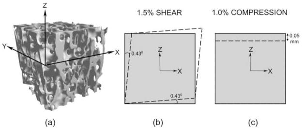
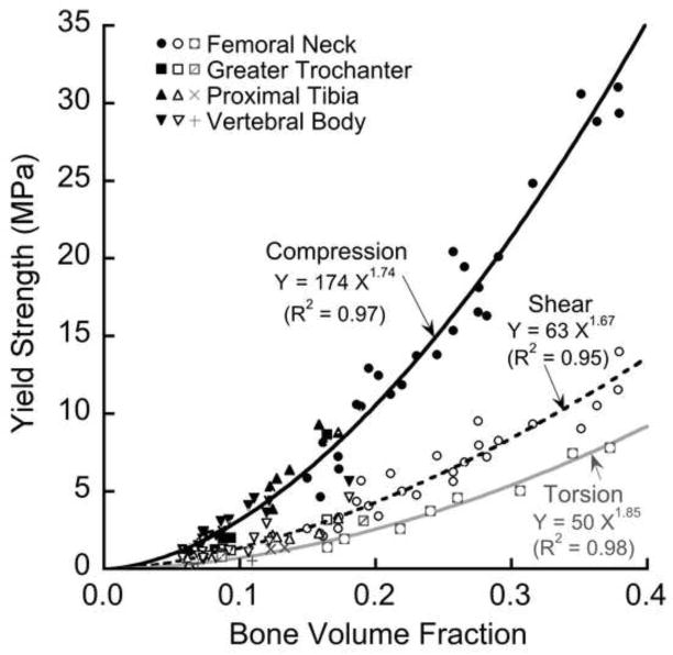
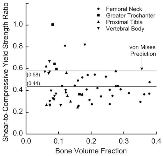
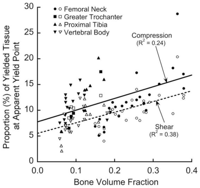
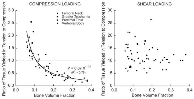
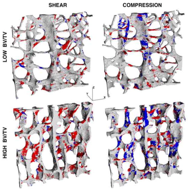
The shear strength of human trabecular bone may influence overall bone strength under fall loading conditions and failure at bone-implant interfaces. Here, we sought to compare shear and compressive yield strengths of human trabecular bone and elucidate the underlying failure mechanisms. We analyzed 54 specimens (5-mm cubes), all aligned with the main trabecular orientation and spanning four anatomic sites, 44 different cadavers, and a wide range of bone volume fraction (0.06-0.38). Micro-CT-based non-linear finite element analysis was used to assess the compressive and shear strengths and the spatial distribution of yielded tissue; the tissue-level constitutive model allowed for kinematic non-linearity and yielding with strength asymmetry. We found that the computed values of both the shear and compressive strengths depended on bone volume fraction via power law relations having an exponent of 1.7 (R(2)=0.95 shear; R(2)=0.97 compression). The ratio of shear to compressive strengths (mean±SD, 0.44±0.16) did not depend on bone volume fraction (p=0.24) but did depend on microarchitecture, most notably the intra-trabecular standard deviation in trabecular spacing (R(2)=0.23, p<0.005). For shear, the main tissue-level failure mode was tensile yield of the obliquely oriented trabeculae. By contrast, for compression, specimens having low bone volume fraction failed primarily by large-deformation-related tensile yield of horizontal trabeculae and those having high bone volume failed primarily by compressive yield of vertical trabeculae. We conclude that human trabecular bone is generally much weaker in shear than compression at the apparent level, reflecting different failure mechanisms at the tissue level.
Copyright © 2012 Elsevier Ltd. All rights reserved.
Conflict of interest statement
Dr. Keaveny has a financial interest in O.N. Diagnostics and both he and the company may benefit from the results of this research. All other authors have no conflict of interest.
Figures
Similar articles
-
Theoretical bounds for the influence of tissue-level ductility on the apparent-level strength of human trabecular bone.J Biomech. 2013 Apr 26;46(7):1293-9. doi: 10.1016/j.jbiomech.2013.02.011. Epub 2013 Mar 14. J Biomech. 2013. PMID: 23497799 Free PMC article.
-
Influence of bone volume fraction and architecture on computed large-deformation failure mechanisms in human trabecular bone.Bone. 2006 Dec;39(6):1218-25. doi: 10.1016/j.bone.2006.06.016. Epub 2006 Aug 10. Bone. 2006. PMID: 16904959
-
Sensitivity of damage predictions to tissue level yield properties and apparent loading conditions.J Biomech. 2001 May;34(5):699-706. doi: 10.1016/s0021-9290(01)00003-3. J Biomech. 2001. PMID: 11311712
-
Differences between the tensile and compressive strengths of bovine tibial trabecular bone depend on modulus.J Biomech. 1994 Sep;27(9):1137-46. doi: 10.1016/0021-9290(94)90054-x. J Biomech. 1994. PMID: 7929463 Review.
-
Mechanical strength of trabecular bone at the knee.Dan Med Bull. 1988 Aug;35(4):345-65. Dan Med Bull. 1988. PMID: 3048922 Review.
Cited by
-
Prospect of Metal Ceramic (Titanium-Wollastonite) Composite as Permanent Bone Implants: A Narrative Review.Materials (Basel). 2021 Jan 7;14(2):277. doi: 10.3390/ma14020277. Materials (Basel). 2021. PMID: 33430455 Free PMC article. Review.
-
Finite Element Analysis of Foot and Ankle Impact Injury: Risk Evaluation of Calcaneus and Talus Fracture.PLoS One. 2016 Apr 27;11(4):e0154435. doi: 10.1371/journal.pone.0154435. eCollection 2016. PLoS One. 2016. PMID: 27119740 Free PMC article.
-
The effects of tensile-compressive loading mode and microarchitecture on microdamage in human vertebral cancellous bone.J Biomech. 2014 Nov 28;47(15):3605-12. doi: 10.1016/j.jbiomech.2014.10.011. J Biomech. 2014. PMID: 25458150 Free PMC article.
-
The roles of architecture and estrogen depletion in microdamage risk in trabecular bone.J Biomech. 2016 Oct 3;49(14):3223-3229. doi: 10.1016/j.jbiomech.2016.08.009. Epub 2016 Aug 9. J Biomech. 2016. PMID: 27544617 Free PMC article.
-
The sensitivity of nonlinear computational models of trabecular bone to tissue level constitutive model.Comput Methods Biomech Biomed Engin. 2016;19(5):465-73. doi: 10.1080/10255842.2015.1041022. Epub 2015 May 11. Comput Methods Biomech Biomed Engin. 2016. PMID: 25959510 Free PMC article.
References
-
- Adams MF, Bayraktar HH, Keaveny TM, Papadopoulos P. Ultrascalable implicit finite element analyses in solid mechanics with over a half a billion degrees of freedom. ACM/IEEE Proceedings of SC2004: High Performance Networking and Computing.2004.
-
- Beck JD, Canfield BL, Haddock SM, Chen TJ, Kothari M, Keaveny TM. Three-dimensional imaging of trabecular bone using the computer numerically controlled milling technique. Bone. 1997;21(3):281–287. - PubMed
-
- Bevill G, Eswaran SK, Farahmand F, Keaveny TM. The influence of boundary conditions and loading mode on high-resolution finite element-computed trabecular tissue properties. Bone. 2009a;44(4):573–578. - PubMed
-
- Bevill G, Eswaran SK, Gupta A, Papadopoulos P, Keaveny TM. Influence of bone volume fraction and architecture on computed large-deformation failure mechanisms in human trabecular bone. Bone. 2006;39(6):1218–1225. - PubMed
Publication types
MeSH terms
Grants and funding
LinkOut - more resources
Full Text Sources
