. 2012 Oct;181(4):1236-49.
doi: 10.1016/j.ajpath.2012.06.035. Epub 2012 Aug 10.

Lysophosphatidic acid increases proximal tubule cell secretion of profibrotic cytokines PDGF-B and CTGF through LPA2- and Gαq-mediated Rho and αvβ6 integrin-dependent activation of TGF-β

Affiliations

Lysophosphatidic acid increases proximal tubule cell secretion of profibrotic cytokines PDGF-B and CTGF through LPA2- and Gαq-mediated Rho and αvβ6 integrin-dependent activation of TGF-β

Hui Geng et al. Am J Pathol. 2012 Oct.

Abstract

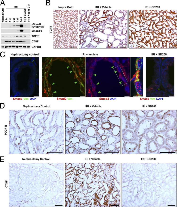
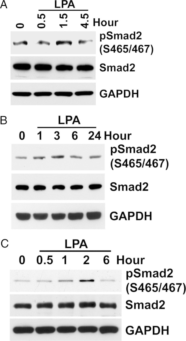
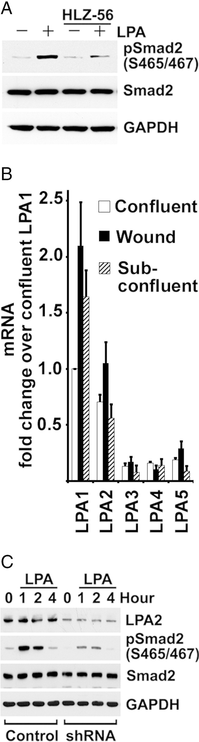
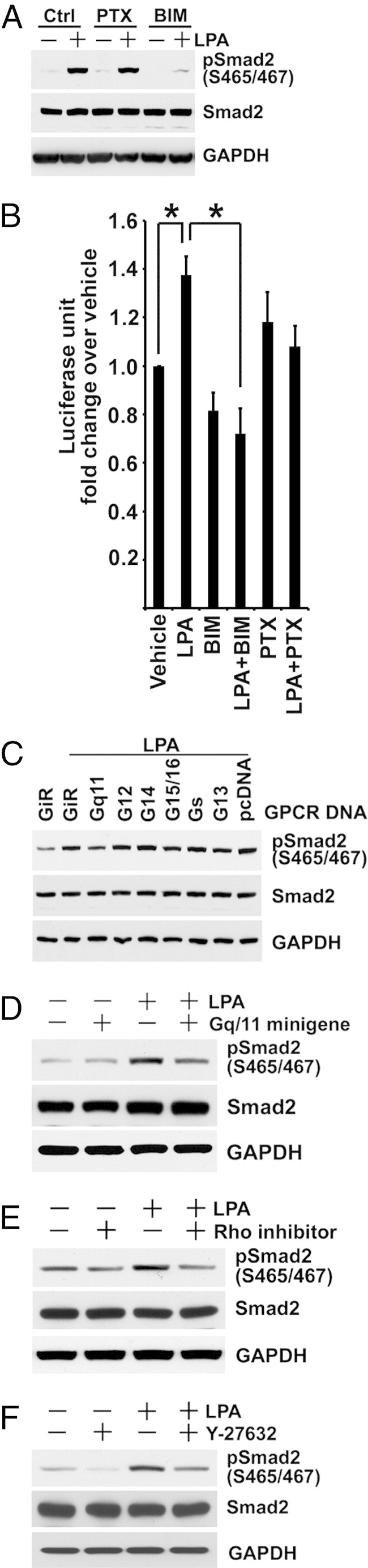
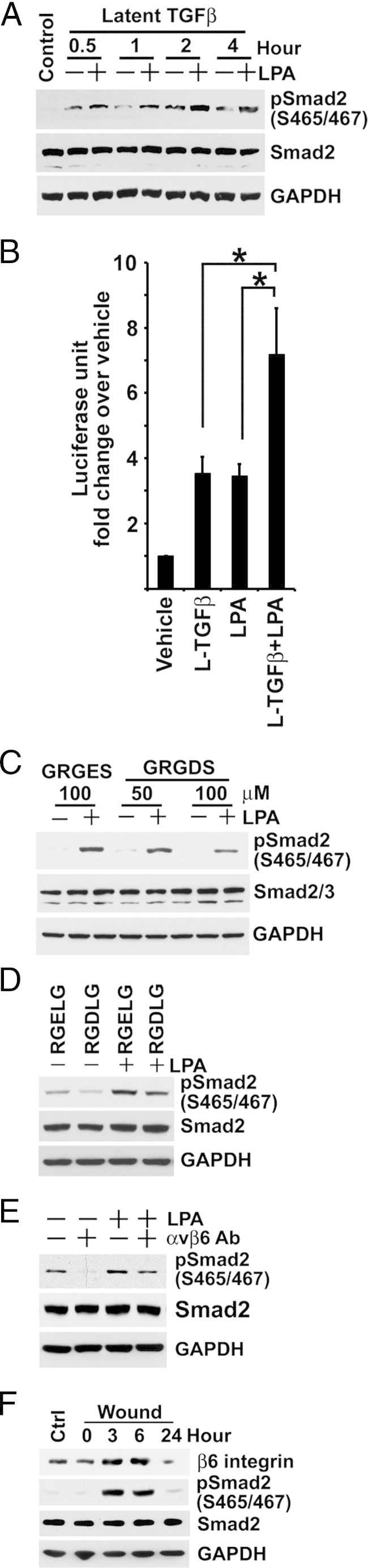
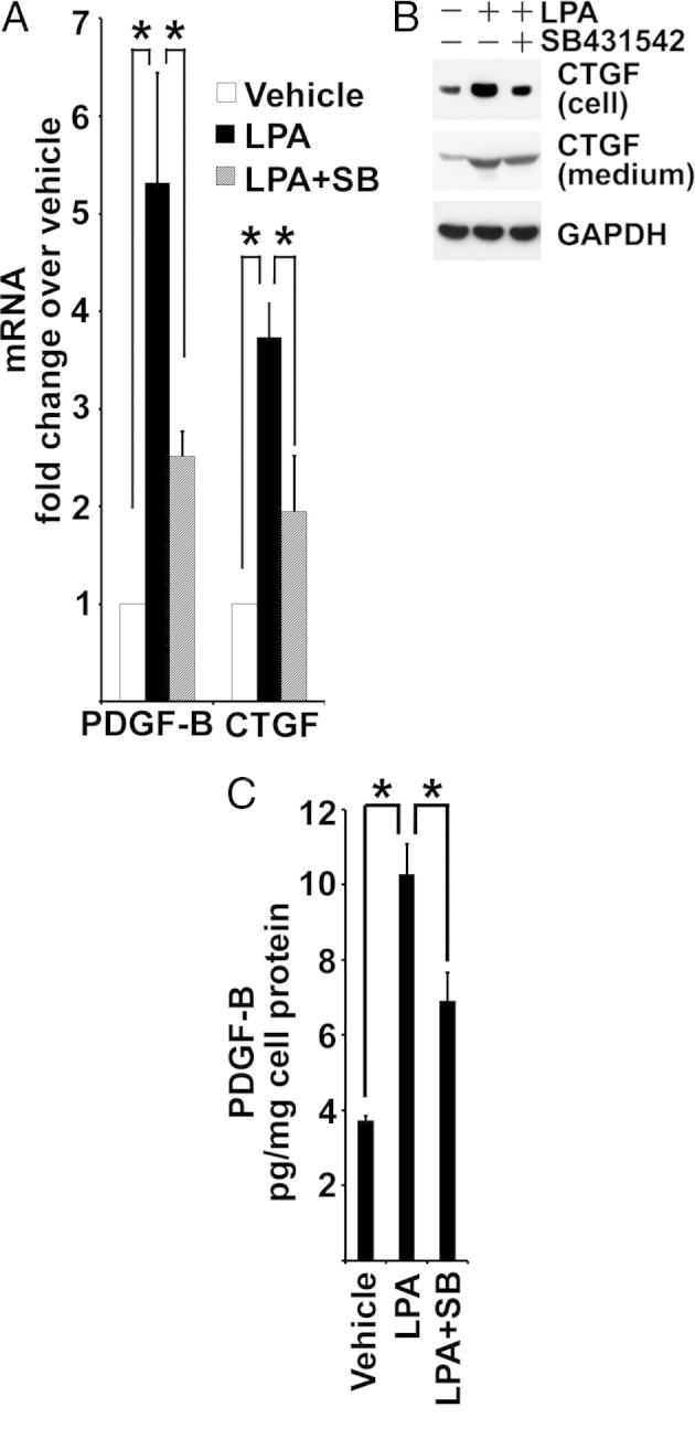
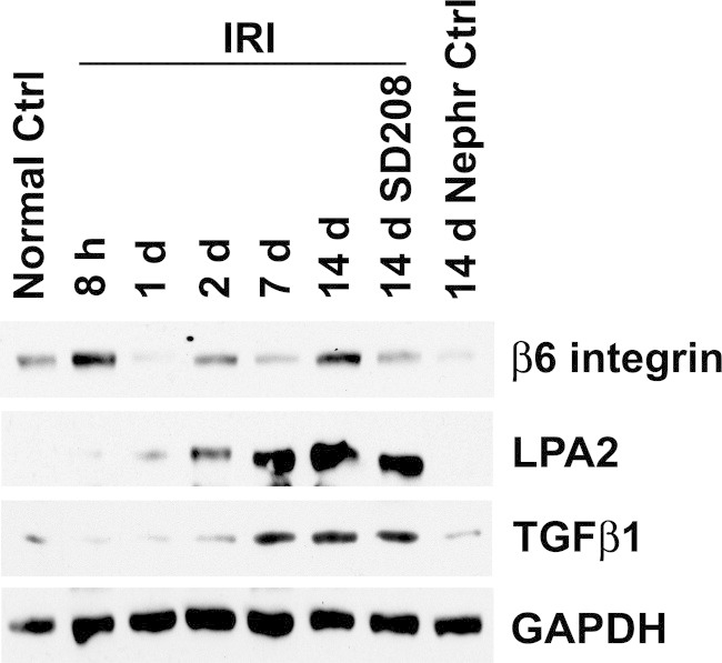
After ischemia-reperfusion injury (IRI), kidney tubules show activated transforming growth factor β (TGF-β) signaling and increased expression of profibrotic peptides, platelet-derived growth factor-B (PDGF-B) and connective tissue growth factor (CTGF). If tubule repair after IRI is incomplete, sustained paracrine activity of these peptides can activate interstitial fibroblast progenitors and cause fibrosis. We show that lysophosphatidic acid (LPA), a ubiquitous phospholipid that is increased at sites of injury and inflammation, signals through LPA2 receptors and Gαq proteins of cultured proximal tubule cells to transactivate latent TGF-β in a Rho/Rho-kinase and αvβ6 integrin-dependent manner. Active TGF-β peptide then initiates signaling to increase the production and secretion of PDGF-B and CTGF. In a rat model of IRI, increased TGF-β signaling that was initiated early during reperfusion did not subside during recovery, but progressively increased, causing tubulointerstitial fibrosis. This was accompanied by correspondingly increased LPA2 and β6 integrin proteins and elevated tubule expression of TGF-β1, together with PDGF-B and CTGF. Treatment with a pharmacological TGF-β type I receptor antagonist suppressed TGF-β signaling, decreased the expression of β6 integrin, PDGF-B, and CTGF, and ameliorated fibrosis. We suggest that LPA-initiated autocrine signaling is a potentially important mechanism that gives rise to paracrine profibrotic signaling in injured kidney tubule cells.


Figures

Figure 1
Figure 1
Ischemia-reperfusion injury stimulates TGF-β signaling and induces profibrotic peptide expression in renal proximal tubules. A: SDS extracts of the outer stripe of the outer medulla from normal control rats without surgery (Normal Ctrl), renal IRI with or without SD208 treatment at the indicated time after surgery, and nephrectomized control (Nephr Ctrl) at day 14 are immunoblotted for phospho-Smad2, total Smad2/3, TGF-β1, and CTGF. B, D, and E: IHC staining of TGF-β1, PDGF, and CTGF in kidneys 14 days after right unilateral nephrectomy (Nephr Cntrl), right unilateral nephrectomy, and left kidney IRI with vehicle treatment (IRI + Vehicle) or SD208 treatment (IRI + SD208). C: Double- or triple-color confocal IF 14 days after surgery. Kidneys from nephrectomy control, 14-day IRI + Vehicle, and 14-day IRI + SD208 rats are stained for Smad2 (red), vimentin (Vim; green), and DAPI (blue). Arrowheads, Vimentin-positive regenerating proximal tubules with nuclear Smad2. Scale bars: 100 μm (IHC images); 5 μm (IF images). GAPDH, glyceraldehyde-3-phosphate dehydrogenase.
Figure 2
Figure 2
Serum lipids stimulate TGF-β signaling. A: Stably incorporated p3TP-Luc TGF-β reporter BUMPT cells (BM-Lux cells) are grown to be confluent monolayers in full medium. Luciferase activity is measured in cells that were in medium with serum (10% FCS) or without serum (FCS free) for 6 hours after wounding. Luciferase activities normalized by cell protein are shown as mean ± SE relative fold change (n = 6). *, **P < 0.01. B: Confluent quiescent BUMPT cells are wounded and incubated in DMEM with or without 10% FCS for 3 and 6 hours. SDS extracts are immunoblotted for phospho-Smad2 (S465/467) and Smad2. C: Confluent BM-Lux cells are wounded and incubated in DMEM with 10% regular FCS (R-FCS) or C-FCS with or without FCS lipid extract for 6 hours (n = 4). *P < 0.05. D: Confluent quiescent BUMPT cells are wounded and incubated in medium with R-FCS, C-FCS, FCS lipid extract, or 10 μmol/L LPA for 6 hours. SDS extracts are immunoblotted for phospho-Smad2 and Smad2. E: After overnight serum starvation in DMEM/F-12 with supplements of 5 μg/mL transferrin, 6.7 μg/mL sodium selenite, and 4 μg/mL dexamethasone, subconfluent BUMPT cells are treated with medium with 10% FCS, without FCS, or with FCS lipid extract for 1 hour. SDS extracts of cells are immunoblotted for phosphorylated and total Smad2. GAPDH, glyceraldehyde-3-phosphate dehydrogenase.
Figure 3
Figure 3
LPA is the major bioactive serum-borne lipid that stimulates TGF-β signaling. A: FCS-starved BUMPT cells are treated with 10 μmol/L LPA, 10 μmol/L S1P, or 10 μmol/L sphingosylphosphorylcholine (SPC) for 45 minutes. Cell lysates are immunoblotted for phospho-Smad2 and Smad2. B: Confluent BM-Lux cells are wounded and treated with 10% regular FCS (R-FCS) or C-FCS with or without 10 μmol/L S1P for 6 hours (n = 6). *P < 0.05. C: Confluent BM-Lux cells are wounded and treated with R-FCS or C-FCS with or without 10 μmol/L LPA for 4 hours (n = 6). *P < 0.01, **P < 0.05. D: Confluent BM-Lux cells are wounded in 10% FCS with or without 10 μmol/L HLZ-56 (HLZ) for 6 hours (n = 6). *P < 0.05. GAPDH, glyceraldehyde-3-phosphate dehydrogenase.
Figure 4
Figure 4
LPA stimulates TGF-β signaling in a dose- and time-dependent manner. A: Overnight FCS-starved subconfluent BUMPT cells are treated with 1.25, 2.5, 5, and 10 μmol/L LPA in FCS-free medium for 1 hour. Cell lysates are immunoblotted for phospho-Smad2 and Smad2. B: Overnight FCS-starved subconfluent BUMPT cells are treated with 10 μmol/L LPA in FCS-free medium for 15 minutes (0.25 hours), 30 minutes (0.5 hours), and 1, 2, and 6 hours. Cell lysates are immunoblotted. C: Subconfluent BUMPT cells are serum starved overnight, pre-incubated without or with 2 μmol/L SB431542 in serum-free medium, and treated with 10 μmol/L LPA for 1 hour. Cells are extracted for Western blot analysis. D: BUMPT cells grown on a membrane filter are pre-incubated with 10 μg/mL TGF-β–neutralizing antibody (TGF-β Ab) or nonimmune IgG in FCS-free medium overnight, and then treated with 10 μmol/L LPA for 1 hour. SDS extracts are immunoblotted for phospho-Smad2 and Smad2. E: Subconfluent BM-Lux cells with stably transfected p3TP-Luc are serum starved overnight, pre-incubated without or with 2 μmol/L SB431542 (SB) in serum-free medium, and then treated with 10 μmol/L LPA for 4 hours. Cells are extracted for luciferase assay (n = 7). * P < 0.01. F: Serum-starved BUMPT cells with stably incorporated (CAGA)12 X TGF-β reporter are pre-incubated with 2 μmol/L SB431542 (SB) and treated with 10 μmol/L LPA for 6 hours in serum-free medium. Luciferase activity is measured, and the values are normalized by cell protein (n = 3). *P < 0.05. GAPDH, glyceraldehyde-3-phosphate dehydrogenase.
Figure 5
Figure 5
LPA stimulates TGF-β signaling in mouse proximal tubule primary cultures and cultured human proximal tubule cell lines. A: First-passage mouse primary culture of proximal tubule cells grown overnight in serum-free DMEM/F-12 with supplements of 5 μg/mL transferrin, 6.7 μg/mL sodium selenite, and 4 μg/mL dexamethasome. SDS extracts from the indicated time of 10 μmol/L LPA treatment are immunoblotted for Smad2. B and C: Subconfluent human proximal tubule cell lines, HK2 and HPCT, are treated with 10 μmol/L LPA after serum starvation overnight. Cell lysates are extracted at the indicated times and immunoblotted. As shown for BUMPT cells in Figure 4, LPA treatment results in increased phosphorylation of Smad2 in a time-dependent manner, after a variable chronology of phospho-Smad2 increase and a subsequent decrease. GAPDH, glyceraldehyde-3-phosphate dehydrogenase.
Figure 6
Figure 6
LPA-induced TGF-β activation is via the LPA2 receptor. A: Serum-starved subconfluent BUMPT cells are pre-incubated with 10 μmol/L HLZ-56 for 3 hours and treated with 10 μmol/L LPA for 1 hour. Cell lysates are subjected to Western blot analysis. B: mRNAs are extracted from confluent, subconfluent, and 6 hours of wound BUMPT cells, and analyzed by quantitative PCR. The mRNA fold change over confluent is shown as mean ± SE (n = 4). For comparisons between LPAs 1 and 2 and LPAs 3, 4, and 5, (P < 0.01). C: Subconfluent BUMPT cells with stably transfected LPA2 shRNA or scrambled control are serum starved overnight and treated with 10 μmol/L LPA in serum-free medium. Cells are extracted for Western blot analysis at indicated times. GAPDH, glyceraldehyde-3-phosphate dehydrogenase.
Figure 7
Figure 7
LPA induces TGF-β activation via Gαq-mediated Rho/ROCK pathway. A: After serum-starved subconfluent BUMPT cells are pre-incubated with 10 μmol/L BIM46174 (BIM) or 0.2 μg/mL pertussis toxin (PTX) for 3 hours, cells are treated with 10 μmol/L LPA for 1 hour. Cell lysates are subjected to Western blot analysis. B: BUMPT cells stably transfected with (CAGA)12 X-Luc reporter are treated as described for Figure 7A. Cell extracts after 6 hours of LPA treatment are measured for luciferase activity (n = 3). *P < 0.01. C: BUMPT cells are transfected with Gα C-terminal minigenes or a scrambled control (GiR) using Fugene HD reagent at a 2:5 ratio of DNA:FuGene. After 24 hours, cells are treated with 10 μmol/L LPA in serum-free medium for 1 hour. SDS extracts of cells are immunoblotted for Smad2. D: BUMPT cells are transfected with Gαq minigene or control. After 48 hours, cells are treated with 10 μmol/L LPA for 1 hour in serum-free medium. SDS extracts of cells are immunoblotted for Smad2. E and F: BUMPT cells are grown and serum starved as in Figure 7A. Cells are pre-incubated without or with 2 μg/mL Rho inhibitor C3 transferase or 20 μmol/L Y-27632 for 2 hours, and then treated with 10 μmol/L LPA for 1 hour. The SDS extracts of cells are immunoblotted. GAPDH, glyceraldehyde-3-phosphate dehydrogenase.
Figure 8
Figure 8
LPA induces integrin αvβ6-mediated latent TGF-β activation. A: Serum-starved subconfluent BUMPT cells are treated with 6.5 ng/mL latent TGF-β with or without 10 μmol/L LPA for the indicated times. Cell lysates are subjected to Western blot analysis for phospho-Smad2 and Smad2. B: Serum-starved subconfluent BM-Lux cells with stably transfected p3TP-Luc are extracted for luciferase activity after 6 hours of treatment with or without latent TGF-β (L-TGF-β) and LPA, as in Figure 8A (n = 5). *P < 0.05. C: Serum-starved subconfluent BUMPT cells are pre-incubated with Gly-Arg-Gly-Asp-Ser (GRGDS) or control peptide Gly-Arg-Gly-Glu-Ser (GRGES) at the indicated concentration for 3 hours, and then treated with 10 μmol/L LPA for 1 hour. SDS extracts of cells are immunoblotted for phospho-Smad2 and Smad2. D: BUMPT cells are plated and serum starved, as previously described. After cells are pre-incubated with 100 μmol/L Arg-Gly-Asp-Leu-Gly (RGDLG) or control peptide Arg-Gly-Glu-Leu-Gly (RGELG) for 3 hours, they are treated with 10 μmol/L LPA for 1 hour. SDS extracts of cells are immunoblotted for phospho-Smad2 and Smad2. E: After subconfluent BUMPT cells are incubated with 10 μg/mL neutralizing 6.3G9 αvβ6 monoclonal antibody (αvβ6 Ab) or nonimmune IgG in serum-free medium overnight, they are treated with 10 μmol/L LPA for 1 hour. SDS extracts of cells are immunoblotted for phospho-Smad2 and Smad2. F: Confluent growth-arrested BUMPT cell monolayers are wounded, and the remaining cell strips are allowed to migrate and proliferate in 10% FCS DMEM for the indicated times. SDS extracts of cells are immunoblotted under nonreducing conditions for β6 integrin with ch2A1 antibody and under reducing conditions for phospho-Smad2 and Smad2. Ctrl, confluent control; GAPDH, glyceraldehyde-3-phosphate dehydrogenase.
Figure 9
Figure 9
LPA stimulates TGF-β–dependent production of PDGF-B and CTGF. A: Subconfluent BUMPT cells are serum starved in DMEM/F-12 with supplements of 5 μg/mL transferrin, 6.7 μg/mL sodium selenite, and 4 μg/mL dexamethasone overnight. Cells are pre-incubated with 1 μmol/L SB431542 (SB) or vehicle for 2 hours, then treated with or without 10 μmol/L LPA for 1 hour. RNA is extracted for real-time PCR analysis (n = 4). *P < 0.05. B: Subconfluent BUMPT cells are incubated in defined medium overnight, as described for Figure 9A, in the presence of vehicle only, 10 μmol/L LPA, or 10 μmol/L LPA plus 1 μmol/L SB431542. Cells are extracted with SDS buffer. Medium is collected and concentrated using 10-kDa cutoff centrifugal filters and reconstituted with SDS buffer. Cells and medium are immunoblotted for CTGF. C: Concentrated media from BUMPT cells, as described in B, are subjected to PDGF-B analysis by enzyme-linked immunosorbent assay and normalized to total protein extracted from the cells (n = 3). *P < 0.05. GAPDH, glyceraldehyde-3-phosphate dehydrogenase.
Figure 10
Figure 10
LPA2 and β6 integrin protein levels are increased during renal ischemia-reperfusion injury. Rat kidney SDS extracts, as described for Figure 1A, are immunoblotted for β6 integrin and LPA2. The TGF-β1 blot is the same as shown in Figure 1A. Ctrl, control; GAPDH, glyceraldehyde-3-phosphate dehydrogenase.

Comment in

Similar articles

Cited by

References

    1. Spurgeon K.R., Donohoe D.L., Basile D.P. Transforming growth factor-beta in acute renal failure: receptor expression, effects on proliferation, cellularity, and vascularization after recovery from injury. Am J Physiol Renal Physiol. 2005;288:F568–F577. - PubMed
    1. Nath K.A., Croatt A.J., Warner G.M., Grande J.P. Genetic deficiency of Smad3 protects against murine ischemic acute kidney injury. Am J Physiol Renal Physiol. 2011;301:F436–F442. - PMC - PubMed
    1. Geng H., Lan R., Wang G., Siddiqi A.R., Naski M.C., Brooks A.I., Barnes J.L., Saikumar P., Weinberg J.M., Venkatachalam M.A. Inhibition of autoregulated TGFbeta signaling simultaneously enhances proliferation and differentiation of kidney epithelium and promotes repair following renal ischemia. Am J Pathol. 2009;174:1291–1308. - PMC - PubMed
    1. Yang L., Besschetnova T.Y., Brooks C.R., Shah J.V., Bonventre J.V. Epithelial cell cycle arrest in G2/M mediates kidney fibrosis after injury. Nat Med. 2010;16:535–543. 1p following 143. - PMC - PubMed
    1. Basile D.P., Martin D.R., Hammerman M.R. Extracellular matrix-related genes in kidney after ischemic injury: potential role for TGF-beta in repair. Am J Physiol. 1998;275:F894–F903. - PubMed

Publication types

MeSH terms