. 2012 Oct;40(19):9903-16.
doi: 10.1093/nar/gks735. Epub 2012 Aug 9.

A novel Plasmodium falciparum SR protein is an alternative splicing factor required for the parasites' proliferation in human erythrocytes

Affiliations

A novel Plasmodium falciparum SR protein is an alternative splicing factor required for the parasites' proliferation in human erythrocytes

Shiri Eshar et al. Nucleic Acids Res. 2012 Oct.

Abstract

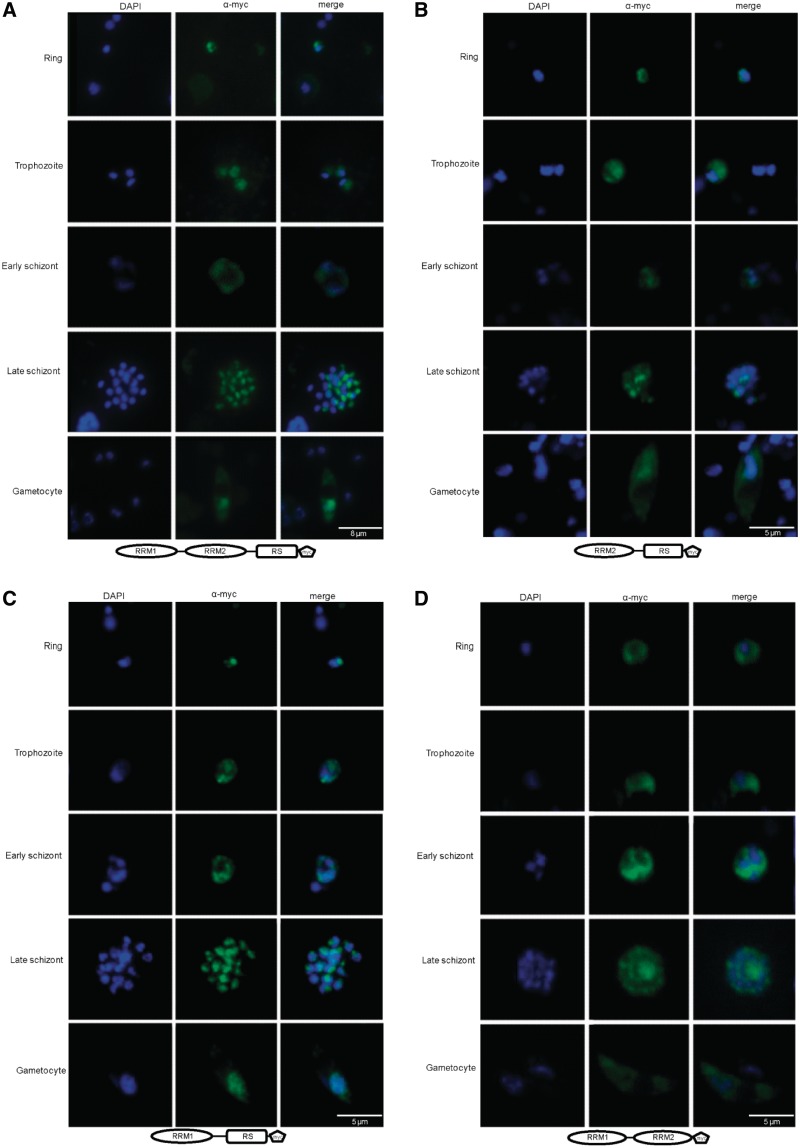
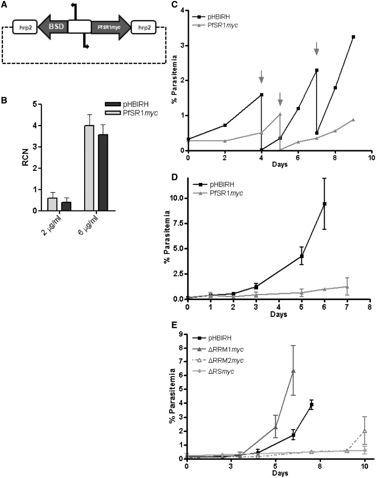
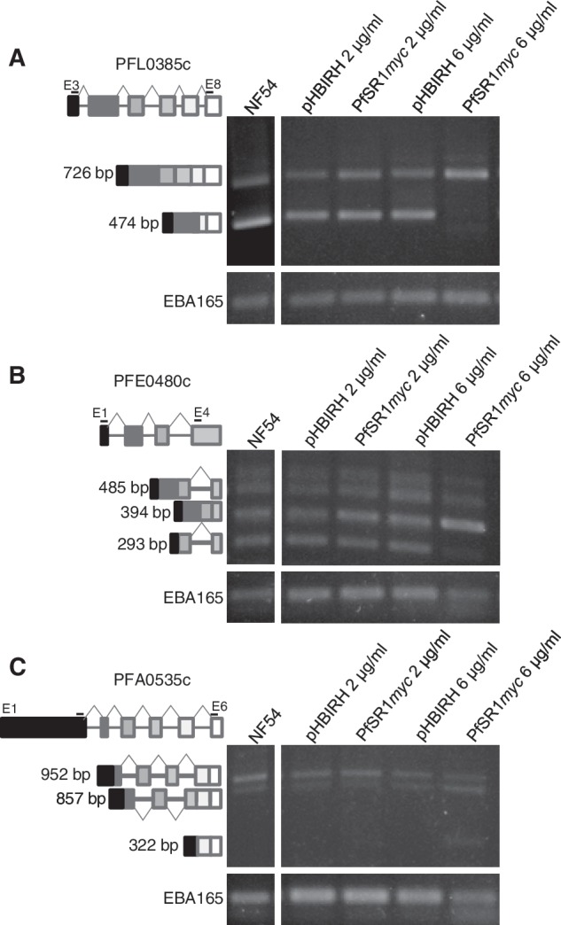
Malaria parasites have a complex life cycle, during which they undergo significant biological changes to adapt to different hosts and changing environments. Plasmodium falciparum, the species responsible for the deadliest form of human malaria, maintains this complex life cycle with a relatively small number of genes. Alternative splicing (AS) is an important post-transcriptional mechanisms that enables eukaryotic organisms to expand their protein repertoire out of relatively small number of genes. SR proteins are major regulators of AS in higher eukaryotes. Nevertheless, the regulation of splicing as well as the AS machinery in Plasmodium spp. are still elusive. Here, we show that PfSR1, a putative P. falciparum SR protein, can mediate RNA splicing in vitro. In addition, we show that PfSR1 functions as an AS factor in mini-gene in vivo systems similar to the mammalian SR protein SRSF1. Expression of PfSR1-myc in P. falciparum shows distinct patterns of cellular localization during intra erythrocytic development. Furthermore, we determine that the predicted RS domain of PfSR1 is essential for its localization to the nucleus. Finally, we demonstrate that proper regulation of pfsr1 is required for parasite proliferation in human RBCs and over-expression of pfsr1 influences AS activity of P. falciparum genes in vivo.


Figures

Figure 1.
Figure 1.
Putative SR and SR-like proteins in P. falciparum. (A) Schematic representation of the domain structure of P. falciparum putative proteins that are predicted to contain RNA Recognition Motif (RRM, ellipses) and SR domain (RS, rectangles). Pervious predictions of homologs are shown with references (B) A phylogenetic tree built with the Neighbor-Joining algorithm based on the entire amino acid sequence of the predicted P. falciparum SR proteins. As shown the closest homologs of SRSF1 are PfSR1 (PFE0865c) and PF10_0217, respectively. (C) Homology modeling of the first RRM of PfSR1 based on the human SRSF1 RRM1. The crystal structure of the human SRSF1 was retrieved from the Protein Data Bank (http://www.rcsb.org/pdb/home/home.do, PDB-1x4aA). The two RRMs have 51% identity over 80 amino acids. The Z-score for the model quality calculated using ProSA (https://prosa.services.came.sbg.ac.at/prosa.php) is 5.5.
Figure 2.
Figure 2.
PfSR1 is a functional SR protein that can mediate pre-mRNA splicing. Recombinant PfSR1 can complement S100 extracts and rescue the splicing activity similar to SRSF1. (A) Schematic of the expression vector used to express the recombinant PfSR1 and SRSF1 in 293 T cells. cDNA of both genes was cloned into the expression vector fused to a His-tag. (B) SDS–PAGE presenting the purified SRSF1 and PfSR1 loaded on gel in increasing volumes (1, 2 and 4 µl, respectively). (C) Complementation of splicing activity of PfSR1 and SRSF1 in S100 extracts (in vitro splicing assay). Increasing amounts of purified SRSF1 or PfSR1 were added to S100 extracts in the presence of radiolabeled Ftz reporter pre-mRNA. Lane 1: labeled Ftz pre-mRNA only; Lane 2: nuclear extract (NE) + Ftz; Lane 3: S100 extract + Ftz; Lane 4: NE + S100 + Ftz; Lanes 5–6: S100 + Ftz with increasing amounts of SRSF1; Lanes 7 and 8: S100 + Ftz with increasing amounts of PfSR1. Schematics of pre-mRNA, spliced mRNA and the spliced intron are shown in the left.
Figure 3.
Figure 3.
PfSR1 is an AS factor with similar activity to SRSF1. (A) Schematic illustration of the experimental procedure designed to demonstrate AS activity of PfSR1. HEK 293 T cells were cotransfected with plasmids that carried a mini-gene as well as an expression vector (pcDNA3). Forty-eight hours post-transfection the cells were harvested and their RNA and protein content were analyzed. (B) Western blot analysis using anti-T7 antibody showing the expression of the tagged SRSF1 (Lane 2) and PfSR1 (Lane 3). An empty pcDNA3 vector was used as a negative control (Lane 1). (C) RT–PCR amplification of total RNA extracted from HEK 293 T cells that were cotransfected with the E1A mini-gene obtained differential splicing patterns mediated by PfSR1 and SRSF1 compared with the control. Schematic of the mini-gene and the differentially spliced isoforms is shown on the left. GAPDH was used as loading control and is presented in the lower panel. (D) Quantification of the changes in the proportion of each spliced isoform of E1A obtained after transfection with PfSR1 and SRSF1 compared with the control. The density of each band is presented as a proportion of the total signal obtained. The averages and standard errors of three different experiments are presented. (E) RT–PCR amplification of total RNA extracted from 293 T cells that were cotransfected with the β-globin mini-gene obtained differential splicing patterns mediated by PfSR1 and SRSF1 compared with the control. Schematic of the mini-gene and the differentially spliced isoforms is shown on the left. GAPDH was used as loading control and is presented in the lower panel. (F) Quantification of the changes in the proportion of each spliced isoform of β-globin obtained after transfection with PfSR1 and SRSF1 compared with the control. The density of each band is presented as a proportion of the total signal obtained. The averages and standard errors of two different experiments are presented.
Figure 4.
Figure 4.
The RS domain localizes PfSR1 to the nucleus. IFAs showing the localization of the entire PfSR1 as well as different PfSR1 mutants lacking either the RRM or the RS domains (ΔRRM1, ΔRRM2 and ΔRS, respectively) during IDC. The different forms of PfSR1 were ectopically express fused to a myc epitope tag and detected using anti-myc antibodies. PfSR1 (green); DAPI staining (blue); developmental stages are indicated on the left. (A) Cellular localization of PfSR1-myc. (B) Cellular localization of the PfSR1ΔRRM1-myc. (C) Cellular localization of the PfSR1ΔRRM2-myc. (D) Cellular localization of the PfSR1ΔRS-myc. The entire PfSR1 and two ΔRRM mutants shuttle between the nucleus and cytoplasm, whereas the ΔRS mutant localizes to the cytoplasm.
Figure 5.
Figure 5.
Proper regulation of the pfsr1 gene is essential for parasites’ proliferation in human RBCs. Over-expression of pfsr1 gene inhibits parasite proliferation. (A) Schematic map of the plasmid (pHBIPfSR1myc) used for ectopically over-expression using increasing concentrations of blasticidin as described (28). Steady-state mRNA levels of pfsr1 and renilla luciferase genes were measured using qRT–PCR from tightly synchronized parasite cultures 36-h post-invasion (hpi). Values are presented as relative copy number to the housekeeping genes arginyl-tRNA synthetase (PFL0900c). (B) Transcription levels of pfsr1 and renilla luciferase expressed from the transfected episomes by parasites growing under 2 and 6 µg/ml blasticidin selection. (C) Growth curves of the parasite populations over-expressing pfsr1 (light gray) compared with those that similarly express renilla luciferase (black) from the pHBIPfSR1myc and the pHBIRH plasmids, respectively, under 2 μg/ml blasticidin. Arrows indicate days in which the culture was cut down to avoid over-parasitemia. Each curve represents the average of three different cultures grown in parallel. (D) Growth curves of the parasite populations over-expressing pfsr1 (light gray) compared with those that similarly express renilla luciferase (black) from the pHBIPfSR1myc and the pHBIRH plasmids, respectively, under 6 μg/ml blasticidin. Day 0 represents the day in which blasticidin concentration was increased. Each curve represents the average of three different parasitemia measurements. (E) Growth curves of the parasite populations over-expressing the different PfSR1 mutants (gray) compared with those that similarly express renilla luciferase (black) under 2 μg/ml blasticidin.
Figure 6.
Figure 6.
Over-expression of the pfsr1 influences AS activity in P. falciparum. RT–PCR amplifications on cDNA of three P. falciparum endogenous genes reveals specific changes in their splicing patterns mediated by PfSR1. cDNA from NF54, wile-type untrasfected parasites was used as control. For each gene RT–PCR amplifications of mRNA extracted from parasites transfected with either pHBIRH or pHBISR1myc and selected on 2 and 6 μg/ml blasticidin are presented. Primers used for these amplifications are listed in Supplementary Table S1. (A) The spliced isoforms obtained for PFL0385c. (B) The spliced isoforms obtained for PFE0480c and (C) The splicing isoforms obtained PFA0535c. Schematic of the genes and the differentially spliced isoforms is shown on the left. Exons are illustrated in boxes and introns in lines. Positions of the PCR primers that were used to amplify each gene are indicated. EBA165 (PFD1155w) was used as loading control and is presented in the lower panels. All of the detected isoforms were sub-cloned and sequenced.

References

    1. WHO. 2010 World Malaria Report (2010)
    1. Bozdech Z, Llinas M, Pulliam BL, Wong ED, Zhu JC, Derisi JL. The transcriptome of the intraerythrocytic developmental cycle of Plasmodium falciparum. PloS Biol. 2003;1:85–100. - PMC - PubMed
    1. Gardner MJ, Hall N, Fung E, White O, Berriman M, Hyman RW, Carlton JM, Pain A, Nelson KE, Bowman S, et al. Genome sequence of the human malaria parasite Plasmodium falciparum. Nature. 2002;419:498–511. - PMC - PubMed
    1. Keren H, Lev-Maor G, Ast G. Alternative splicing and evolution: diversification, exon definition and function. Nat. Rev. Genet. 2010;11:345–355. - PubMed
    1. Knapp B, Nau U, Hundt E, Kupper HA. Demonstration of alternative splicing of a pre-mRNA expressed in the blood stage form of Plasmodium falciparum. J. Biol. Chem. 1991;266:7148–7154. - PubMed

Publication types

MeSH terms