. 2012 Sep;19(9):925-9.
doi: 10.1038/nsmb.2358. Epub 2012 Aug 12.

The structure of purified kinetochores reveals multiple microtubule-attachment sites

Affiliations

The structure of purified kinetochores reveals multiple microtubule-attachment sites

Shane Gonen et al. Nat Struct Mol Biol. 2012 Sep.

Abstract

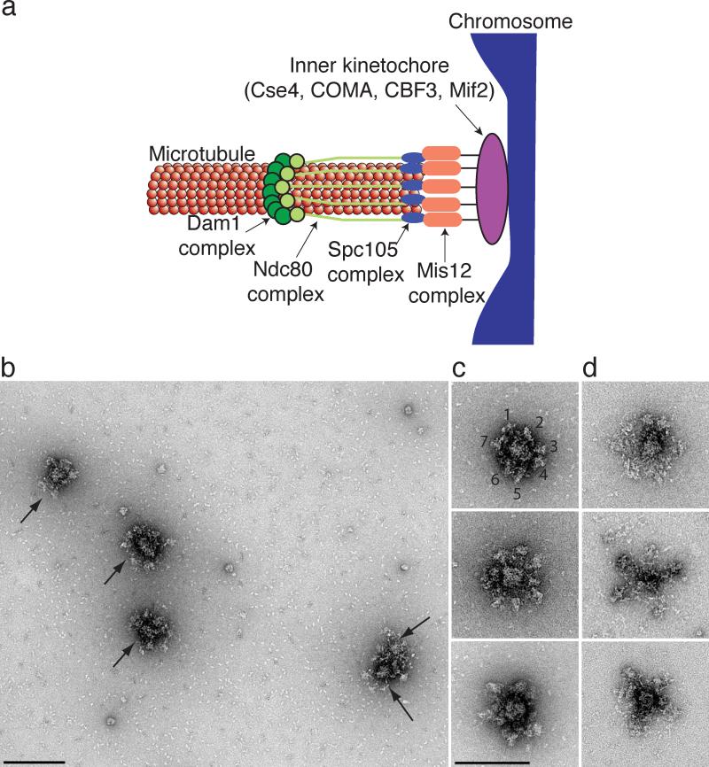
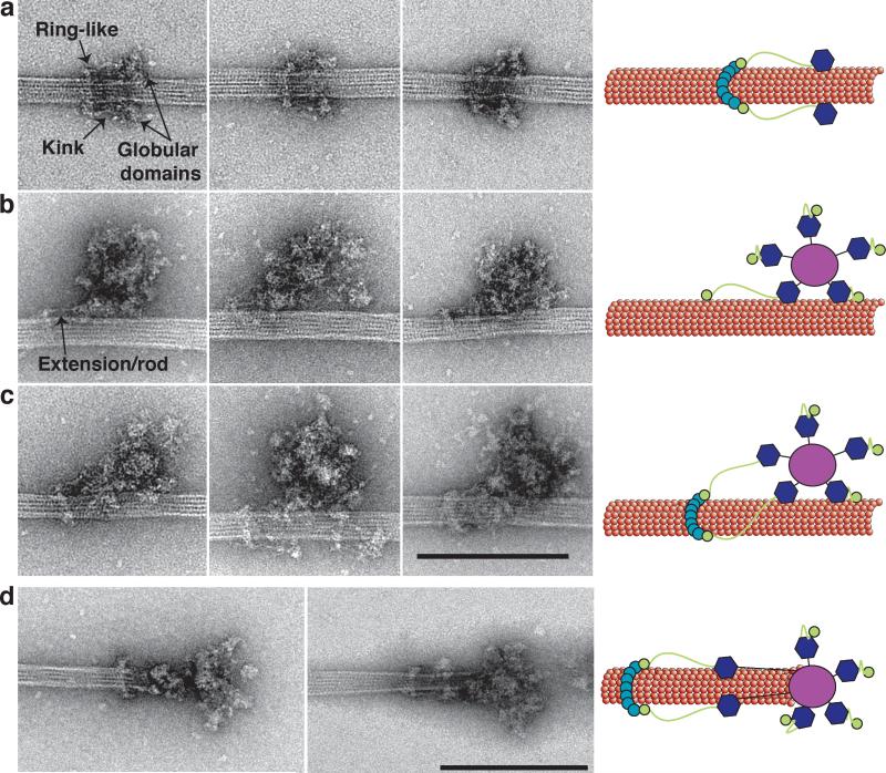
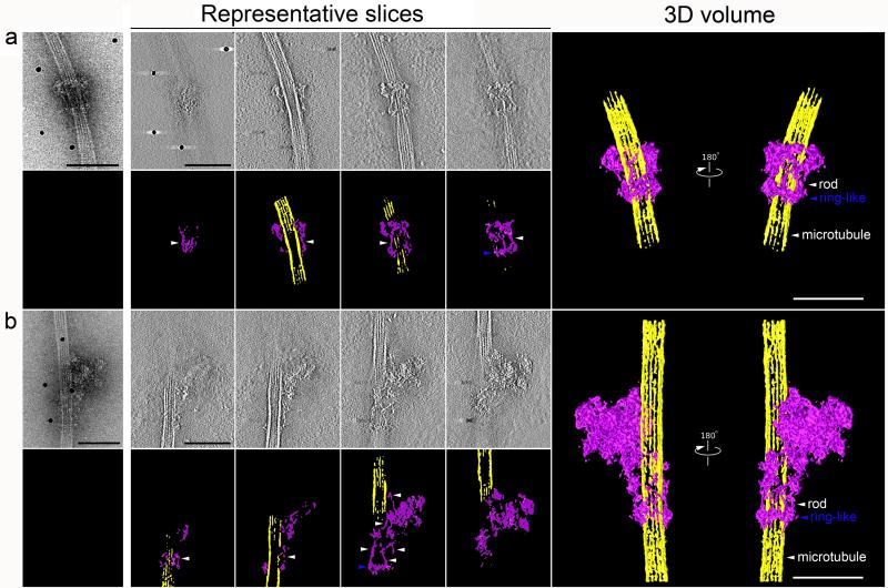
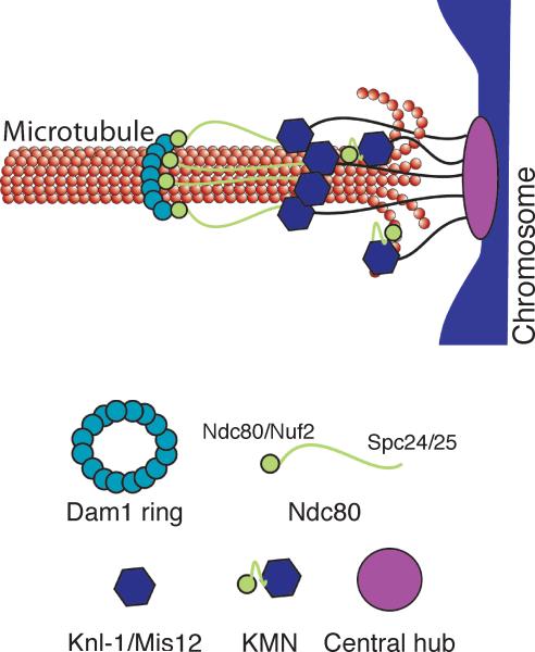
Chromosomes must be accurately partitioned to daughter cells to prevent aneuploidy, a hallmark of many tumors and birth defects. Kinetochores are the macromolecular machines that segregate chromosomes by maintaining load-bearing attachments to the dynamic tips of microtubules. Here, we present the structure of isolated budding-yeast kinetochore particles, as visualized by EM and electron tomography of negatively stained preparations. The kinetochore appears as an ~126-nm particle containing a large central hub surrounded by multiple outer globular domains. In the presence of microtubules, some particles also have a ring that encircles the microtubule. Our data, showing that kinetochores bind to microtubules via multivalent attachments, lay the foundation to uncover the key mechanical and regulatory mechanisms by which kinetochores control chromosome segregation and cell division.


Figures

Figure 1
Figure 1. Kinetochore particles contain a central hub surrounded by a number of globular domains
(a) A model for the budding yeast kinetochore shows that multiple copies of the Dam1, Ndc80, KNL-1 (Spc105) and Mis12 kinetochore subcomplexes mediate binding of the chromosome (blue) to the microtubule. The inner kinetochore contains one or more copies of the Cse4, Mif2, CBF3 and COMA subcomplexes. (b) A field of kinetochore particles in microtubule polymerization buffer was visualized by EM of negatively stained preparations. Five particles (arrows) and other small material are apparent. Note that two particles are touching. Scale bar = 200 nm. (c) Images of representative compact kinetochore particles in microtubule polymerization buffer with lower salt. The globular domains on a single particle in the top panel are numbered. (d). The particles are more extended in higher salt buffer used for purification. Scale bar = 150 nm. Additional particles are presented in Supplementary Figure 1.
Figure 2
Figure 2. Kinetochore particles bound to taxol-stabilized microtubules
(a) Representative images of fragments of kinetochore particles (56 nm long) bound to taxol-stabilized microtubules reveal a rod with a kink (arrow) connected to a ring on one end and a globular domain on the opposite end. (b) Large kinetochore particles (126 nm long) bind to microtubules through globular domains and an additional extension/rod (arrow) that emanates from one of the globular domains. (c) Large kinetochore particles bind to microtubules through multiple globular domains and contain an extension that connects to a ring. Scale bar is 200 nm. (d) Two selected images of kinetochores at the tip of taxol-stabilized microtubules. Globular domains extending 50 nm from the central hub bind to the microtubules and are connected to a distal ring 50 nm further. Scale bar is 200 nm. Cartoons on the left schematize the key features of the images.
Figure 3
Figure 3. Three-dimensional structures of two types of kinetochore particles bound to a microtubule
a and b, projection images of the two types of kinetochore assemblies on taxol-stabilized microtubules selected for tomographic reconstruction. Representative slices through the each particle are presented with the corresponding segmentation analysis in color underneath. Finally, 3D reconstruction of this complex by electron tomography is presented on the right. All slices are arranged from left to right traversing through the z-axis from bottom to top with the bottom defined at the grid surface (carbon layer). Both structures contain a ring-like structure (blue arrowhead) and multiple rods (white arrowhead). Scale bars for all panels represent 100 nm. Movies are presented in Supplementary Material.
Figure 4
Figure 4. Schematic of the proposed model of kinetochore architecture
The central globular domain binds to the centromeric locus of the chromosome and globular domains containing the KMN complex extend to attach to the microtubule. The Ndc80 subcomplex makes an additional extension to contact a distal ring composed of the Dam1 subcomplex.

Comment in

References

    1. Pfau SJ, Amon A. Chromosomal instability and aneuploidy in cancer: from yeast to man. EMBO Rep. 2012;13:515–27. - PMC - PubMed
    1. Compton DA. Mechanisms of aneuploidy. Curr Opin Cell Biol. 2011;23:109–13. - PMC - PubMed
    1. Santaguida S, Musacchio A. The life and miracles of kinetochores. EMBO J. 2009;28:2511–31. - PMC - PubMed
    1. Cheeseman IM, Desai A. Molecular architecture of the kinetochore-microtubule interface. Nat Rev Mol Cell Biol. 2008;9:33–46. - PubMed
    1. Takeuchi K, Fukagawa T. Molecular architecture of vertebrate kinetochores. Exp Cell Res. 2012;318:1367–74. - PubMed

Publication types

MeSH terms

Substances