. 2012 Oct-Dec;6(4):290-7.
doi: 10.4161/fly.21534. Epub 2012 Aug 13.

Drosulfakinin activates CCKLR-17D1 and promotes larval locomotion and escape response in Drosophila

Affiliations

Drosulfakinin activates CCKLR-17D1 and promotes larval locomotion and escape response in Drosophila

Xu Chen et al. Fly (Austin). 2012 Oct-Dec.

Abstract

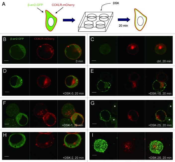
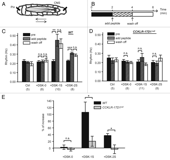
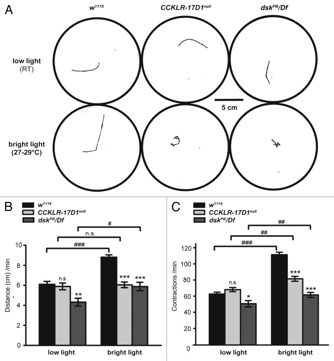
Neuropeptides are ubiquitous in both mammals and invertebrates and play essential roles in regulation and modulation of many developmental and physiological processes through activation of G-protein-coupled-receptors (GPCRs). However, the mechanisms by which many of the neuropeptides regulate specific neural function and behaviors remain undefined. Here we investigate the functions of Drosulfakinin (DSK), the Drosophila homolog of vertebrate neuropeptide cholecystokinin (CCK), which is the most abundant neuropeptide in the central nervous system. We provide biochemical evidence that sulfated DSK-1 and DSK-2 activate the CCKLR-17D1 receptor in a cell culture assay. We further examine the role of DSK and CCKLR-17D1 in the regulation of larval locomotion, both in a semi-intact larval preparation and in intact larvae under intense light exposure. Our results suggest that DSK/CCKLR-17D1 signaling promote larval body wall muscle contraction and is necessary for mediating locomotor behavior in stress-induced escape response.


Figures

None
Figure 1. Translocation of βarr2-GFP in response to DSK-1S and DSK-2S, but not DSK-0, in HEK-293 cells expressing CCKLR-17D1. (A) Dual-label system for visualization of βarr2-GFP translocation in GPCR-expressing cells. mCherry-tagged CCKLR-17D1 enables visualization of cells with GPCR expression and proper localization to the cell membrane. (B–H) Confocal images (0.1 μM optical slice) of HEK-293 cells transiently expressing both CCKLR-17D1-mCherry and βarr2-GFP. (B) Before addition of peptide, CCKLR-17D1-mCherry localizes to the cell membrane and βarr2-GFP is diffuse in the cytoplasm. (C) Control cells with addition of peptide-free solution (sham experiment) for 20 min. In cells that are exposed to DSK-0 (D), DSK-1 (F) or DSK-2 (H) for 20 min, βarr2-GFP remains diffuse in the cytoplasm, similar to control cells. When cells are exposed to DSK-1S (E) and DSK-2S (G) for 20 min, βarr2-GFP displays clear translocation to the membrane. Cells marked with asterisks in this panel are examples of cells that express βarr2-GFP but fail to express CCKLR-17D1-mCherry. As shown, these cells do not respond to the addition of DSK-2S or DSK-1S (not shown) peptides indicating that the response is dependent on the presence of CCKLR-17D1-mCherry rather than an endogenous receptor present in HEK-293 cells. (I) Z-stack of confocal images of HEK-293 cells exposed to DSK-2S for 20 min. Peptides were applied at 1 μM.
None
Figure 2. DSK-1S and DSK-2S promote larval fictive locomotion. (A) Diagram of restrained semi-intact third instar wandering larva (arrow indicates direction of peristaltic wave of locomotor muscle contractions). (B) Diagram of the experimental scheme. (C) Frequency of fictive locomotion in semi-intact wild-type larvae significantly increases following addition of DSK-1S or DSK-2S into hemolymph-like saline HL-3 and remains high after wash out. Frequency of fictive locomotion does not change after addition of DSK-0. In the control (ctrl) experiment, peptide-free solution is added at 2 min. (n) is the number of larvae scored. Error bars denote SEM ***, p < 0.001. **, p < 0.01. n.s., not significant (paired Student’s t-test). (D) Contraction frequency in semi-intact CCKLR-17D1null mutant larvae does not change significantly after addition of DSK-0, DSK-1S, or DSK-2S (paired Student’s t-test). (E) Percent increase in contraction frequency after addition of peptide. Percent increase is calculated for each individual larva as (frqpost – frqpre)/frqpre. Error bars denote SEM *, p < 0.05. n.s., not significant (unpaired Student’s t-test).
None
Figure 3.CCKLR-17D1 and dsk mutants exhibit deficits in stress-induced escape behavior. (A) Representative traces of tracks made by crawling larvae of the indicated genotype during a 1 min recording period under dim light (20–22°C) and bright light (27–30°C). (B) Distance traveled during a 1 min recording period under low light and bright light. The distance traveled by control larvae is significantly increased under bright light (p < 0.001). In contrast, dskPB/Df mutants exhibit a relatively smaller increase (p < 0.05) and CCKLR-17D1null mutants exhibit no significant increase under bright light. Compared with control larvae, both CCKLR-17D1null and dskPB/Df mutants travel significantly shorter distance under bright light conditions (p < 0.001). (C) Number of body wall contractions during a 1 min recording period under low light and bright light. The number of contractions in control larvae is significantly increased under bright light (p < 0.001). Both CCKLR-17D1null and dskPB/Df mutant larvae also show a significant increase in the number of contractions under bright light but not to the same extent as control larvae and the number of contractions in both mutants differs significantly from controls (p < 0.001). Number of larvae assayed: w1118 dark (28), CCKLR-17D1null dark (27), dskPB/Df dark (11), w1118 light (34), CCKLR-17D1null light (27), dskPB/Df light (10). Error bars denote SEM *** or ###, p < 0.001. ** or ##, p < 0.01. * or #, p < 0.05. n.s, not significant (unpaired Student’s t-test was used in all cases since each larva was tested only once. Pairwise comparisons are either with w1118 control under the same condition or as indicated by brackets).

References

    1. Nässel DR. Neuropeptides in the nervous system of Drosophila and other insects: multiple roles as neuromodulators and neurohormones. Prog Neurobiol. 2002;68:1–84. doi: 10.1016/S0301-0082(02)00057-6. - DOI - PubMed
    1. Hewes RS, Taghert PH. Neuropeptides and neuropeptide receptors in the Drosophila melanogaster genome. Genome Res. 2001;11:1126–42. doi: 10.1101/gr.169901. - DOI - PMC - PubMed
    1. Hauser F, Williamson M, Cazzamali G, Grimmelikhuijzen CJ. Identifying neuropeptide and protein hormone receptors in Drosophila melanogaster by exploiting genomic data. Brief Funct Genomic Proteomic. 2006;4:321–30. doi: 10.1093/bfgp/eli003. - DOI - PubMed
    1. Fink H, Rex A, Voits M, Voigt JP. Major biological actions of CCK--a critical evaluation of research findings. Exp Brain Res. 1998;123:77–83. doi: 10.1007/s002210050546. - DOI - PubMed
    1. Price DA, Greenberg MJ. Purification and characterization of a cardioexcitatory neuropeptide from the central ganglia of a bivalve mollusc. Prep Biochem. 1977;7:261–81. doi: 10.1080/00327487708061643. - DOI - PubMed

Publication types

MeSH terms

LinkOut - more resources