. 2012 Sep 20:1474:29-39.
doi: 10.1016/j.brainres.2012.07.057. Epub 2012 Aug 4.

zRICH, a protein induced during optic nerve regeneration in zebrafish, promotes neuritogenesis and interacts with tubulin

Affiliations

zRICH, a protein induced during optic nerve regeneration in zebrafish, promotes neuritogenesis and interacts with tubulin

Satya S Pathi et al. Brain Res. .

Abstract

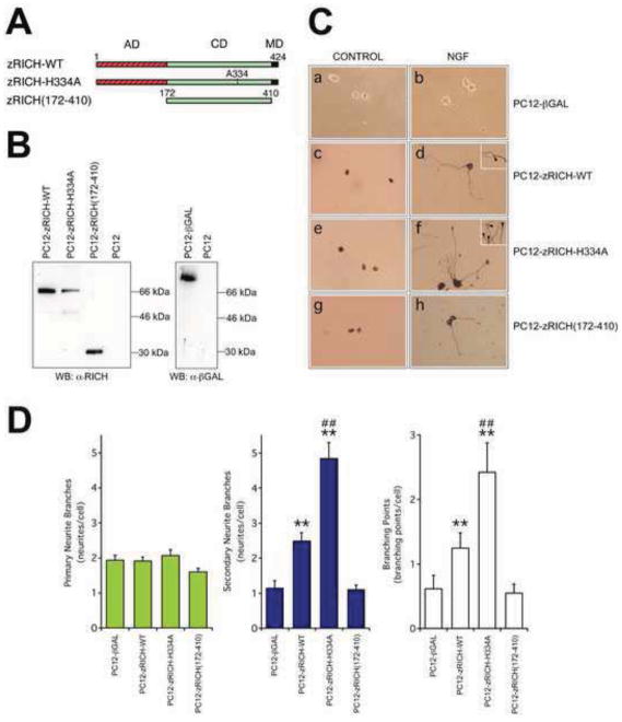
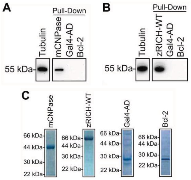
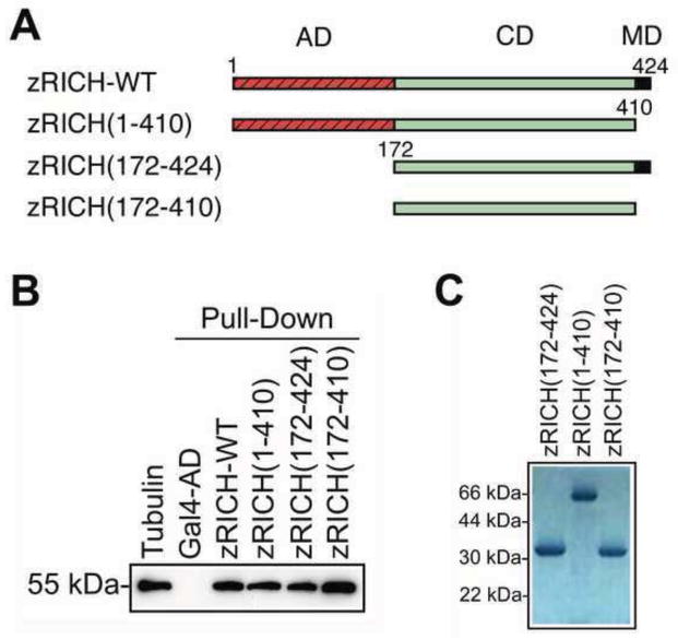
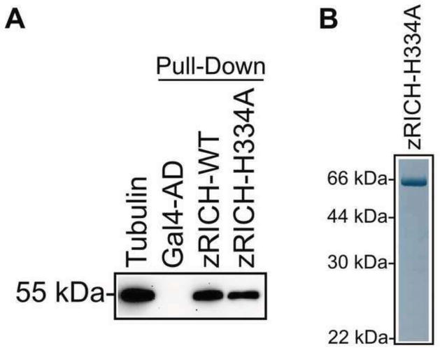
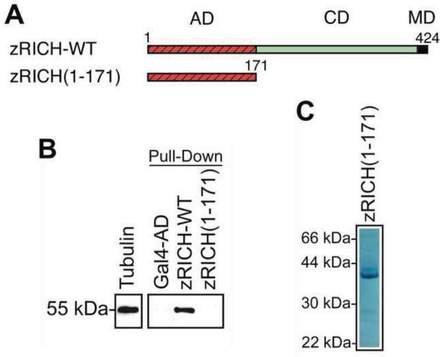
Mammals do not regenerate axons in their central nervous system (CNS) spontaneously. This phenomenon is the cause of numerous medical conditions after damage to nerve fibers in the CNS of humans. The study of the mechanisms of nerve regeneration in other vertebrate animals able to spontaneously regenerate axons in their CNS is essential for understanding nerve regeneration from a scientific point of view, and for developing therapeutic approaches to enhance nerve regeneration in the CNS of humans. RICH proteins are a novel group of proteins implicated in nerve regeneration in the CNS of teleost fish, yet their mechanisms of action are not well understood. A number of mutant versions of the zebrafish RICH (zRICH) protein were generated and characterized at biochemical and cellular levels in our laboratory. With the aim of understanding the effects of RICH proteins in neuronal axon outgrowth, stable transfectants derived from the neuronal model PC12 cell line expressing zRICH Wild-Type or mutant versions of zRICH were studied. Results from differentiation experiments suggest that RICH proteins enhance neuronal plasticity by facilitating neurite branching. Biochemical co-purification results have demonstrated that zRICH binds to the cytoskeletal protein tubulin. The central domain of the protein is sufficient for tubulin binding, but a mutant version of the protein lacking the terminal domains, which cannot bind to the plasma membrane, was not able to enhance neurite branching. RICH proteins may facilitate axon regeneration by regulating the axonal cytoskeleton and facilitating the formation of new neurite branches.


Figures

Figure 1
Figure 1
Effects of zRICH proteins on neuronal plasticity of transfected PC12 cells. A: Structure of the proteins expressed in PC12 cells. There are three domains in Wild-Type zRICH protein, an amino-terminal acidic domain, a central CNPase homology domain, and a small carboxyl-terminal membrane localization domain (which contains an isoprenylation motif). The mutant protein zRICH-H334A has a highly conserved histidine amino acid residue (H334), which is essential for catalysis, substituted by an alanine. Both the acidic domain and the membrane localization domain are missing in the zRICH(172-410) mutant version of the protein. Numbers indicated are amino acid positions related to the Wild-Type protein. (AD: acidic domain, shown in red with black stripes; CD: CNPase homology domain, shown in green; MD: membrane localization domain containing isoprenylation-motif, shown in black). B: Western blot analysis of PC12 stable transfectants. The proteins in lysates from cells transfected with eukaryotic expression plasmids were separated by SDS-PAGE and blotted onto a nitrocellulose membrane. Immunodetection was performed with polyclonal anti-RICH antibody (left membrane) or anti-β-Galactosidase antibody (right membrane). All the three different versions of zRICH and the β-Galactosidase proteins were expressed in the corresponding stable transfectants, as shown by the presence of specific immunoreactive bands. Lysates from untransfected parental PC12 cells were used as negative controls. C: Differentiation characteristics of PC12 cells expressing zRICH proteins. Stable transfectant PC12 cells expressing zRICH-WT, zRICH-H334A, or zRICH(172-410) versions of zRICH protein were detected by immunocytochemistry with anti-RICH antibody. Expression of high levels of zRICH-WT or mutant versions of the protein in PC12 cells did not result in neurite extension in the absence of NGF (panels c, e, g). NGF treatment triggered neurite branching in the PC12 stable transfectants expressing the zRICH proteins (panels d, f, h). Cells expressing zRICH-WT or catalytically inactive mutant zRICH-H334A often displayed more extensive branching than cells expressing the cytosolic mutant zRICH(172-410). The insets in panels d and f show a magnified view of neurites with arrowheads marking the location of branching points. PC12-βgal stable transfectants expressing β-galactosidase were used as controls (panels a, b). D: Morphometric analysis of neurites in differentiated stable transfectants. From left to right, the graphs represent the number of primary neurites, the number of secondary neurite segments, and the number of branching points per cell, respectively. Compared to the control PC12 expressing β-galactosidase, cells expressing zRICH-WT showed significant increases in branching points and formation of secondary neurites, but not in the number of primary neurites. The neurite branching effect of the catalytically deficient zRICH-H334A mutant was even more pronounced. Conversely, the cytosolic mutant zRICH(172-410) was not able to enhance neurite branching, and in fact the cells displayed slight decreases in neurite formation. The bars show the average ± SEM; n = 49 PC12-βgal, 70 PC12-zRICH-WT, 57 PC12-zRICH-H334A, and 75 PC12-zRICH(172-410) cells. Statistical analysis (Tukey-HSD): * p < 0.05, ** p < 0.01, groups were compared with PC12-βgal control; additionally, PC12-zRICH-H334A cells were compared with PC12-zRICH-WT cells (# p < 0.05, ## p <0.01).
Figure 2
Figure 2
Interaction of tubulin with zRICH-WT. A: Co-affinity purification (pull-down) of tubulin with mCNPase. Purified brain tubulin incubated with the indicated purified recombinant proteins were subjected to an affinity pull-down procedure. Immunodetection was performed with anti-α-tubulin antibody. Tubulin co-purified specifically with mCNPase, but was not pulled-down by the control recombinant proteins Gal4-AD or Bcl-2. Results are representative of three independent experiments. B: Co-affinity purification (pull-down) of tubulin with zRICH-WT. Purified brain tubulin incubated with the indicated purified recombinant proteins were subjected to an affinity pull-down procedure. Tubulin co-purified specifically with zRICH-WT, but not with negative control proteins, suggesting direct interaction between these two proteins. Results are representative of three independent experiments. C: Electrophoretic analysis of purified recombinant proteins used in pull-down assays. The position and size of the molecular weight standards is indicated. (Please note that zRICH-WT migrates anomalously approximately 20 kDa above the similarly sized mCNPase due to its highly acidic N-terminal domain).
Figure 3
Figure 3
Interaction of tubulin with the catalytic domain of zRICH. A: Structure of the zRICH domain deletion mutants. The zRICH(1-410) deletion mutant is missing the small carboxyl-terminal membrane localization domain. The zRICH(172-424) deletion mutant is lacking the amino-terminal acidic domain. The zRICH(172-410) is missing both terminal domains, retaining only the central CNPase homology domain. Numbers indicated are amino acid positions related to the Wild-Type protein. (AD: acidic domain, shown in red with black stripes; CD: CNPase homology domain, shown in green; MD: membrane localization domain containing isoprenylation-motif, shown in black). B: Co-affinity purification (pull-down) of tubulin with zRICH deletion mutants. Purified brain tubulin and purified recombinant proteins were subjected to affinity pull-down assays. Tubulin co-purified specifically with zRICH-WT, and with all three deletion mutants zRICH(1-410), zRICH(172-424), and zRICH(172-410), indicating that the central catalytic domain is sufficient for interaction with tubulin. Specificity of the pull-down assay was confirmed with a negative control performed with the recombinant protein Gal4-AD. Results are representative of three independent experiments. C: Electrophoretic analysis of purified recombinant zRICH(172-424), zRICH(1-410) and zRICH(172-410) proteins. The position and size of the molecular weight standards is indicated.
Figure 4
Figure 4
Interaction of tubulin with zRICH is independent of phosphodiesterase enzymatic activity. A: Co-affinity purification (pull-down) of tubulin with zRICH-H334A. Purified brain tubulin and purified recombinant proteins were subjected to affinity pull-down assays. Tubulin co-purified specifically with zRICH-WT and with the catalytically inactive zRICH-H334A mutant version of the protein. The results indicate that catalytic activity is not required for interaction of tubulin with the central domain of zRICH. A negative control for the affinity pull-down assay was performed with the recombinant protein Gal4-AD. Results are representative of three independent experiments. B: Electrophoretic analysis of purified recombinant zRICH-H334A. The position and size of the molecular weight standards is indicated.
Figure 5
Figure 5
The acidic domain of zRICH is not sufficient for co-purification of tubulin. A: Structure of the zRICH(1-171) mutant. A new deletion mutant of zRICH comprising exclusively the amino-terminal acidic domain was generated. The zRICH(1-171) deletion mutant is missing both the central catalytic domain and the small carboxyl-terminal membrane localization domain. Numbers indicated are amino acid positions related to the Wild-Type protein. (AD: acidic domain, shown in red with black stripes; CD: CNPase homology domain, shown in green; MD: membrane localization domain containing isoprenylation-motif, shown in black). B: Tubulin does not co-purify with zRICH(1-171) in pull-down assays. Purified brain tubulin and purified recombinant proteins were subjected to affinity pull-down assays. Tubulin co-purified specifically with zRICH-WT, but not with the deletion mutant zRICH(1-171), indicating that the amino-terminal acidic domain of zRICH is not a major determinant of the interaction of zRICH with tubulin. Specificity of the pull-down assay was confirmed with a negative control performed with the recombinant protein Gal4-AD. Results are representative of three independent experiments. C: Electrophoretic analysis of purified recombinant zRICH(1-171). The position and size of the molecular weight standards is indicated.

Similar articles

Cited by

References

    1. Agranoff BW, Ford-Holevinski TS. Biochemical aspects of the regenerating goldfish visual system. In: Elam JS, Cancalon P, editors. Axonal transport in neuronal growth and regeneration. Plenum Press; New York: 1984. pp. 69–86.
    1. Aigner L, Arber S, Kapfhammer JP, Laux T, Schneider C, Botteri F, Brenner HR, Caroni P. Overexpression of the neural growth-associated protein GAP-43 induces nerve sprouting in the adult nervous system of transgenic mice. Cell. 1995;83:269–278. - PubMed
    1. Asch WS, Leake D, Canger AK, Passini MA, Argenton F, Schechter N. Cloning of zebrafish neurofilament cDNAs for plasticin and gefiltin: increased mRNA expression in ganglion cells after optic nerve injury. J Neurochem. 1998;71:20–32. - PubMed
    1. Azarashvili T, Stricker R, Reiser G. The mitochondria permeability transition pore complex in the brain with interacting proteins - promising targets for protection in neurodegenerative diseases. Biol Chem. 2010;391:619–629. - PubMed
    1. Bahr M, Bonhoeffer F. Perspectives on axonal regeneration in the mammalian CNS. Trends Neurosci. 1994;17:473–479. - PubMed

Publication types

LinkOut - more resources