. 2012 Oct 4;490(7418):116-20.
doi: 10.1038/nature11378. Epub 2012 Aug 12.

Burkitt lymphoma pathogenesis and therapeutic targets from structural and functional genomics

Affiliations

Burkitt lymphoma pathogenesis and therapeutic targets from structural and functional genomics

Roland Schmitz et al. Nature. .

Abstract

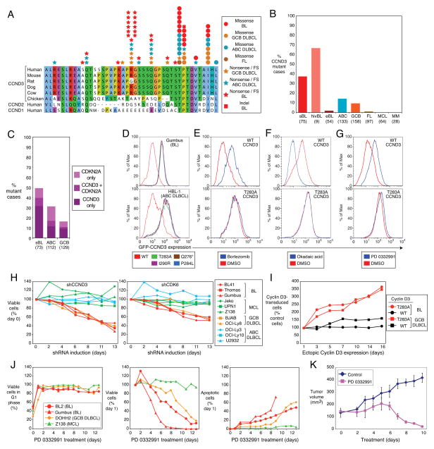
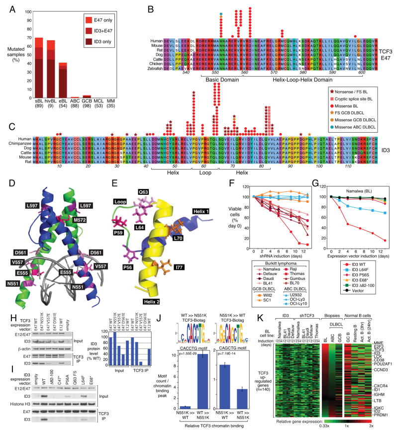
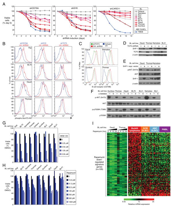
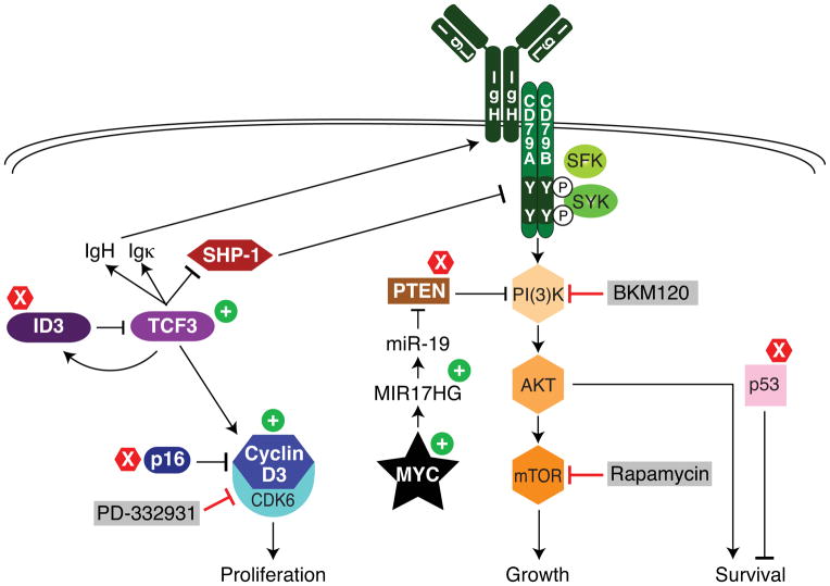
Burkitt's lymphoma (BL) can often be cured by intensive chemotherapy, but the toxicity of such therapy precludes its use in the elderly and in patients with endemic BL in developing countries, necessitating new strategies. The normal germinal centre B cell is the presumed cell of origin for both BL and diffuse large B-cell lymphoma (DLBCL), yet gene expression analysis suggests that these malignancies may use different oncogenic pathways. BL is subdivided into a sporadic subtype that is diagnosed in developed countries, the Epstein-Barr-virus-associated endemic subtype, and an HIV-associated subtype, but it is unclear whether these subtypes use similar or divergent oncogenic mechanisms. Here we used high-throughput RNA sequencing and RNA interference screening to discover essential regulatory pathways in BL that cooperate with MYC, the defining oncogene of this cancer. In 70% of sporadic BL cases, mutations affecting the transcription factor TCF3 (E2A) or its negative regulator ID3 fostered TCF3 dependency. TCF3 activated the pro-survival phosphatidylinositol-3-OH kinase pathway in BL, in part by augmenting tonic B-cell receptor signalling. In 38% of sporadic BL cases, oncogenic CCND3 mutations produced highly stable cyclin D3 isoforms that drive cell cycle progression. These findings suggest opportunities to improve therapy for patients with BL.


Figures

Figure 1
Figure 1. OncogenicCCND3 mutations in Burkitt lymphoma
a, CCND3 mutations in BL, GCB DLBCL, ABC DLBCL and follicular lymphoma (FL) are shown with respect to amino acid positions 250–292 of protein accession NP_001751. b, Frequencies of CCND3 mutations in different lymphoma subtypes. c, Occurrence of CDKN2A and CCND3 mutations in BL, ABC DLBCL and GCB DLBCL Number of samples analyzed are indicated in parentheses. d, CCND3 mutations increase protein stability. FACS analysis of lymphoma cell lines transduced with mutant or wild type GFP-CCND3 fusion proteins. Cells expressing the different cyclin D3 isoforms had equivalent expression of Lyt2-reporter encoded by the same mRNA as the GFP-CCND3 proteins. e, Mutant cyclin D3 proteins are not degraded by the proteasome. GFP-CCND3-transduced HBL1 cells were cultured overnight in the presence of the proteasomal inhibitor bortezomib (20 nM) and analyzed by FACS. f, Mutant cyclin D3 proteins are not destabilized in response to phosphatase inhibition. BL41 cells expressing GFP-CCND3 proteins were treated for 30 min with the pan-phosphatase inhibitor okadaic acid (750 nM) and analyzed by FACS. g, Stability of mutant cyclin D3 proteins is not regulated in the cell cycle. BL41 cells expressing GFP-CCND3 isoforms were treated overnight with the CDK4/6 inhibitor PD 0332991 (1.5 μM), causing arrest at the G1 phase of the cell cycle. FACS analysis indicated that wild type, but not mutant cyclin D3 fusion proteins were stabilized in G1 phase. h, CCND3 and CDK6 shRNAs have selective toxicity for BL and GCB DLBCL cell lines. Shown is the fraction of GFP+, shRNA-expressing cells relative to the GFP, shRNA-negative fraction at the indicated times, normalized to the day 0 values. i, Mutant CCND3 confers a proliferation advantage in lymphoma cells. Endogenous CCND3 was knocked down by induction of a CCND3 shRNA in Gumbus (BL) and BJAB (GCB DLBCL) cells, while different isoforms of GFP-CCND3 were ectopically expressed. The relative number of GFP-CCND3 expressing cells is plotted versus time after shRNA and GFP-CCND3 induction, normalized to day 0. j, Cell cycle block in G1 phase is lethal to BL and GCB DLBCL cell lines. BL, GCB DLBCL and mantle cell lymphoma (MCL) cell lines were treated with PD 0332991 (1 μM) over indicated time course. G1 phase arrest of viable cells was confirmed using Propidium Iodide (PI) staining and analyzed by FACS (left panel); viable cells were counted using the trypan blue method (middle panel); apoptotic cells were quantified by cleaved PARP/active caspase 3 FACS-staining (right panel). Survival of cells and the fraction of apoptotic cells after PD 0332991 treatment is shown normalized to day 1. Time courses of G1 phase arrest and apoptotic cells were discontinued after day 9 for Gumbus cell line due to the lack of viable cells. All analyses were performed in triplicate. k, Therapeutic potential of the CDK4/6 inhibitor PD 0332991 revealed using a BL xenograft model. Immunodeficient mice bearing established subcutaneous BL xenografts were treated with PD 0332991 (150 mg/kg/day p.o.) for the indicated times. Tumor volumes were estimated by quantitative imaging of luciferase luminescence. Error bars are s.e.m. (n=3). FS: frameshift
Figure 2
Figure 2. Transcriptional activity of TCF3 is essential for Burkitt lymphoma viability
a, Fre quencies of TCF3 and ID3 mutations in different lymphoma subtypes. b, TCF3 missense mutations in the E47 isoform in BL, ABC DLBCL and GCB DLBCL. Amino acid positions 532–604 of E47 are shown according to protein accession NP_001129611. c, ID3 mutations in BL. Amino acid positions of full length ID3 protein are shown according to protein accession NP_002158. d, Location of TCF3 mutations in the three-dimensional structure of the dimeric E47 basic-helix-loop-helix domain from reference . e, Location of ID3 mutations in the three-dimensional structure the ID3 helix-loop-helix domain (pdb accession 2LFH). f, Selective toxicity of TCF3 shRNA for BL cell lines. A vector co-expressing a TCF3 shRNA and GFP was transduced into the indicated cell lines. shRNA expression was induced for the indicated times and the fraction of live GFP+, shRNA+ cells was normalized to the value on day 0. Data represent at least four independent experiments. g, Toxicity of wild type but not mutant ID3 isoforms in ID3-mutant Namalwa cell line. A vector co-expressing ID3 and GFP was transduced into the indicated cell lines. ID3 expression was induced for the indicated times and the fraction of live GFP+, ID3+ cells was normalized to the value on day 0. h, TCF3 E47 mutants with reduced ability to bind ID3. Wild type or mutant TCF3 isoforms were coexpressed with wild type ID3 in 293T cells, and the indicated proteins were assessed either by Western blot analysis in total cellular extracts or after immunoprecipatation (IP) with anti-TCF3 antibodies. ID3 protein levels based on densitometric quantitation of the Western blot results were normalized to E47 protein levels and graphed in the right panel. i, BL-derived mutant ID3 proteins accumulate to lower levels than wild type ID3 and have decreased ability to bind to TCF3. Mutant or wild type ID3 isoforms were expressed in the ID3-deficient Namalwa cell line and analyzed by Western blot for total ID3 protein or ID3 protein immunoprecipitated with TCF3. See text for details. j. DNA sequence motifs enriched under TCF3 genomic binding peaks. Shown at the top are the motifs that were most enriched under peaks that were bound > 4-fold more or less by N551K TCF3 compared to wild type TCF3. The mean number (+/−SEM) of the indicated motifs per peak is plotted below. k, A TCF3 gene expression signature is highly expressed in BL and in normal germinal center cells. Changes of gene expression were profiled in ID3-mutant BL cell lines following TCF3 knockdown or wild type ID3 overexpression. Shown are genes that were downregulated by at least 0.33 log2 in >70% of samples following TCF3 knockdown or following ID3 overexpression. Previously published gene expression profiling datasets from BL, GCB DLBCL, and ABC DLBCL were used. Expression in normal B cell subpopulations was based on RNA-seq. Genes were ranked according to the difference in expression between BL and ABC DLBCL. FS: frameshift, Δ : Deletion
Figure 3
Figure 3. PI(3) kinase activity in Burkitt lymphoma
a, CD79A and SYK knockdown is toxic for a subset of BL cell lines. Vectors co-expressing either CD79A or SYK shRNAs together with GFP were transduced into the indicated cell lines. shRNA expression was induced for the indicated times and the fraction of live GFP+, shRNA+ cells was normalized to the value on day 0. BCR-dependent BL lines are depicted using red colors. The BCR-dependent ABC DLBCL line TMD8 is also shown. Data represent at least three independent experiments. b, Knockdown of CD79A, SYK or TCF3 reduces PI(3) kinase activity. The indicated BL cell lines were transduced with a control shRNA or shRNAs targeting CD79A, SYK, or TCF3. shRNA expression was induced for 2 days, and cells were analyzed by FACS for phospho-S473-AKT staining as a measure of PI(3) kinase activity. c, TCF3 regulates expression of the B cell receptor (BCR) in BL. An shRNA targeting TCF3 or a control shRNA was induced in the indicated BL cell lines for 1 day. Surface expression of the BCR was quantified by FACS analysis of the BCR subunit CD79B. d, TCF3 suppresses PTPN6 (SHP-1) expression. The indicated BL cells were transduced with an shRNA targeting TCF3 and analyzed by Western blot analysis for the indicated proteins. e, Ectopic expression of SHP-1 suppresses phospho-S473-AKT in BL cell lines. Indicated cell lines were transduced with SHP-1 expression vector (+) or an empty vector control (−), whereupon cells were subjected to Western Blot analysis of indicated proteins. f, BL cell lines have constitutively high levels of PI(3) kinase activity. Western blot analysis of BL cell lines revealed high levels of phospho-S473-AKT and phospho-T309-p70S6K, which were reduced by treatment with the PI(3) kinase inhibitor LY294002, as indicated. g, Inhibition of PI(3) kinase activity is toxic to BL cell lines. Shown are viable cells, as quantified by MTS assay, in cultures of BL lines treated with indicated concentrations of the pan-class I PI(3) kinase inhibitor BKM120. h, Inhibition of mTOR activity is lethal to most BL cell lines. Shown are viable cells, as quantified by an MTS assay, in cells treated with the indicated concentrations of rapamycin. i, Genes down regulated by Rapamycin are highly expressed in BL. Changes of gene expression were profiled in BL cell lines following Rapamycin (100 pM) addition for indicated time points. Shown are genes that were downregulated by at least 0.4 log2 in at least 3 of 4 time points in both cell lines. Previously published gene expression profiling datasets from BL, GCB DLBCL, and ABC DLBCL were used.
Figure 4
Figure 4. Schematic of recurrent oncogenic pathways in Burkitt lymphoma
Shown is a model of BL pathogenesis highlighting pathways that regulate proliferation, growth and survival. Gain-of-function mutations are indicated by plus signs and loss-of-mutations by X signs. Grey boxes indicate drugs that can block these deregulated pathways. See text for details.

Comment in

References

    1. Yustein JT, Dang CV. Biology and treatment of Burkitt’s lymphoma. Curr Opin Hematol. 2007;14:375–381. - PubMed
    1. Dalla-Favera R, et al. Human c-myc onc gene is located on the region of chromosome 8 that is translocated in Burkitt lymphoma cells. Proc Natl Acad Sci U S A. 1982;79:7824–7827. - PMC - PubMed
    1. Taub R, et al. Translocation of the c-myc gene into the immunoglobulin heavy chain locus in human Burkitt lymphoma and murine plasmacytoma cells. Proc Natl Acad Sci U S A. 1982;79:7837–7841. - PMC - PubMed
    1. Adams JM, Gerondakis S, Webb E, Corcoran LM, Cory S. Cellular myc oncogene is altered by chromosome translocation to an immunoglobulin locus in murine plasmacytomas and is rearranged similarly in human Burkitt lymphomas. Proc Natl Acad Sci U S A. 1983;80:1982–1986. - PMC - PubMed
    1. Dave SS, et al. Molecular diagnosis of Burkitt’s lymphoma. N Engl J Med. 2006;354:2431–2442. - PubMed

Publication types

MeSH terms

Associated data