. 2012 Nov 9;423(5):664-76.
doi: 10.1016/j.jmb.2012.07.027. Epub 2012 Aug 9.

Binding and translocation of termination factor rho studied at the single-molecule level

Affiliations

Binding and translocation of termination factor rho studied at the single-molecule level

Daniel J Koslover et al. J Mol Biol. .

Abstract

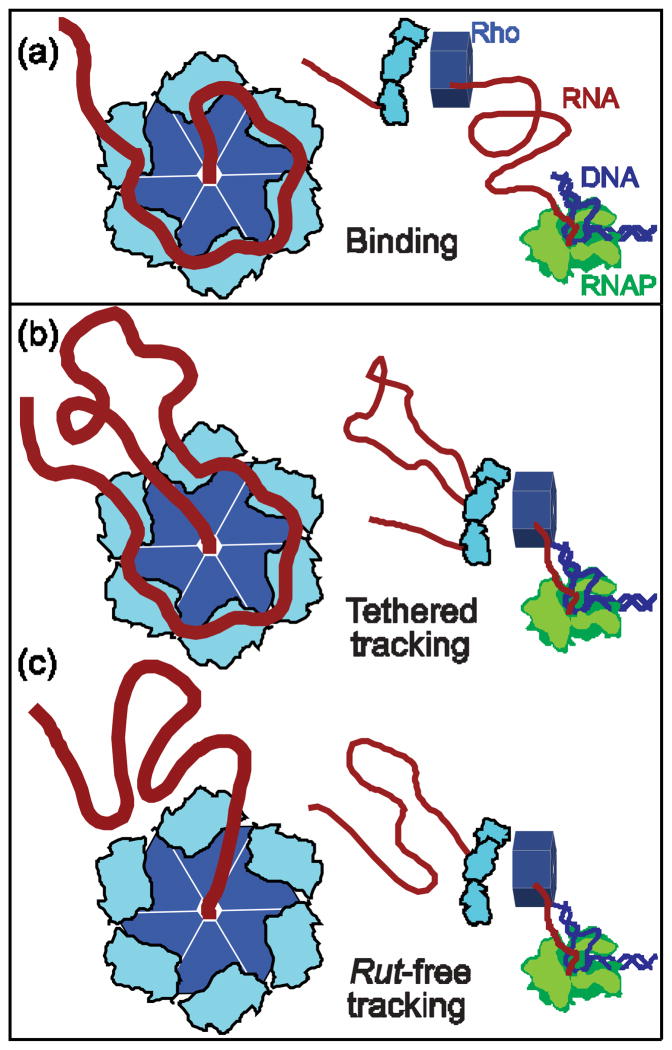
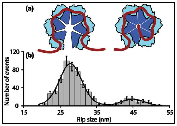
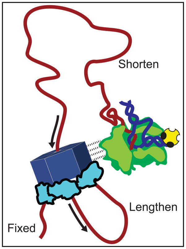
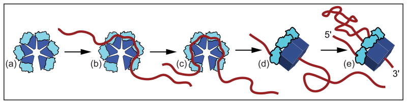
Rho termination factor is an essential hexameric helicase responsible for terminating 20-50% of all mRNA synthesis in Escherichia coli. We used single-molecule force spectroscopy to investigate Rho-RNA binding interactions at the Rho utilization site of the λtR1 terminator. Our results are consistent with Rho complexes adopting two states: one that binds 57 ± 2nt of RNA across all six of the Rho primary binding sites, and another that binds 85 ± 2nt at the six primary sites plus a single secondary site situated at the center of the hexamer. The single-molecule data serve to establish that Rho translocates 5'→3' toward RNA polymerase (RNAP) by a tethered-tracking mechanism, looping out the intervening RNA between the Rho utilization site and RNAP. These findings lead to a general model for Rho binding and translocation and establish a novel experimental approach that should facilitate additional single-molecule studies of RNA-binding proteins.


Figures

Figure 1
Figure 1. Proposed Models of Rho Translocation
(a) A Rho hexamer (left; light and dark blue) binds RNA (red) at a rut site. It subsequently translocates towards a downstream molecule of RNAP (right; green). Rho binds via six N-terminal primary domains (light blue) and a single secondary binding site (center). (b) The tethered-tracking mechanism. Rho maintains binding interactions with the rut site via its primary binding domains while moving 5′ to 3′, threading the downstream nucleotides through the secondary site, and consequently forming a growing loop of RNA, until it reaches the position of RNAP. (c) The rut-free tracking mechanism. The primary binding domains release the rut site once translocation begins, and Rho interacts with RNA exclusively via its secondary site. No loop is formed in the RNA as Rho moves towards RNAP.
Figure 2
Figure 2. Single-Molecule Dumbbell Assay
(a) Experimental geometry of the dumbbell assay (not to scale). Tension was applied by moving apart the two trapped beads. The beads (light blue) were maintained in separate optical traps (pink), with a dsDNA handle (dark blue) attached to one and an RNAP molecule (green) to the other by biotin-avidin linkages (yellow). The handle was hybridized to the 5′ end of the RNA transcript (red) via a 25- nt overhang. The RNA was transcribed in situ from a template that carried the rut site of the? tR1 terminator followed by a downstream sequence of variable length (30 bp, 75 bp or 150 bp) ending in a transcriptional roadblock (yellow) that stalls RNAP. The assay permits Rho to bind to the nascent RNA and, in the presence of ATP, translocate towards RNAP. (b) Representative force-extension curves (FECs). Most FECs (green) resemble those obtained in the absence of Rho and exhibited a small rip due to unfolding of the boxB hairpin in the rut site. FECs displaying larger, high- force rips were observed only in the presence of Rho(blue), and correspond to Rho release from the RNA.
Figure 3
Figure 3. Distributions of Rho Unbinding Rips
(a) Diagram of the RB30, RB75, and RB150 transcripts, drawn to approximate scale along the pulling coordinate (scale bar, 10 nt). Two segments of RNA (red) flank an insert from ?phage(light blue) carrying the rut site of ?tR1 (including the boxB hairpin). The transcripts were transcribed in situ from templates carrying 30, 75, or 150 bp downstream of the insert prior to a 3′ roadblock (yellow). Transcripts were hybridized to DNA handles (dark blue) via their 5′ ends (Fig. 2); ~25 nt at the 3′ end of each transcript lies under the footprint of RNAP (green) and is inaccessible to Rho. (B–D′) Histograms of the rip sizes for RNA release obtained for the three templates in the presence of AMP-PNP(left) or ATP (right). Population peaks are color-coded as follows: primary rips, with sizes of ~28 nm or multiples thereof (blue bars); secondary rips, with sizes of ~45 nm (green bars); translocation rips, with sizes that depend upon transcript length (red bars). (b) RB30 with AMP-PNP. Two populations, at 29 ±1 nm and 46 ±1 nm(mean ± SEM). Data from 8 molecules (N =33). (b′) RB30 with ATP. Two populations, at 30 ±1 nm and 46 ±1 nm. Data from 8 molecules (N =37). One population, at 27 ± 1 nm, was obtained for the RB30 boxB deletion construct (Fig. S1) (c) RB75 with AMP-PNP. Three populations, at 28 ±1 nm, 45 ±1 nm and 57 ±1 nm. Data from 22 molecules (N =65). (c′) RB75 with ATP. Four populations, three of which were observed in the presence of AMP-PNP (at 28 ±1 nm, 45 ±1 nm, and 57 ±1 nm), as well as a new peak at 73 ±1 nm. Data from 33 molecules (N =96). (d) RB150 with AMP-PNP. Three evenly spaced populations at 27 ±1 nm, 55 ±1 nm and 84 ±1 nm. Data from 17 molecules (N =54). (d′) RB150 with ATP. Four populations, three of which were observed in the presence of AMP-PNP, (30 ±1 nm, 56 ±1 nm and 84 ±1 nm) as well as a new peak at 111 ±1 nm. Data from 37 molecules (N =114).
Figure 4
Figure 4. Blocking Oligomers Inhibit Multiple Rho Binding but Permit Translocation
Histogram of unbinding rip sizes in the presence of blocking oligomers and ATP on RB75 (inset: scale diagram of RB75 showing the 20 nt DNA oligomer (black) hybridized). Three populations were observed, at 28 ±1 nm, 45 ±1 nm and 71 ±2 nm (color-coding as in Fig. 3). The frequency of multiple Rho unbinding events (blue bars) was greatly diminished and the number of secondary rips (green bars) enhanced compared to conditions without blocking oligomers (see Fig. 3c′), whereas translocation rips (red bars) occurred with the same frequency (11 ±3 % with blocking oligomers vs. 10 ±3 % without). Data from 43 molecules (N =98).
Figure 5
Figure 5. Two RNA Binding States of Rho
(a) Diagram depicting the two Rho binding states consistent with the observed rips. Left: RNA bound to six Rho primary sites in its “open” configuration. Right: RNA bound to six primary sites plus the central secondary site in the “closed” configuration. (b) Global histogram (grey bars) of the sizes of RNA-release rips observed in the presence of ATP or AMP-PNP for all templates studied (N =570). Solid line: Fit to sum of two Gaussians, with peaks at 28 ±1 nm and 45 ±1 nm (mean ± S.E.M.). Data from populations centered at 56 nm and 84 nm(Fig. 3) were included in the global dataset by dividing the measured values by 2 or 3, respectively (see text).
Figure 6
Figure 6. Transient Intermediate States in Rho Unbinding Rips
(a) Histogram showing the first of two sub-rips occasionally observed within the 28 nm primary rip. The data indicate two transient intermediates, one characterized by an initial sub-rip of 9.6 ±0.3 nm (N =38) and the other by an initial sub-rip of 16.0 ±0.4 nm (N =27). The first intermediate likely corresponds to the release of RNA by one of the six primary domains; the second corresponds to release from two (see text; also Fig. S2). Rip sizes were obtained by measuring the fractional size of each sub-rip and multiplying this value by the population-average rip size (27.7 ±0.4 nm). (b) A representative FEC that exhibits a transient intermediate rip (blue trace). The extension for the intermediate was fit to a double-WLC model (dotted line). The FEC for a molecule with no Rho bound is shown for comparison (green trace).
Figure 7
Figure 7. Proposed Model for Direct Interaction of Rho and RNAP
Epshtein et al. proposed that Rho directly associates with RNAP throughout transcription (dashed lines). In this model, such an association would generate a loop of RNA, composed of sequences downstream of the rut site and upstream of RNAP, that shortens continuously during translocation. Furthermore, in the tethered-tracking mechanism, a second loop is expected to form, composed of sequences between the primary and secondary Rho binding sites, that lengthens continuously at the identical rate. Our results disfavor any strong, direct interaction between Rho and RNAP.
Figure 8
Figure 8. General Model of Rho Binding and Translocation
In its open (lock-washer) configuration, Rho(a) binds RNA via the rut site, transiently occupying one or more binding states (b) before associating with 57 ±2 nt of RNA via all six primary domains (c). RNA is then passed into the center of the hexamer and Rho adopts the closed (ring) configuration, binding the transcript via its secondary site as well, with an overall RNA footprint of 85 ±2 nt (d). Finally, Rho translocates via a tethered-tracking mechanism (e), hydrolyzing ATP and proceeding downstream towards RNAP, looping out the intervening RNA sequence (see also Fig. S3).

Comment in

  • Getting a grip on the terminator.
    Stone MD. Stone MD. J Mol Biol. 2012 Nov 9;423(5):661-3. doi: 10.1016/j.jmb.2012.09.004. Epub 2012 Sep 10. J Mol Biol. 2012. PMID: 22975072 No abstract available.

Similar articles

Cited by

References

    1. Larson MH, Greenleaf WJ, Landick R, Block SM. Applied force reveals mechanistic and energetic details of transcription termination. Cell. 2008;132:971–82. - PMC - PubMed
    1. Nudler E, Gottesman ME. Transcription termination and anti- termination in E. coli. Genes Cells. 2002;7:755–68. - PubMed
    1. Richardson JP. Rho-dependent termination and ATPases in transcript termination. Biochim Biophys Acta. 2002;1577:251–260. - PubMed
    1. Peters JM, Vangeloff AD, Landick R. Bacterial Transcription Terminators: The RNA 3′-end Chronicles. J Mol Biol 2011 - PMC - PubMed
    1. Ciampi MS. Rho-dependent terminators and transcription termination. Microbiology. 2006;152:2515–28. - PubMed

Publication types

LinkOut - more resources