. 2012 Sep;122(9):3355-67.
doi: 10.1172/JCI57407. Epub 2012 Aug 13.

PTH-independent regulation of blood calcium concentration by the calcium-sensing receptor

Affiliations

PTH-independent regulation of blood calcium concentration by the calcium-sensing receptor

Alexandre Loupy et al. J Clin Invest. 2012 Sep.

Abstract

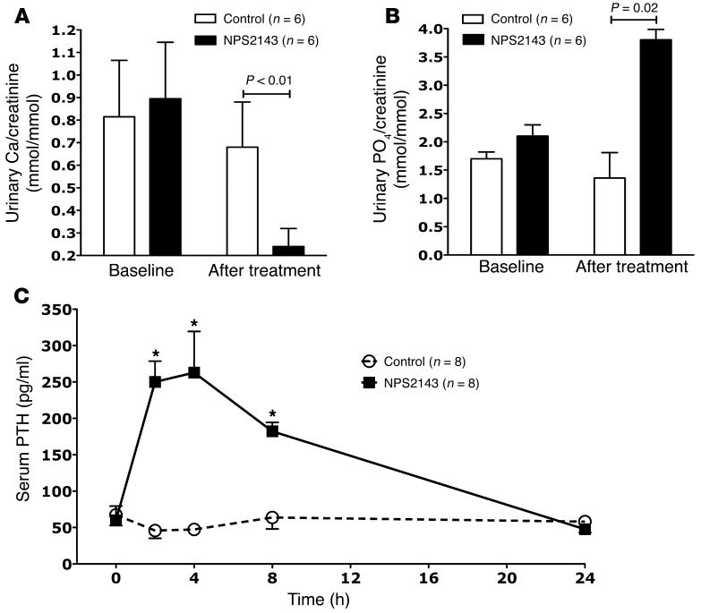
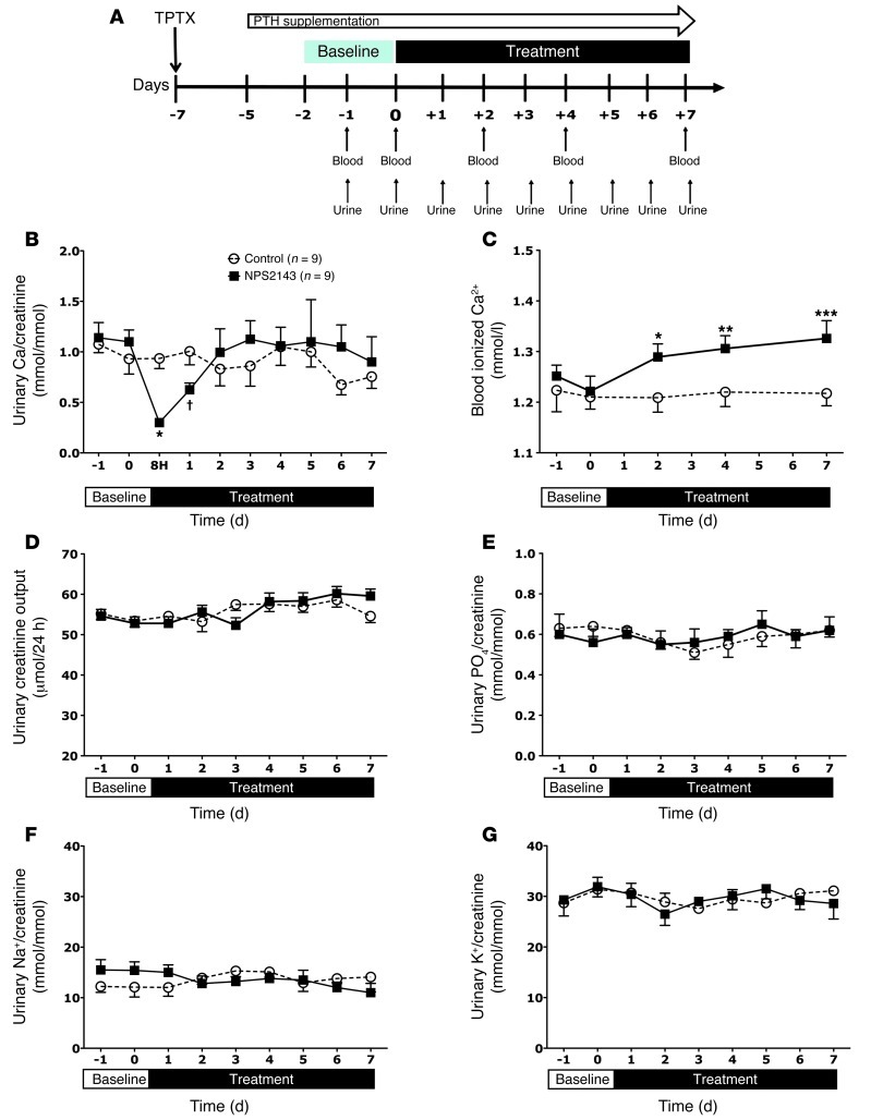
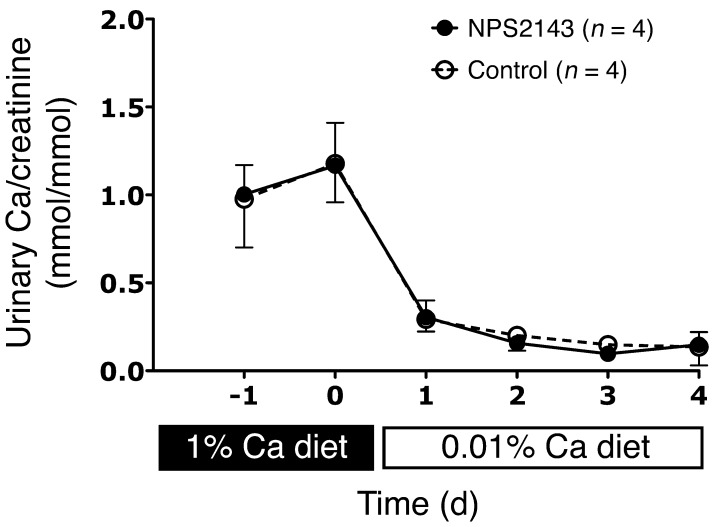
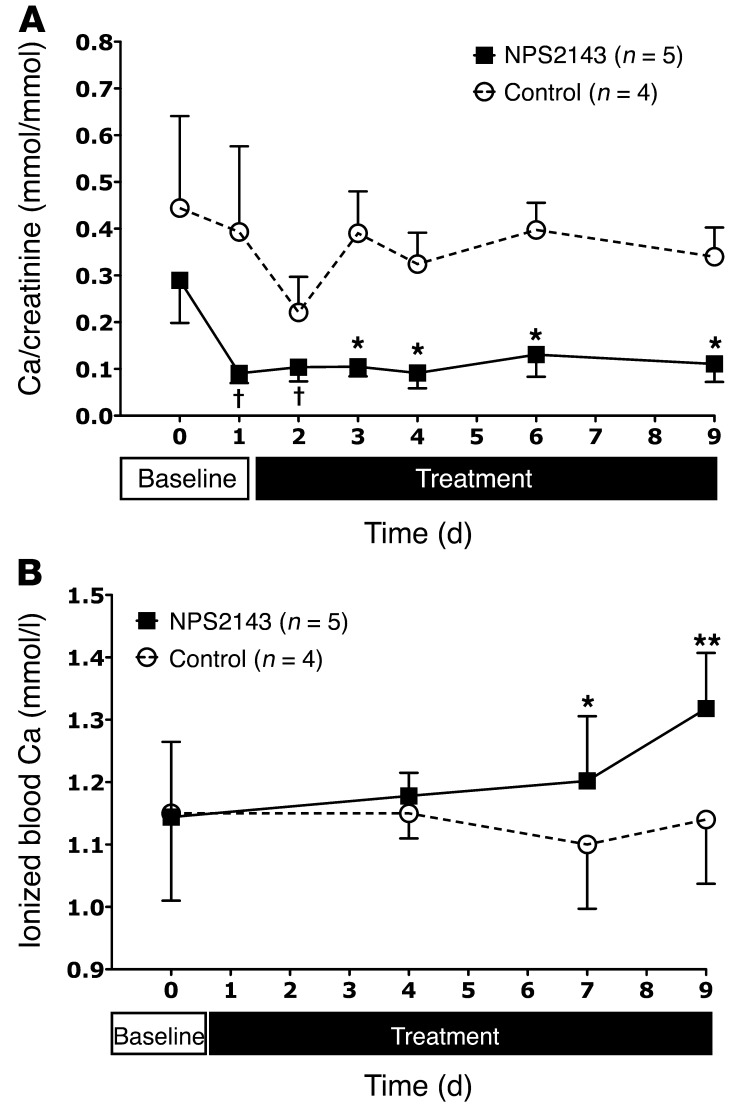
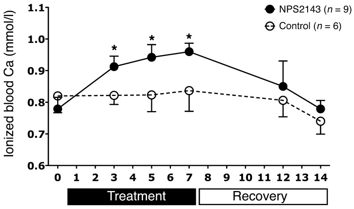
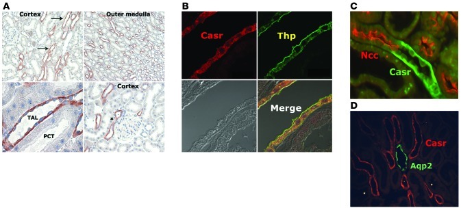
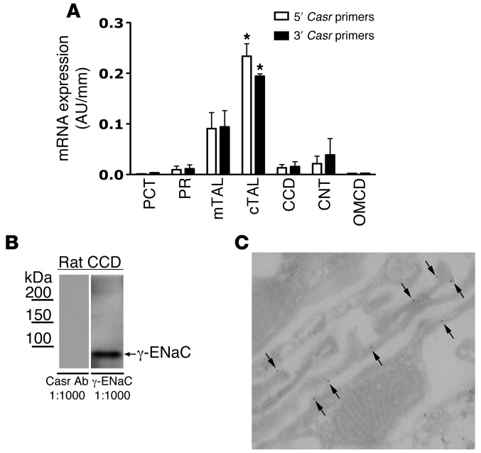
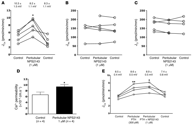
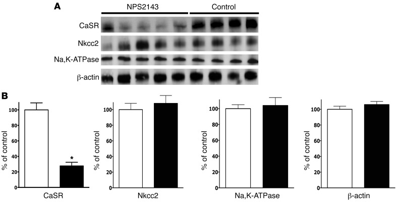
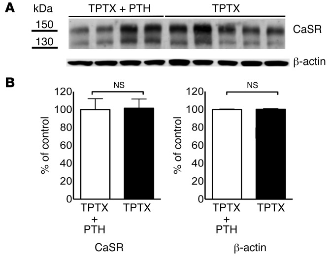
Tight regulation of calcium levels is required for many critical biological functions. The Ca2+-sensing receptor (CaSR) expressed by parathyroid cells controls blood calcium concentration by regulating parathyroid hormone (PTH) secretion. However, CaSR is also expressed in other organs, such as the kidney, but the importance of extraparathyroid CaSR in calcium metabolism remains unknown. Here, we investigated the role of extraparathyroid CaSR using thyroparathyroidectomized, PTH-supplemented rats. Chronic inhibition of CaSR selectively increased renal tubular calcium absorption and blood calcium concentration independent of PTH secretion change and without altering intestinal calcium absorption. CaSR inhibition increased blood calcium concentration in animals pretreated with a bisphosphonate, indicating that the increase did not result from release of bone calcium. Kidney CaSR was expressed primarily in the thick ascending limb of the loop of Henle (TAL). As measured by in vitro microperfusion of cortical TAL, CaSR inhibitors increased calcium reabsorption and paracellular pathway permeability but did not change NaCl reabsorption. We conclude that CaSR is a direct determinant of blood calcium concentration, independent of PTH, and modulates renal tubular calcium transport in the TAL via the permeability of the paracellular pathway. These findings suggest that CaSR inhibitors may provide a new specific treatment for disorders related to impaired PTH secretion, such as primary hypoparathyroidism.


Figures

Figure 1
Figure 1. Effects of a single administration of the CaSR inhibitor NPS2143 in intact rats.
(A) Urinary Ca excretion (expressed as a ratio to urinary creatinine excretion) before (Baseline) and during the 8 hours following a single oral dose of NPS2143 (After treatment) (100 μmol.kg BW–1) or vehicle in intact rats. n = 6 in each group. (B) Urinary phosphate excretion (expressed as a ratio to urinary creatinine excretion) before and during the 8 hours following a single oral dose of NPS2143 or vehicle in intact rats. n = 6 in each group. (C) Plasma PTH concentration before (time 0) and during the 24 hours following a single oral dose of NPS2143 (black symbols) or vehicle (open symbols) in intact rats. n = 8 in each group. *P < 0.01 versus controls.
Figure 2
Figure 2. Effects of 1-week daily administration of NPS2143 in TPTX, PTH-supplemented rats.
(A) Summary of the protocol. (B) Urinary Ca excretion (expressed as a ratio to urinary creatinine excretion) at baseline and during 1 week of daily administration of NPS2143 (or placebo). In NPS2143-treated rats (1.5 mg/d intraperitoneally), urinary Ca excretion significantly decreased and was lower than in control rats (P < 0.01) on the first day of NPS2143 administration. Subsequently, urinary Ca excretion increased back to baseline values. n = 9 per group. *P < 0.01, P = 0.03 versus control group. (C) Blood ionized Ca concentration at baseline and on days 2, 4, and 7 of daily administration of NPS2143 (or placebo); in treated rats, blood ionized Ca concentration significantly increased by the second day of NPS2143 administration and remained higher than in control rats throughout the period. *P = 0.05, **P = 0.02, ***P = 0.01. (DG) Twenty-four-hour urinary creatinine excretion and urinary phosphate, Na, and K excretion (expressed as a ratio to urinary creatinine excretion) at baseline and during 1 week of daily administration of NPS2143 (or placebo). No statistical difference between treated and control rats was seen for any variable. n = 9 per group.
Figure 3
Figure 3. Effects of a low-Ca diet in NPS2143-treated or control TPTX, PTH-supplemented rats.
Shown is the effect of a low-Ca (0.01%, w/w) diet as compared with a normal Ca diet on urinary Ca excretion in TPTX, PTH-supplemented rats treated with a CaSR inhibitor or vehicle alone. The rats received either NPS2143 (1.5 mg daily) or vehicle for 4 days before the switch while on the regular Ca diet. The switch to the low-Ca diet induced a similar decrease in urinary Ca/creatinine ratio in NPS2143-treated and control rats (P = NS). This result indicates that net bone Ca release was not affected by treatment with the CaSR inhibitor. n = 4 in each group.
Figure 4
Figure 4. Effect of NPS2143 in TPTX, PTH-supplemented rats treated with the anti-bone-resorptive agent sodium pamidronate.
All rats were treated with subcutaneous injections of pamidronate (2.5 μg.g BW–1, daily for 4 days prior to the experiment and throughout the remainder of the experiment). At baseline, the two groups of rats exhibited similar blood ionized Ca concentration and urinary Ca excretion. (A) During treatment with NPS2143, urinary Ca excretion decreased by the first day of treatment; throughout the remainder of the experiment, the NPS-treated rats exhibited significantly lower urine Ca excretion as compared with control rats. *P = 0.01 versus controls; P = 0.02 versus baseline. (B) In contrast to control rats in which blood Ca concentration did not change during the experiment, blood ionized Ca significantly increased in NPS2143-treated rats by day 7 (*P = 0.023 and **P = 0.012 at day 7 and day 9, respectively, as compared with day 0). This result indicates that even when bone resorption is inhibited, NPS2143 induces an increase in renal tubular Ca absorption and a positive Ca balance, resulting in a secondary increase in blood Ca concentration. n = 5 in the NPS2143-treated group and n = 4 in the control group.
Figure 5
Figure 5. Effects of 1-week daily administration of NPS2143 in TPTX rats without PTH supplementation.
The time course of blood Ca concentration during 1-week treatment of TPTX rats with either vehicle or NPS2143 is summarized. At baseline, both groups had low blood Ca concentration. In the control group, serum Ca concentration remained unchanged throughout the experiment, while a significant and sustained increase in blood Ca concentration was observed in the NPS2143-treated group (*P < 0.02 as compared with day 0). After a 7-day recovery period, serum Ca concentration returned to baseline values. n = 9 and n = 6 in the NPS2143-treated and control groups, respectively.
Figure 6
Figure 6. Immunolocalization of CaSR in the rat kidney.
(A) Immunolocalization of the CaSR protein in the renal cortex and outer medulla. Immunoperoxidase staining in rat kidney sections showed a cytoplasmic and basolateral labeling of tubular cells in the cortex (original magnification, ×200 and ×400) and the inner and outer stripes of the outer medulla (×100). The stained tubules were present in the medullary rays (arrows) and in the juxtaglomerular apparatus (asterisk). Proximal tubules and glomeruli showed no detectable CaSR staining (×1,000). (B) CaSR is expressed in the rat TAL. Shown is double staining with Abs directed against CaSR (red) and Tamm-Horsfall protein (Thp), specifically expressed in the TAL (green). When comparing with tubular segments that stained for Tamm-Horsfall protein, CaSR staining was basolateral (original magnification, ×2,000). (C) CaSR is not detectable in the rat distal convoluted tubule (DCT). Sections of rat kidney double stained for CaSR and the thiazide-sensitive NaCl cotransporter (Ncc) localized in the DCT. Basolateral CaSR staining was interrupted when the tubule showed apical staining for Ncc, thus representing the distal limit for CaSR staining (original magnification, ×1,500). (D) CaSR is not detectable in the rat CCD. Sections of rat kidney double stained for CaSR and aquaporin-2 (Aqp2). In the merged image, tubular segments that stained for aquaporin-2 did not show CaSR staining and vice versa (original magnification, ×1,000).
Figure 7
Figure 7. Localization of CaSR in the rat kidney.
(A) Quantification of Casr transcripts along the rat nephron. RT-PCR analysis of mRNAs encoding CaSR was performed on microdissected renal tubules. Casr transcripts were detected at a significant level in the medullary TAL (mTAL) and cTAL. No significant transcript expression was detected in the PCT, PR, CCD, or OMCD. A non-significant level of Casr mRNAs was found in the CNT. *P < 0.001 between cTAL and mTAL Casr mRNA expression. (B) Western blotting for CaSR in rat microdissected CCD, using 1:1,000 anti-CaSR Ab (MA1-934). No signal was observed using anti-CaSR Ab. γ-ENaC (specific for CCD) was used as a positive control. (C) CaSR localization in the kidney by EM. CaSR immunocytochemistry of ultrathin cryosections from rat kidney with gold labeling for CaSR in the TAL showing basolateral gold labeling (arrow). No apical TAL labeling and no CCD labeling were noted (original magnification, ×100,000).
Figure 8
Figure 8. Effect of NPS2143 on transepithelial ion transport in the rat cTAL.
cTALs were dissected from rat kidney and perfused in vitro as explained in Methods. Peritubular addition of 1 μM NPS2143 elicited a significant and reversible increase in transepithelial Ca absorption (*P = 0.03), but did not change Vte (A) or transepithelial sodium or chloride absorption (B and C, respectively) (P = NS). n = 5. (D) 1 μM NPS2143 elicited a significant increase in the paracellular pathway permeability to Ca in rat cTAL microperfused in vitro (*P = 0.0027). n = 4. (E) As compared with the control period, PTH (300 pM in the peritubular fluid) elicited a significant increase in transepithelial Ca absorption (**P = 0.024); peritubular addition of 1 μM NPS2143, in the presence of PTH elicited a further increase in transepithelial Ca absorption (*P = 0.045 as compared with the PTH period) that was significantly lower than in the absence of peritubular PTH (n = 4). Vte was not changed throughout the experiment.
Figure 9
Figure 9. Effect of a 7-day treatment with NPS2143 on CaSR, Nkcc2, and Na+,K+-ATPase α1 subunit protein abundance in the rat kidney cortex.
(A) Immunoblots of membrane fractions from the rat kidney cortex obtained from 5 NPS2143-treated and 4 control rats. Immunoblots were reacted with Abs directed against CaSR, Nkcc2, the α1 subunit of the Na+, K+-ATPase, and β-actin. (B) Western blot analyses showing the abundance of CaSR, Nkcc2, and the α1 subunit of the Na+,K+-ATPase in NPS2143-treated rats (black bars) and in control rats (white bars). CaSR expression was quantified by densitometric analysis and expressed as percentage of control values. *P < 0.001 versus controls. Nkcc2, Na+, K+-ATPase α1 subunit, and β-actin expression was not modified by the treatment.
Figure 10
Figure 10. Effect of a 7-day hypocalcemia on CaSR protein abundance in the rat kidney cortex.
(A) Immunoblots of membrane fractions from the rat kidney cortex obtained from 4 TPTX, PTH-supplemented and 5 TPTX rats. Immunoblots were reacted with Abs directed against CaSR. (B) Western blot analyses showing the abundance of CaSR in TPTX, PTH-supplemented and TPTX, non-PTH-supplemented rats. CaSR expression was quantified by densitometric analysis and expressed as percentage of control values.

Comment in

Similar articles

Cited by

References

    1. Jaeger P, et al. Animal model of primary hyperparathyroidism. Am J Physiol. 1987;252(6 pt 1):E790–E798. - PubMed
    1. Brown EM, et al. Cloning and characterization of an extracellular Ca(2+)-sensing receptor from bovine parathyroid. Nature. 1993;366(6455):575–580. doi: 10.1038/366575a0. - DOI - PubMed
    1. Riccardi D, Brown EM. Physiology and pathophysiology of the calcium-sensing receptor in the kidney. Am J Physiol Renal Physiol. 2010;298(3):F485–F499. doi: 10.1152/ajprenal.00608.2009. - DOI - PMC - PubMed
    1. Brown EM. Clinical lessons from the calcium-sensing receptor. Nat Clin Pract Endocrinol Metab. 2007;3(2):122–133. doi: 10.1038/ncpendmet0388. - DOI - PubMed
    1. Peacock M, Bilezikian JP, Klassen PS, Guo MD, Turner SA, Shoback D. Cinacalcet hydrochloride maintains long-term normocalcemia in patients with primary hyperparathyroidism. J Clin Endocrinol Metab. 2005;90(1):135–141. doi: 10.1210/jc.2004-0842. - DOI - PubMed

Publication types

MeSH terms