. 2012 Oct 15;303(8):L692-702.
doi: 10.1152/ajplung.00390.2011. Epub 2012 Aug 10.

Neuronal Wiskott-Aldrich syndrome protein (N-WASP) is critical for formation of α-smooth muscle actin filaments during myofibroblast differentiation

Affiliations

Neuronal Wiskott-Aldrich syndrome protein (N-WASP) is critical for formation of α-smooth muscle actin filaments during myofibroblast differentiation

Guo-Qiang Cai et al. Am J Physiol Lung Cell Mol Physiol. .

Abstract

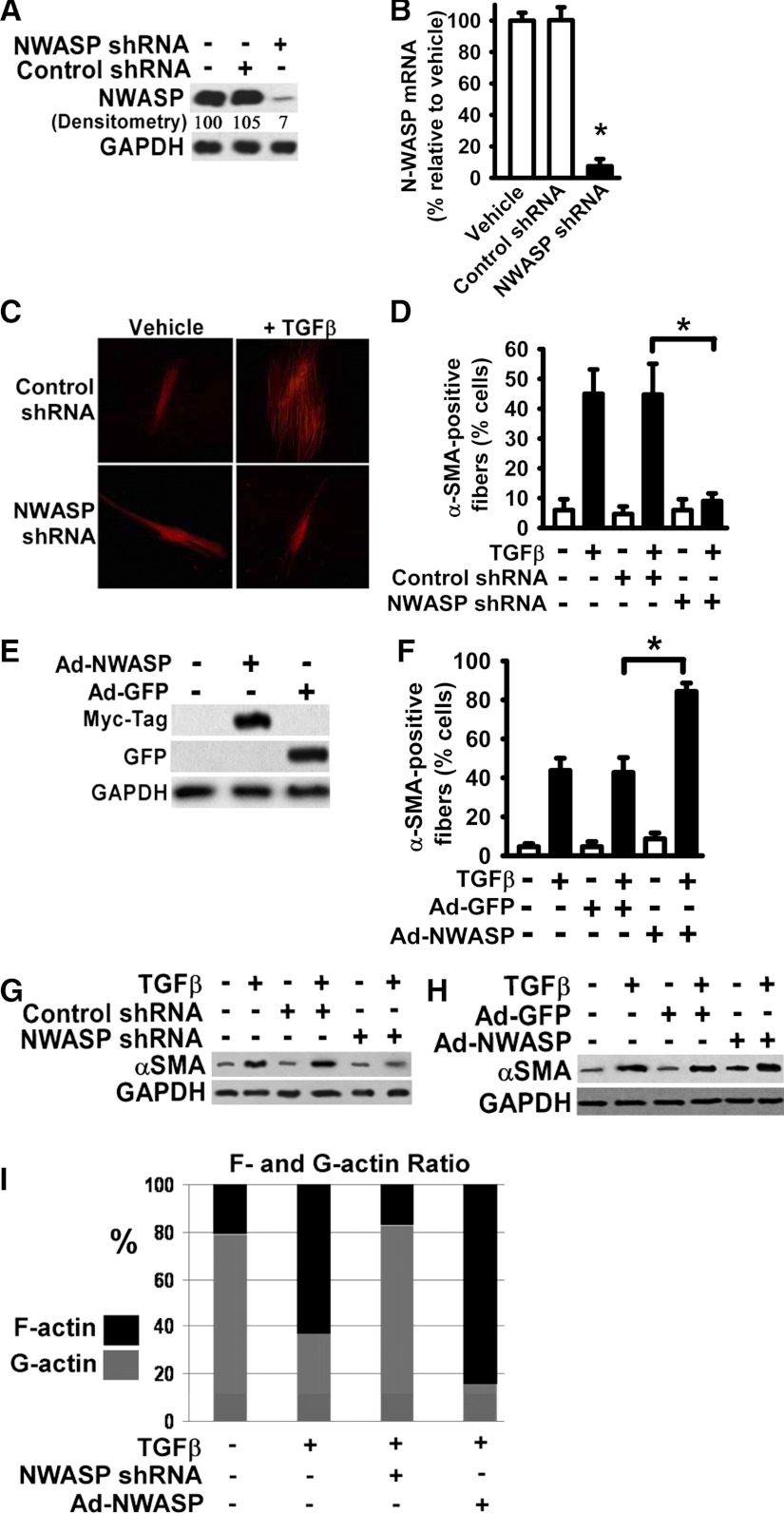
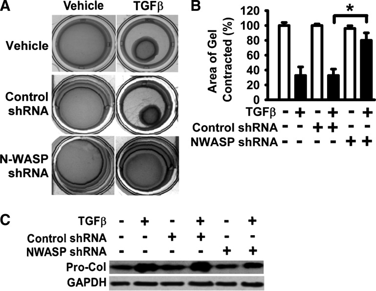
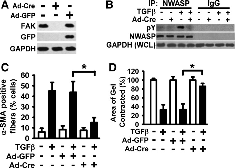
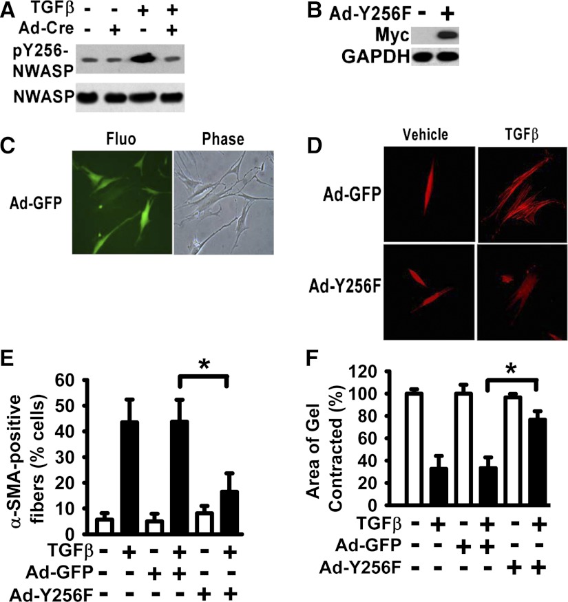
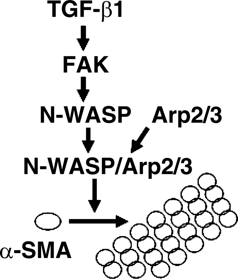
Myofibroblasts are implicated in pathological stromal responses associated with lung fibrosis. One prominent phenotypic marker of fully differentiated myofibroblasts is the polymerized, thick cytoplasmic filaments containing newly synthesized α-smooth muscle actin (α-SMA). These α-SMA-containing cytoplasmic filaments are important for myofibroblast contractility during tissue remodeling. However, the molecular mechanisms regulating the formation and maturation of α-SMA-containing filaments have not been defined. This study demonstrates a critical role for neuronal Wiskott-Aldrich syndrome protein (N-WASP) in regulating the formation of α-SMA-containing cytoplasmic filaments during myofibroblast differentiation and in myofibroblast contractility. Focal adhesion kinase (FAK) is activated by transforming growth factor-β1 (TGF-β1) and is required for phosphorylation of tyrosine residue 256 (Y256) of N-WASP. Phosphorylation of Y256 of N-WASP is essential for TGF-β1-induced formation of α-SMA-containing cytoplasmic filaments in primary human lung fibroblasts. In addition, we demonstrate that actin-related protein (Arp) 2/3 complex is downstream of N-WASP and mediates the maturation of α-SMA-containing cytoplasmic filaments. Together, this study supports a critical role of N-WASP in integrating FAK and Arp2/3 signaling to mediate formation of α-SMA-containing cytoplasmic filaments during myofibroblast differentiation and maturation.


Figures

Fig. 1.
Fig. 1.
Neuronal Wiskott-Aldrich syndrome protein (N-WASP) downregulation abrogated the transforming growth factor (TGF)-β1-induced α-smooth muscle actin (α-SMA)-containing cytoplasmic filaments in human lung fibroblasts. A: normal human lung fibroblasts were infected with lentiviral vectors containing N-WASP shRNA or control nontargeting shRNA, and stable clones were selected according to puromycin resistance as described in materials and methods. Fibroblasts were harvested and lysed. Equivalent amount of whole cell detergent lysates were Western blotted with the indicated antibodies. GAPDH is a loading control. Densitometry of band intensity relative to GAPDH is shown (vehicle-treated cells in lane 1 is as 100%). B: lung fibroblasts stably expressing N-WASP shRNA or nontargeting control shRNA or vehicle-treated lung fibroblasts were subjected to total RNA extraction and followed by quantitative RT-PCR to determine the level of N-WASP mRNA. Data are represented as means ± SE. *P < 0.01 for cells with N-WASP shRNA compared with cells with vehicle only. C: lung fibroblasts stably expressing N-WASP shRNA or control shRNA were cultured in serum-free media (SFM) with 1% BSA, and treated without or with TGF-β1 (10 ng/ml). Cells were fixed after 48 h and immunofluorescently stained with Cy-3-labeled monoclonal antibody toward α-SMA, and fluorescent microscopic digital images were taken (×200). Representative pictures were shown. D: quantification of the percentage of cells with α-SMA-containing filaments. The data are presented as the means ± SE. Open bars are vehicle-treated cells. Solid bars are TGF-β1-treated cells. *P < 0.01. E: lung fibroblasts were infected with adenoviral vectors expressing either myc-tagged N-WASP cDNA (Ad-NWASP) or green fluorescent protein (GFP) cDNA (Ad-GFP), lysed, and Western blotted with indicated antibodies. F: lung fibroblasts infected with Ad-NWASP or control Ad-GFP were treated with or without TGF-β1 (10 ng/ml), and stained with Cy-3-labeled antibody toward α-SMA as in C. The percentages of cells with α-SMA-containing filaments were quantified and presented as in D. * P < 0.01. G–H: lung fibroblasts stably expressing N-WASP shRNA (as in C) or infected with Ad-NWASP (as in F) were treated with or without TGF-β1 (10 ng/ml) for 48 h, lysed, and Western blotted with indicated antibodies. I: effect of N-WASP downregulation (by shRNA) or overexpression (by Ad-NWASP) on actin dynamics were determined (in cells as described in G–H) by measurements of F-/G-actin content as described in materials and methods.
Fig. 2.
Fig. 2.
N-WASP downregulation blocked the ability of myofibroblasts to contract the collagen gels. A: lung fibroblasts stably expressing N-WASP shRNA or control shRNA were cultured in SFM with 1% BSA, treated with or without TGF-β1 (10 ng/ml), and followed by the collagen gel contraction assays for 60 h (at 37°C, 5% CO2) as described in materials and methods. Representative digital images were shown. B: areas of collagen gels from digitized images were measured. The ratio of collagen gel area after contraction against the original collagen gel area (before contraction and equivalent to the culture-well area) was calculated. Data are presented as the percentage of gel area relative to control (vehicle only, set as 100%). *P < 0.01. C: lung fibroblasts stably expressing N-WASP shRNA or control shRNA were cultured in SFM with 1% BSA and treated without or with TGF-β1 (10 ng/ml). Procollagen 1A1 expression was examined at 48 h by Western blot analysis.
Fig. 3.
Fig. 3.
Focal adhesion kinase (FAK) expression is essential for TGF-β1-induced tyrosine phosphorylation of N-WASP and the formation of α-SMA-containing filaments. A: primary murine lung fibroblasts were derived from FAK-floxed mice and treated with or without Ad-Cre (adenoviral vector expressing Cre recombinase) in SFM with 1% BSA. Fibroblasts were lysed, and equivalent amount of whole cell detergent lysates were Western blotted with the indicated antibodies. GAPDH is a loading control. B: cells in A were treated with or without TGF-β1 (10 ng/ml) in SFM with 1% BSA for 6 h (37°C, 5% CO2) and detergent lysed, and equivalent amount of whole cell lysates were immunoprecipitated with an anti-N-WASP antibody (NWASP IP) or control rabbit IgG (IgG IP) as indicated. The immunoprecipitates (IP) were analyzed by Western Blot with anti-phospho-tyrosine (top, pY) or anti-N-WASP (middle, NWASP). For loading controls, equivalent amounts of whole cell detergent lysates (WCL) were also analyzed with anti-GAPDH (bottom). C: lung fibroblasts as described in A were treated with or without TGF-β1 (10 ng/ml) in SFM with 1% BSA for 48 h, fixed, and stained with Cy-3-labeled antibody to visualize α-SMA-containing filaments. The percentage of cells with α-SMA-containing filaments was quantified as in Fig. 1D. Ad-GFP infection was a control for adenoviral vector. Data are presented as the means ± SE. *P < 0.01. D: lung fibroblasts were treated as in C and subjected to collagen gel contraction assay for 60 h (at 37°C, 5% CO2). Collagen gel contraction was determined, and data are presented as shown in Fig. 2. *P < 0.01.
Fig. 4.
Fig. 4.
Expression of N-WASP Y256F mutant significantly inhibited the formation of α-SMA-containing filaments and cell contraction in lung fibroblasts treated with TGF-β1. A: lung fibroblasts derived from FAK-floxed mice were treated with or without Ad-Cre for 24 h and treated with or without TGF-β1 (10 ng/ml) in SFM with 1% BSA for 6 h (37°C, 5% CO2) as shown in Fig. 3. Cells were lysed, and equivalent amount of whole cell lysates were subjected to Western Blot analysis with indicated antibodies. B: human lung fibroblasts were infected with adenoviral vector containing the myc-tagged Y256F N-WASP mutant (Ad-Y256F), and equivalent amount of whole cell lysates were subjected to Western Blot analysis with indicated antibodies. C: human lung fibroblasts were infected with adenoviral vector containing the GFP (Ad-GFP) for 48 h. Fluorescent image for GFP expression (left), phase contrast image (right) (×200). D: lung fibroblasts were infected with Ad-Y256F or Ad-GFP, treated with or without TGF-β1 (10 ng/ml) for 48 h, fixed, and stained with Cy-3-labeled antibody toward α-SMA as shown in Fig. 1 (×200). E: percentage of cells with α-SMA-containing filaments in D was quantified as in Fig. 1. Data are presented as the means ± SE. *P < 0.01. F: lung fibroblasts were treated as in E and subjected to collagen gel contraction assay as shown in Fig. 2. *P < 0.01.
Fig. 5.
Fig. 5.
Actin-related protein (Arp) 3 is required for TGF-β1-induced formation of α-SMA-containing filaments during myofibroblast differentiation and cell contraction. A: lung fibroblasts were infected with lentiviral vectors containing Arp3-specific shRNA or control nontargeting shRNA, and stable clones were selected by puromycin resistance as in Fig. 1. Cells were lysed, and equivalent amount of whole cell lysates were subjected to Western Blot analysis with indicated antibodies. B: lung fibroblasts stably expressing Arp3 shRNA or control nontargeting shRNA were treated with or without TGF-β1 (10 ng/ml) for 48 h, fixed, and stained with Cy-3-labeled antibody toward α-SMA as shown in Fig. 1. The percentage of cells with α-SMA-containing filaments was quantified and presented as the means ± SE. *P < 0.01. C: lung fibroblasts stably expressing Arp3 shRNA or control nontargeting shRNA were subjected to collagen gel contraction assays for 60 h as shown in Fig. 2. *P < 0.01. D: in vitro pyrene actin polymerization assays were performed as described in materials and methods. Polymerization of fluorescent pyrene-labeled α-SMA monomer was determined in the presence of α-SMA monomer and varying combinations of Arp2/3, FAK, N-WASP, and N-WASP mutant (N-WASP Y256F). Nonpolymerized α-SMA monomer (Actin) was used as a negative control. The VCA domain of N-WASP (VCA) served as a positive control.
Fig. 6.
Fig. 6.
A working model for N-WASP in integrating FAK and Arp2/3 signaling to mediate formation of α-SMA-containing cytoplasmic filaments during myofibroblast differentiation and maturation. TGF-β1 is well accepted for its ability to induce myofibroblast differentiation. The formation of α-SMA-containing filaments is the hallmark of myofibroblast differentiation and maturation and also important for myofibroblast functions. The current study shows that N-WASP plays a key role in regulating the formation of α-SMA-containing cytoplasmic filaments during myofibroblast differentiation and maturation. TGF-β1 induces FAK activation, and activated FAK functions as an upstream regulator of N-WASP and mediates phosphorylation of Y256 of N-WASP. Phosphorylation of Y256 of N-WASP is required for N-WASP-mediated formation of α-SMA-containing filaments. Arp2/3 complex is recruited by N-WASP and is required for TGF-β1-induced formation of α-SMA-containing filaments. Together, this study supports a critical role of N-WASP in integrating FAK and Arp2/3 signaling to mediate formation of α-SMA-containing filaments during myofibroblast differentiation and maturation.

Similar articles

Cited by

References

    1. Arora PD, McCulloch CA. Dependence of collagen remodelling on alpha-smooth muscle actin expression by fibroblasts. J Cell Physiol 159: 161–175, 1994 - PubMed
    1. Beggs HE, Schahin-Reed D, Zang K, Goebbels S, Nave KA, Gorski J, Jones KR, Sretavan D, Reichardt LF. FAK deficiency in cells contributing to the basal lamina results in cortical abnormalities resembling congenital muscular dystrophies. Neuron 40: 501–514, 2003 - PMC - PubMed
    1. Bompard G, Caron E. Regulation of WASP/WAVE proteins: making a long story short. J Cell Biol 166: 957–962, 2004 - PMC - PubMed
    1. Cai GQ, Zheng A, Tang Q, White ES, Chou CF, Gladson CL, Olman MA, Ding Q. Downregulation of FAK-related non-kinase mediates the migratory phenotype of human fibrotic lung fibroblasts. Exp Cell Res 316: 1600–1609, 2010 - PMC - PubMed
    1. Chan MW, Chaudary F, Lee W, Copeland JW, McCulloch CA. Force-induced myofibroblast differentiation through collagen receptors is dependent on mammalian diaphanous (mDia). J Biol Chem 285: 9273–9281, 2010 - PMC - PubMed

Publication types

MeSH terms