. 2012 Nov;192(3):905-28.
doi: 10.1534/genetics.112.143271. Epub 2012 Aug 10.

SACY-1 DEAD-Box helicase links the somatic control of oocyte meiotic maturation to the sperm-to-oocyte switch and gamete maintenance in Caenorhabditis elegans

Affiliations

SACY-1 DEAD-Box helicase links the somatic control of oocyte meiotic maturation to the sperm-to-oocyte switch and gamete maintenance in Caenorhabditis elegans

Seongseop Kim et al. Genetics. 2012 Nov.

Abstract

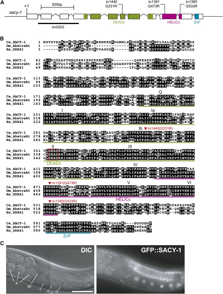
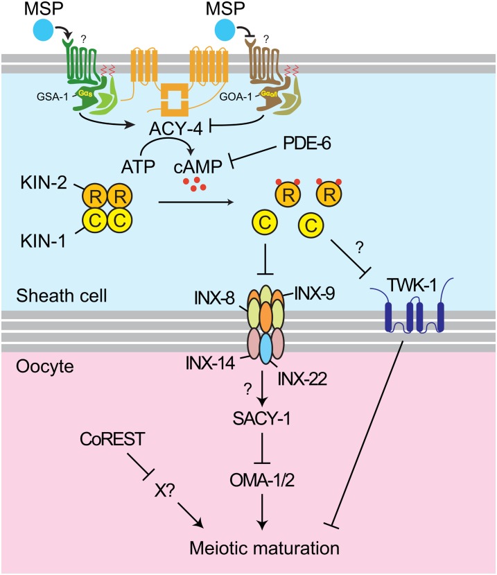
In sexually reproducing animals, oocytes arrest at diplotene or diakinesis and resume meiosis (meiotic maturation) in response to hormones. In Caenorhabditis elegans, major sperm protein triggers meiotic resumption through a mechanism involving somatic Gα(s)-adenylate cyclase signaling and soma-to-germline gap-junctional communication. Using genetic mosaic analysis, we show that the major effector of Gα(s)-adenylate cyclase signaling, protein kinase A (PKA), is required in gonadal sheath cells for oocyte meiotic maturation and dispensable in the germ line. This result rules out a model in which cyclic nucleotides must transit through sheath-oocyte gap junctions to activate PKA in the germ line, as proposed in vertebrate systems. We conducted a genetic screen to identify regulators of oocyte meiotic maturation functioning downstream of Gα(s)-adenylate cyclase-PKA signaling. We molecularly identified 10 regulatory loci, which include essential and nonessential factors. sacy-1, which encodes a highly conserved DEAD-box helicase, is an essential germline factor that negatively regulates meiotic maturation. SACY-1 is a multifunctional protein that establishes a mechanistic link connecting the somatic control of meiotic maturation to germline sex determination and gamete maintenance. Modulatory factors include multiple subunits of a CoREST-like complex and the TWK-1 two-pore potassium channel. These factors are not absolutely required for meiotic maturation or its negative regulation in the absence of sperm, but function cumulatively to enable somatic control of meiotic maturation. This work provides insights into the genetic control of meiotic maturation signaling in C. elegans, and the conserved factors identified here might inform analysis in other systems through either homology or analogy.


Figures

Figure 1
Figure 1
kin-1 is required in the gonadal sheath cells for oocyte meiotic maturation. (A) An adult hermaphrodite gonad arm. Germline stem cells proliferate near the distal tip cell (DTC) and then enter meiosis as they move proximally. Oocytes grow by receiving cytoplasmic flow (arrows) and progress to the diakinesis stage of meiotic prophase I prior to undergoing meiotic maturation (at the −1 position) in response to MSP secreted from sperm in the spermatheca (sp). Five pairs of gonadal sheath cells surround the germ line in each gonad arm. (B) DIC images showing that loss of kin-1(+) function in the somatic gonad prevents meiotic maturation, causing oocytes to stack up in the gonad arm despite the presence of sperm. This genetic mosaic resulted from a complex loss within the Z1 lineage, such that the anterior sheath cells and the anterior DTC were mutant for kin-1, but a few spermathecal cells were kin-1(+) (the mosaic is displayed with anterior to the right). By contrast, wild-type and kin-1(ok338); tnEx109 gonad arms are fertile; embryos are observed in the uterus (ut). Bar, 50 μm. (C) Genetic mosaic analysis of kin-1 in meiotic maturation. Derivation of the somatic gonad and the germ line, the points in the lineage where the kin-1(+) array was lost and the resulting phenotypes are indicated (3378 animals were screened). Circles represent single mosaic animals, with array losses at the indicated position. Squares and stars indicate animals with complex losses affecting the somatic gonad (Figure S2). We did not recover losses of the kin-1(+) array in EMS or MS, similar to what was observed in the genetic mosaic analysis of gsa-1 (Govindan et al. 2009).
Figure 2
Figure 2
Genetic screen for acy-4(lf) suppressor mutations. (A) acy-4(ok1806) animals possessing an extrachromosomal acy-4(+) array (green) were mutagenized, and F1 progeny were cultured individually. Cultures containing fertile animals not possessing the extrachromosomal array (nongreen animals) were sought in the F2 generation. (B) Brood sizes of Sacy mutants, measured in the acy-4(ok1806) background. Brood sizes of pde-6 alleles were measured in a unc-46(e177) acy-4(ok1806) background. At least 10 hermaphrodites were scored for each genotype. Error bars represent one standard deviation.
Figure 3
Figure 3
Molecular identification of Sacy mutations in pde-6, twk-1, and CoREST genes. Newly identified Sacy mutations (tn alleles) and independently isolated mutant alleles that suppress acy-4(lf) sterility are shown (asterisks indicate premature stop codons).
Figure 4
Figure 4
sacy-1 mutations suppress acy-4(lf) sterility. (A) sacy-1 alleles isolated as acy-4(lf) suppressor mutations are shown. The sacy-1(tm5503) deletion is underlined. (B) C. elegans SACY-1 is highly conserved. ClustalW alignment of SACY-1, Drosophila Abstrakt (Irion and Leptin 1999; Schmucker et al. 2000), and human DDX41. SACY-1 and Abstrakt share 54% (323/603) identity and 70% (424/603) similarity; SACY-1 and DDX41 share 60% (318/533) identity and 75% (401/533) similarity. sacy-1 mutant alleles (triangles) and the DEAD box (boxed in red) are indicated. Ce_SACY-1 (NP_491962.1), Dm_Abstrakt (NP_524220.1), and Hs_DDX41 (NP_057306.2) were used for the analysis. Conserved domains [DEAD-box domain (DEADc), helicase domain (HELICc), and zinc finger domain (ZnF)] and motifs (Q, I, Ia, Ib, II, III, IV, V, and VI) are indicated (Henn et al. 2012). (C) Rescuing GFP::SACY-1 fusion (tnEx159) is broadly expressed in the nuclei and cytoplasm of most or all cells. Embryos (e), spermatheca (sp), oocytes (−1, −2, and −3). Bar, 50 μm.
Figure 5
Figure 5
acy-4(lf) suppressor mutations in sacy-1 reduce but do not eliminate gene function. (A) sacy-1 RNAi suppresses acy-4(lf) sterility. sacy-1 dsRNAs were injected into the acy-4(ok1806) hermaphrodites bearing an acy-4–rescuing array and non–array-bearing F1 progeny were scored by DIC microscopy. sacy-1 RNAi also suppresses acy-4(lf) sterility in the rrf-1(pk1417) background, suggesting that sacy-1 likely functions in the germ line. The numbers of gonad arms scored are indicated. (B) sacy-1–feeding RNAi induces embryonic lethality. L1-stage larvae were fed bacterial food expressing the indicated dsRNAs and embryonic lethality was scored in the F1 generation. The embryonic lethality is enhanced in sacy-1 mutants, as compared to the wild type. Three independent experiments were conducted and the error bars represent one standard deviation. At least 400 embryos were analyzed for each experimental condition. *P < 0.05, **P < 0.01, ***P < 0.001 compared to the empty vector RNAi controls using Student’s t-test. (C) GFP::SACY-1 (tnEx159) partially rescues sacy-1(tn1391) for suppression of acy-4(lf) sterility. tnEx159[gfp::sacy-1 unc-119(+)] itself does not reduce fertility because sacy-1(tn1391); acy-4(ok1806); tnEx159; tnEx37[acy-4(+) sur-5::gfp] hermaphrodites have a brood size of 105 ± 67 (n = 27). The numbers of animals scored are indicated. (D) GFP::SACY-1 rescues the sterility of the sacy-1(tm5503) deletion allele.
Figure 6
Figure 6
The C. elegans germline sex-determination pathway (genes promoting the male and female fate are shown in blue and black, respectively). The data in Table 3 suggest that sacy-1 promotes the oocyte fate antagonistically to fog-2, which promotes spermatogenesis.
Figure 7
Figure 7
sacy-1 is required for gamete maintenance. (A) sacy-1(tm5503) adult hermaphrodites and males produce gametes that degenerate. Embryos (e), spermatheca (sp), oocytes (−1, −2, and −3), vulva (vu), sperm (s). (B) sacy-1 functions in the germ line to prevent gamete necrosis. GFP::SACY-1 fusion rescues sacy-1(tm5503) sterility (top). A genetic mosaic that lost GFP::SACY-1 in the primordial germ cell P4 exhibits gamete necrosis and is sterile (bottom). (C) sacy-1(tm5503) hermaphrodites produce male and female gametes that ultimately degenerate. The yolk receptor RME-2 and MSP were used for markers of oocyte and sperm fates, respectively. Proximal is to the left. Bars, 50 μm.
Figure 8
Figure 8
Germline feminization delays the onset of oocyte necrosis in sacy-1(tm5503) mutants. (A) Oocytes in fog-3(q470) females stack within the gonad arm, and the uterus and spermatheca (sp) are empty (top). In sacy-1(tm5503) fog-3(q470) females, oocytes undergo meiotic maturation despite the absence of sperm, and the uterus fills with unfertilized oocytes (bottom). Bar, 50 μm. (B) Feminization of the gonad delays the onset of oocyte necrosis. A time-course analysis of gamete degeneration conducted over the first 4 days of adulthood. The numbers of gonad arms scored are indicated. The severity of degeneration was scored using a qualitative scale. Representative images illustrative of the scoring criteria are shown in Figure S4.
Figure 9
Figure 9
The gamete degeneration phenotype in sacy-1(tm5503) is independent of apoptosis. Acridine orange was used to identify germ cells dying by apoptosis or necrosis. Wild-type and sacy-1(tm5503) hermaphrodites exhibit apoptotic germ cells in the gonadal loop region (arrowheads), but ced-3(n717) hermaphrodites do not. sacy-1(tm5503) and sacy-1(tm5503); ced-3(n717) hermaphrodites exhibit acridine orange staining in the proximal gonad arm (arrows) that appears to coincide with degenerating gametes. This proximal acridine orange staining is not observed in wild-type hermaphrodites. Proximal is to the left. Bar, 50 μm.
Figure 10
Figure 10
Expression of TWK-1::GFP in the somatic gonad. Immunostaining of TWK-1::GFP in dissected and fixed gonads using anti-GFP antibodies. TWK-1::GFP is expressed in the distal tip cell (DTC) (A) and the gonadal sheath cells (sh), but not spermathecal cells (sp) (B). Phalloidin was used to detect actin in the proximal gonadal sheath cells and the spermatheca. Identical exposure times were used to acquire GFP images. Proximal oocytes (−1, −2, and −3). Bar, 50 μm.
Figure 11
Figure 11
Model for the control of oocyte meiotic maturation in C. elegans. MSP signaling for oocyte meiotic maturation requires Gαs–ACY-4–PKA activity in the gonadal sheath cells. PDE-6 and TWK-1 may function in the gonadal sheath cells as negative regulators of meiotic maturation. The gonadal sheath cells inhibit meiotic maturation in part via gap-junctional communication involving the innexins INX-8 and INX-9 in the gonadal sheath cells (T. Starich and D. Greenstein, unpublished data) and INX-14 and INX-22 in oocytes (Govindan et al. 2009). SACY-1 is a strong negative regulator of meiotic maturation that functions in the germ line upstream of, or in parallel to, the positive regulators OMA-1 and OMA-2. CoREST-like complex has a function in the germ line that is needed for the dependence of meiotic maturation on the Gαs–ACY-4–PKA sheath cell pathway. For illustrative purposes, TWK-1 and sheath cell MSP binding activity are displayed in a localized fashion, though this is unlikely to be the case (Govidan et al. 2009; this work).

Similar articles

Cited by

References

    1. Anderson E., Albertini D. F., 1976. Gap junctions between the oocyte and companion follicle cells in the mammalian ovary. J. Cell Biol. 71(2): 680–686. - PMC - PubMed
    1. Barton M. K., Schedl T. B., Kimble J., 1987. Gain-of-function mutations of fem-3, a sex-determination gene in Caenorhabditis elegans. Genetics 115(1): 107–119. - PMC - PubMed
    1. Bender A. M., Kirienko N. V., Olson S. K., Esko J. D., Fay D. S., 2007. lin-35/Rb and the CoREST ortholog spr-1 coordinately regulate vulval morphogenesis and gonad development in C. elegans. Dev. Biol. 302(2): 448–462. - PMC - PubMed
    1. Bigelow H., Doitsidou M., Sarin S., Hobert O., 2009. MAQGene: software to facilitate C. elegans mutant genome sequence analysis. Nat. Methods 6(8): 549. - PMC - PubMed
    1. Boxem M., Srinivasan D. G., van den Heuvel S., 1999. The Caenorhabditis elegans gene ncc-1 encodes a cdc2-related kinase required for M phase in meiotic and mitotic cell divisions, but not for S phase. Development 126(10): 2227–2239. - PubMed

Publication types

MeSH terms