

Cervical cord FMRI abnormalities differ between the progressive forms of multiple sclerosis
- PMID: 22887824
- PMCID: PMC6870085
- DOI: 10.1002/hbm.21346
Cervical cord FMRI abnormalities differ between the progressive forms of multiple sclerosis
Abstract
Objective: Aim of this study was to compare tactile-associated cervical cord fMRI activity between primary progressive (PP) and secondary progressive (SP) MS patients and to investigate whether cord recruitment was associated with structural brain and cord damage.
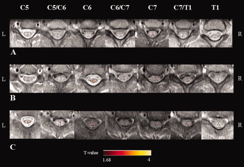
Experimental design: Cervical cord fMRI during a tactile stimulation of the right hand was acquired from 17 healthy controls, 18 SPMS patients, and 16 PPMS patients. Average fMRI activity and its topographical distribution in cord sectors (left vs. right, posterior vs. anterior) were assessed. Correlations between cord recruitment and structural cord and brain MRI were estimated.
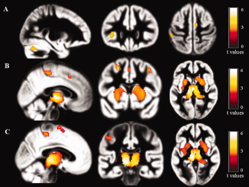
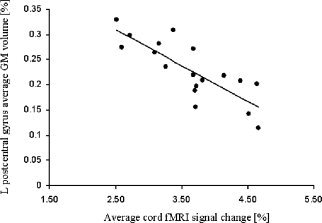
Principal observations: Progressive MS patients showed an increased cord recruitment compared with controls (P = 0.003). Despite a similar structural cord damage, cord activity was increased in SPMS compared to PPMS patients (P = 0.05). Regional analysis showed a non-lateralized pattern of cord recruitment in MS patients. Compared to PPMS, SPMS patients had grey matter (GM) atrophy in several cortical and subcortical regions. In SPMS patients, atrophy of the left postcentral gyrus was correlated with cord activity (r = -0.48, P = 0.04).
Conclusions: Patients with progressive MS had an over-recruitment of the cervical cord, which was more pronounced in SPMS than PPMS, despite similar cord structural damage. The alteration of the complex modulation of spinal cord interneurons possibly due to a loss of supratentorial inhibition secondary to brain injury might contribute to explain the observed functional cord abnormalities.
Copyright © 2011 Wiley Periodicals, Inc.
Figures
References
-
- Agosta F, Absinta M, Sormani MP, Ghezzi A, Bertolotto A, Montanari E, Comi G, Filippi M ( 2007a): In vivo assessment of cervical cord damage in MS patients: A longitudinal diffusion tensor MRI study. Brain 130 ( Part 8): 2211–2219. - PubMed
-
- Agosta F, Pagani E, Caputo D, Filippi M ( 2007b): Associations between cervical cord gray matter damage and disability in patients with multiple sclerosis. Arch Neurol 64: 1302–1305. - PubMed
-
- Agosta F, Valsasina P, Rocca MA, Caputo D, Sala S, Judica E, Stroman PW, Filippi M ( 2008): Evidence for enhanced functional activity of cervical cord in relapsing multiple sclerosis. Magn Reson Med 59: 1035–1042. - PubMed
-
- Agosta F, Valsasina P, Absinta M, Sala S, Caputo D, Filippi M ( 2009a): Primary progressive multiple sclerosis: Tactile‐associated functional MR activity in the cervical spinal cord. Radiology 253: 209–215. - PubMed
MeSH terms
LinkOut - more resources
Full Text Sources
Medical
Miscellaneous
