. 2012 Sep;158A(9):2204-13.
doi: 10.1002/ajmg.a.35532. Epub 2012 Aug 7.

SMARCAL1 deficiency predisposes to non-Hodgkin lymphoma and hypersensitivity to genotoxic agents in vivo

Affiliations

SMARCAL1 deficiency predisposes to non-Hodgkin lymphoma and hypersensitivity to genotoxic agents in vivo

Alireza Baradaran-Heravi et al. Am J Med Genet A. 2012 Sep.

Abstract

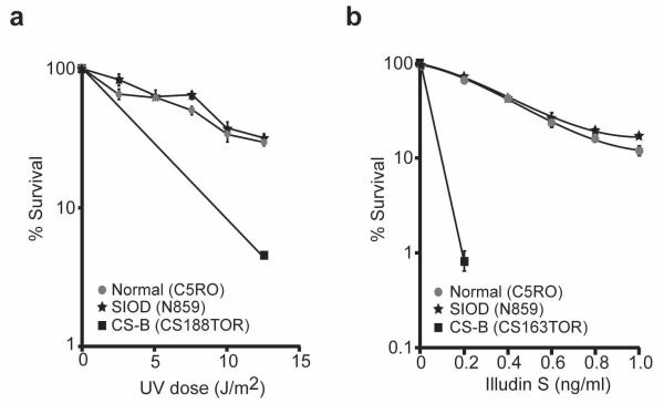
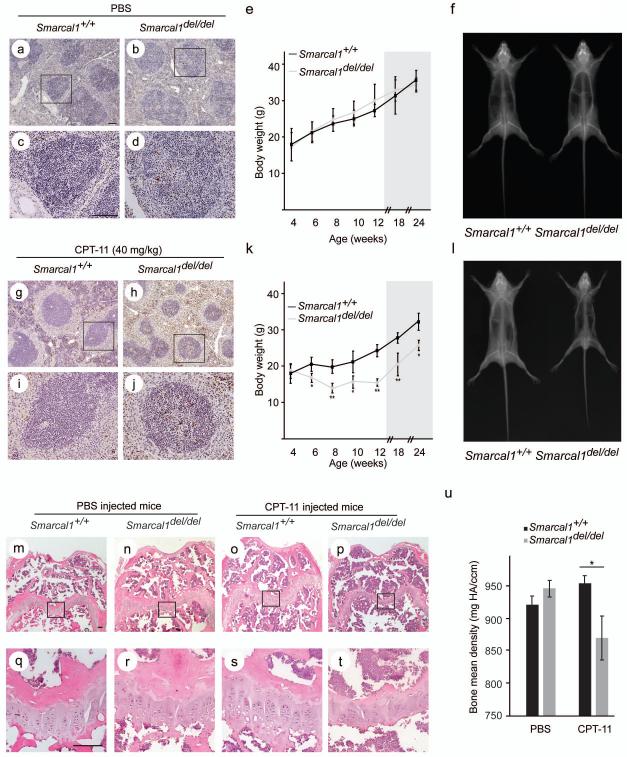
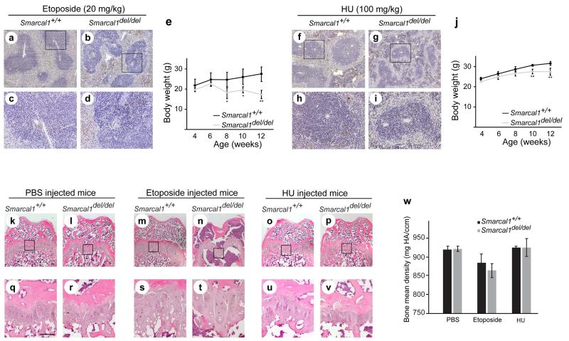
Schimke immuno-osseous dysplasia (SIOD) is a multisystemic disorder with prominent skeletal, renal, immunological, and ectodermal abnormalities. It is caused by mutations of SMARCAL1 (SWI/SNF-related, matrix-associated, actin-dependent regulator of chromatin, subfamily a-like 1), which encodes a DNA stress response protein. To determine the relationship of this function to the SIOD phenotype, we profiled the cancer prevalence in SIOD and assessed if defects of nucleotide excision repair (NER) and nonhomologous end joining (NHEJ), respectively, explained the ectodermal and immunological features of SIOD. Finally, we determined if Smarcal1(del/del) mice had hypersensitivity to irinotecan (CPT-11), etoposide, and hydroxyurea (HU) and whether exposure to these agents induced features of SIOD. Among 71 SIOD patients, three had non-Hodgkin lymphoma (NHL) and one had osteosarcoma. We did not find evidence of defective NER or NHEJ; however, Smarcal1-deficient mice were hypersensitive to several genotoxic agents. Also, CPT-11, etoposide, and HU caused decreased growth and loss of growth plate chondrocytes. These data, which identify an increased prevalence of NHL in SIOD and confirm hypersensitivity to DNA damaging agents in vivo, provide guidance for the management of SIOD patients.


Figures

FIG. 1
FIG. 1
Analysis of SMARCAL1 participation in nucleotide excision repair. a: Survival curve comparing the sensitivity of SIOD (N859), unaffected (C5RO), and Cockayne syndrome B (CS-B, CS188TOR) skin fibroblasts to killing by UV radiation (254 nm). This is a measure of both TC-NER and global NER. b: Survival curve comparing the sensitivity of SIOD (N859), unaffected (C5RO), and CS-B (CS163TOR) skin fibroblasts to killing by illudin-S. This is a measure of TC-NER.
FIG. 2
FIG. 2
Hypersensitivity of Smarcal1del/del mice to CPT-11. a-d: Photomicrographs of TUNEL-positive nuclei in splenic tissue from postnatal day (P) 36 Smarcal1del/del and Smarcal1+/+ mice following 5 daily ip-injections with PBS. Panels c and d are higher magnification photomicrographs of the boxed areas in panels a and b, respectively. e: Graph showing the average weight gain of Smarcal1del/del (n = 4) and Smarcal1+/+ (n = 4) mice receiving daily ip-injections of PBS from P28 through P84 and during 90 days following treatment (shaded area). f: Representative skeletal radiographs of Smarcal1del/del and Smarcal1+/+ mice at P174 following 8 weeks of daily injections with PBS. g-j: Photomicrographs of TUNEL-positive nuclei in splenic tissue from P36 Smarcal1del/del and Smarcal1+/+ mice following 5 daily ip-injections with CPT-11. Panels i and j are higher magnification photomicrographs of the boxed areas in panels g and h, respectively. k: Graph showing the average weight gain of Smarcal1del/del (n = 3) and Smarcal1+/+ (n = 4) mice receiving daily ip-injections of CPT-11 from P28 through P84 and during 90 days following treatment (shaded area). l: Representative skeletal radiographs of Smarcal1del/del and Smarcal1+/+ mice at P173 following 8 weeks of daily injections with CPT-11. m-t: Photomicrographs of representative sections of the distal femoral growth plate of Smarcal1+/+ and Smarcal1del/del male mice treated with PBS or CPT-11 for 8 weeks. The tissue was stained with H&E. Panels q-t are higher magnification photomicrographs of the boxed areas in panels m-p, respectively. Note the poorly organized columns of chondrocytes in the smaller hypocellular growth plates of the CPT-11 treated Smarcal1del/del mice. u: Graph showing the mean bone density of the distal femur of Smarcal1+/+ and Smarcal1del/del mice treated either with PBS or CPT-11. Scale bars = 100 μm. Error bars represent one standard deviation. For statistically significant differences between Smarcal1del/del and Smarcal1+/+ mice, a * indicates P<0.05 and a ** indicates P<0.01. Abbreviations: CPT-11, irinotecan; ip, intraperitoneal; mg HA/ccm, milligrams hydroxyapatite per cubic centimetre.
FIG. 3
FIG. 3
Hypersensitivity of Smarcal1del/del mice to etoposide and HU. a-d: Photomicrographs of TUNEL-positive nuclei in splenic tissue from P36 Smarcal1del/del and Smarcal1+/+ mice following 5 daily ip-injections with etoposide. Panels c and d are higher magnification photomicrographs of the boxed areas in panels a and b, respectively. e: Graph showing the average weight gain of Smarcal1del/del (n = 4) and Smarcal1+/+ (n=3) mice receiving daily ip-injections of etoposide from P28 through P84. f-i: Photomicrographs of TUNEL-positive nuclei in splenic tissue from P36 Smarcal1del/del and Smarcal1+/+ mice following 5 daily ip-injections with hydroxyurea (HU). Panels h and i are higher magnification photomicrographs of the boxed areas in panels f and g, respectively. j: Graph showing the average weight gain of Smarcal1del/del (n = 5) and Smarcal1+/+ (n = 4) mice receiving daily ip-injections of HU from P28 through P84. k-v: Photomicrographs of representative sections of the distal femoral growth plate of Smarcal1+/+ and Smarcal1del/del male mice treated with PBS, etoposide, or HU for 8 weeks. The tissue was stained with H&E. Panels q-v are higher magnification photomicrographs of the boxed areas in panels k-p, respectively. Note the poorly organized columns of chondrocytes in the hypocellular growth plates of the etoposide and HU treated Smarcal1del/del mice. Also note the near absence of trabeculae in the etoposide treated Smarcal1del/del mice. w: Graph showing the bone mean density of the distal femur of Smarcal1+/+ and Smarcal1del/del mice treated with PBS, etoposide or HU. Scale bars = 100 μm. Error bars represent one standard deviation. For statistically significant differences between Smarcal1del/del and Smarcal1+/+ mice, a * indicates P<0.05 and a ** indicates P<0.01. Abbreviations: HU, hydroxyurea; ip, intraperitoneal; mg HA/ccm, milligrams hydroxyapatite per cubic centimetre.

References

    1. Alexander DD, Mink PJ, Adami HO, Chang ET, Cole P, Mandel JS, Trichopoulos D. The non-Hodgkin lymphomas: a review of the epidemiologic literature. Int J Cancer. 2007;120(Suppl 12):1–39. - PubMed
    1. Arama E, Steller H. Detection of apoptosis by terminal deoxynucleotidyl transferase-mediated dUTP nick-end labeling and acridine orange in Drosophila embryos and adult male gonads. Nat Protoc. 2006;1:1725–1731. - PubMed
    1. Bansbach CE, Betous R, Lovejoy CA, Glick GG, Cortez D. The annealing helicase SMARCAL1 maintains genome integrity at stalled replication forks. Genes Dev. 2009;23:2405–2414. - PMC - PubMed
    1. Baradaran-Heravi A, Cho KS, Tolhuis B, Sanyal M, Morozova O, Morimoto M, Elizondo LI, Bridgewater D, Lubieniecka J, Beirnes K, Myung C, Leung D, Fam HK, Choi K, Huang Y, Dionis KY, Zonana J, Keller K, Stenzel P, Mayfield C, Lucke T, Bokenkamp A, Marra MA, van Lohuizen M, Lewis DB, Shaw C, Boerkoel CF. Penetrance of biallelic SMARCAL1 mutations is associated with environmental and genetic disturbances of gene expression. Hum Mol Genet. 2012;21:2572–2587. - PMC - PubMed
    1. Basiratnia M, Baradaran-Heravi A, Yavarian M, Gerami zadeh B, Karimi M. Non-Hodgkin Lymphoma in A Child with Schimke immuno osseous dysplasia. IJMS. 2011;36:222–225. - PMC - PubMed

Publication types