

Long-term safety and efficacy follow-up of prophylactic higher fluence collagen cross-linking in high myopic laser-assisted in situ keratomileusis
- PMID: 22888210
- PMCID: PMC3413339
- DOI: 10.2147/OPTH.S31256
Long-term safety and efficacy follow-up of prophylactic higher fluence collagen cross-linking in high myopic laser-assisted in situ keratomileusis
Abstract
Background: The purpose of this study was to evaluate the safety and efficacy of ultraviolet A irradiation cross-linking on completion for cases of high myopic laser-assisted in situ keratomileusis (LASIK).
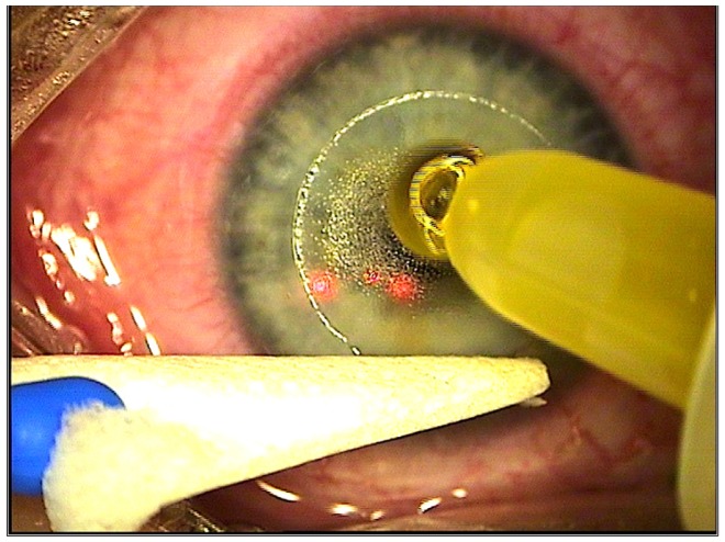
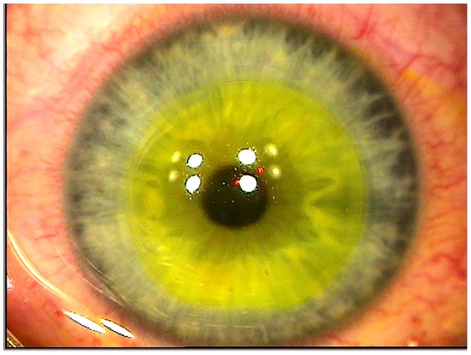
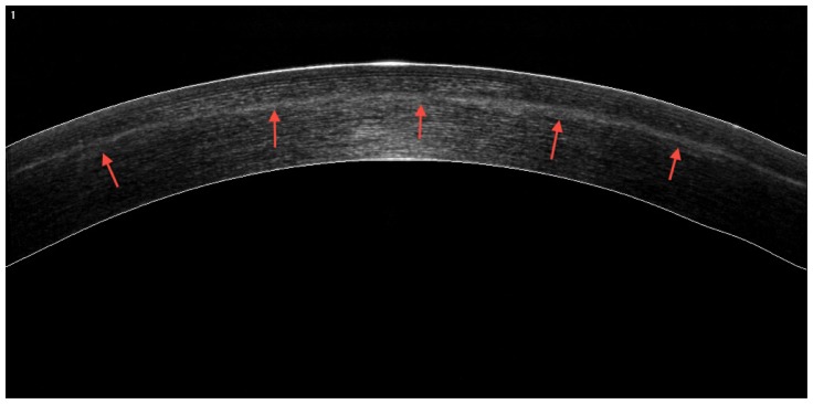
Methods: Forty-three consecutive LASIK cases treated with femtosecond laser flap and the WaveLight excimer platform were evaluated perioperatively for uncorrected visual acuity, best corrected spectacle visual acuity, refraction, keratometry, topography, total and flap pachymetry, corneal optical coherence tomography, and endothelial cell count. All eyes at the completion of LASIK had cross-linking through the repositioned flap, with higher fluence (10 mW/cm(2)) ultraviolet light of an average 370 μm wavelength and 10 mW/cm(2) fluence applied for 3 minutes following an earlier single instillation of 0.1% riboflavin within the flap interface. Mean follow-up duration was 3.5 (range 1.0-4.5) years.
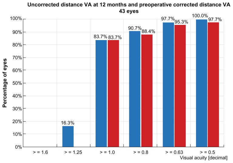
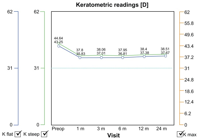
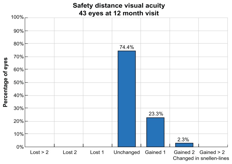
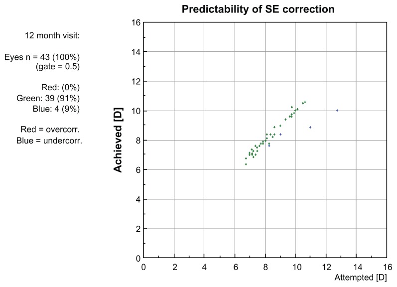
Results: Mean uncorrected visual acuity changed from 0.2 to 1.2, best corrected spectacle visual acuity from 1.1 to 1.2, spherical equivalent from -7.5 diopters (D) to -0.2 D, keratometry from 44.5 D to 38 D, flap pachymetry from 105 μm to, total pachymetry from 525 to 405, and endothelial cell count from 2750 to 2800. None of the cases developed signs of ectasia or significant regression during follow-up.
Conclusion: Prophylactic collagen cross-linking for high-risk LASIK cases appears to be a safe and effective adjunctive treatment for refractive regression and potential ectasia. This application may be viewed as prophylactic customization of the biomechanical behavior of corneal collagen.
Keywords: high-risk; laser-assisted in situ keratomileusis; post-LASIK ectasia; prophylactic collagen cross-linking.
Figures
Similar articles
-
Comparison of prophylactic higher fluence corneal cross-linking to control, in myopic LASIK, one year results.Clin Ophthalmol. 2014 Nov 27;8:2373-81. doi: 10.2147/OPTH.S68372. eCollection 2014. Clin Ophthalmol. 2014. PMID: 25473264 Free PMC article.
-
Combined laser in situ keratomileusis and prophylactic high-fluence corneal collagen crosslinking for high myopia: two-year safety and efficacy.J Cataract Refract Surg. 2015 Jul;41(7):1426-33. doi: 10.1016/j.jcrs.2014.10.045. J Cataract Refract Surg. 2015. PMID: 26287881 Clinical Trial.
-
Short-term Variance of Refractive Outcomes After Simultaneous LASIK and High-Fluence Cross-linking in High Myopic Correction.J Refract Surg. 2016 Oct 1;32(10):664-670. doi: 10.3928/1081597X-20160728-01. J Refract Surg. 2016. PMID: 27722753
-
Same-day intrastromal corneal ring segment and collagen cross-linking for ectasia after laser in situ keratomileusis: long-term results.Am J Ophthalmol. 2014 May;157(5):1070-6. doi: 10.1016/j.ajo.2014.02.011. Epub 2014 Feb 7. Am J Ophthalmol. 2014. PMID: 24513095
-
Comparison of Femto-LASIK With Combined Accelerated Cross-linking to Femto-LASIK in High Myopic Eyes: A Prospective Randomized Trial.Am J Ophthalmol. 2020 Mar;211:42-55. doi: 10.1016/j.ajo.2019.10.024. Epub 2019 Nov 1. Am J Ophthalmol. 2020. PMID: 31678559 Clinical Trial.
Cited by
-
In vivo Confocal Microscopy Report after Lasik with Sequential Accelerated Corneal Collagen Cross-Linking Treatment.Case Rep Ophthalmol. 2014 Apr 12;5(1):125-31. doi: 10.1159/000362327. eCollection 2014 Jan. Case Rep Ophthalmol. 2014. PMID: 24847258 Free PMC article.
-
Evaluation of Biomechanical Changes in Myopia Patients with Unsatisfactory Corneas After Femto Second-Laser In Situ Keratomileusis (FS-LASIK) Concurrent with Accelerated Corneal Collagen Cross-Linking Using Corvis-ST: Two-Year Follow-Up Results.Med Sci Monit. 2017 Jul 27;23:3649-3656. doi: 10.12659/msm.905493. Med Sci Monit. 2017. PMID: 28747621 Free PMC article.
-
Toric topographically customized transepithelial, pulsed, very high-fluence, higher energy and higher riboflavin concentration collagen cross-linking in keratoconus.Case Rep Ophthalmol. 2014 Jun 18;5(2):172-80. doi: 10.1159/000363371. eCollection 2014 May. Case Rep Ophthalmol. 2014. PMID: 25076897 Free PMC article.
-
Comparison of prophylactic higher fluence corneal cross-linking to control, in myopic LASIK, one year results.Clin Ophthalmol. 2014 Nov 27;8:2373-81. doi: 10.2147/OPTH.S68372. eCollection 2014. Clin Ophthalmol. 2014. PMID: 25473264 Free PMC article.
-
Progress of corneal collagen cross-linking combined with refractive surgery.Int J Ophthalmol. 2014 Feb 18;7(1):157-62. doi: 10.3980/j.issn.2222-3959.2014.01.29. eCollection 2014. Int J Ophthalmol. 2014. PMID: 24634883 Free PMC article. Review.
References
-
- Seiler T, Koufala K, Richter G. Iatrogenic keratectasia after laser in situ keratomileusis. J Refract Surg. 1998;14(3):312–317. - PubMed
-
- Geggel HS, Talley AR. Delayed onset keratectasia following laser in situ keratomileusis. J Cataract Refract Surg. 1999;25(4):582–586. - PubMed
-
- Uzbek AK, Kamburoğlu G, Mahmoud AM, Roberts CJ. Change in biomechanical parameters after flap creation using the Intralase femtosecond laser and subsequent excimer laser ablation. Curr Eye Res. 2011;36(7):614–619. - PubMed
-
- Padmanabhan P, Radhakrishnan A, Natarajan R. Pregnancy-triggered iatrogenic (post-laser in situ keratomileusis) corneal ectasia – a case report. Cornea. 2010;29(5):569–572. - PubMed
LinkOut - more resources
Full Text Sources
Other Literature Sources
