. 2012:8:329-42.
doi: 10.2147/TCRM.S32666. Epub 2012 Jul 23.

Training in peroral endoscopic myotomy (POEM) for esophageal achalasia

Affiliations

Training in peroral endoscopic myotomy (POEM) for esophageal achalasia

Nicholas Eleftheriadis et al. Ther Clin Risk Manag. 2012.

Abstract

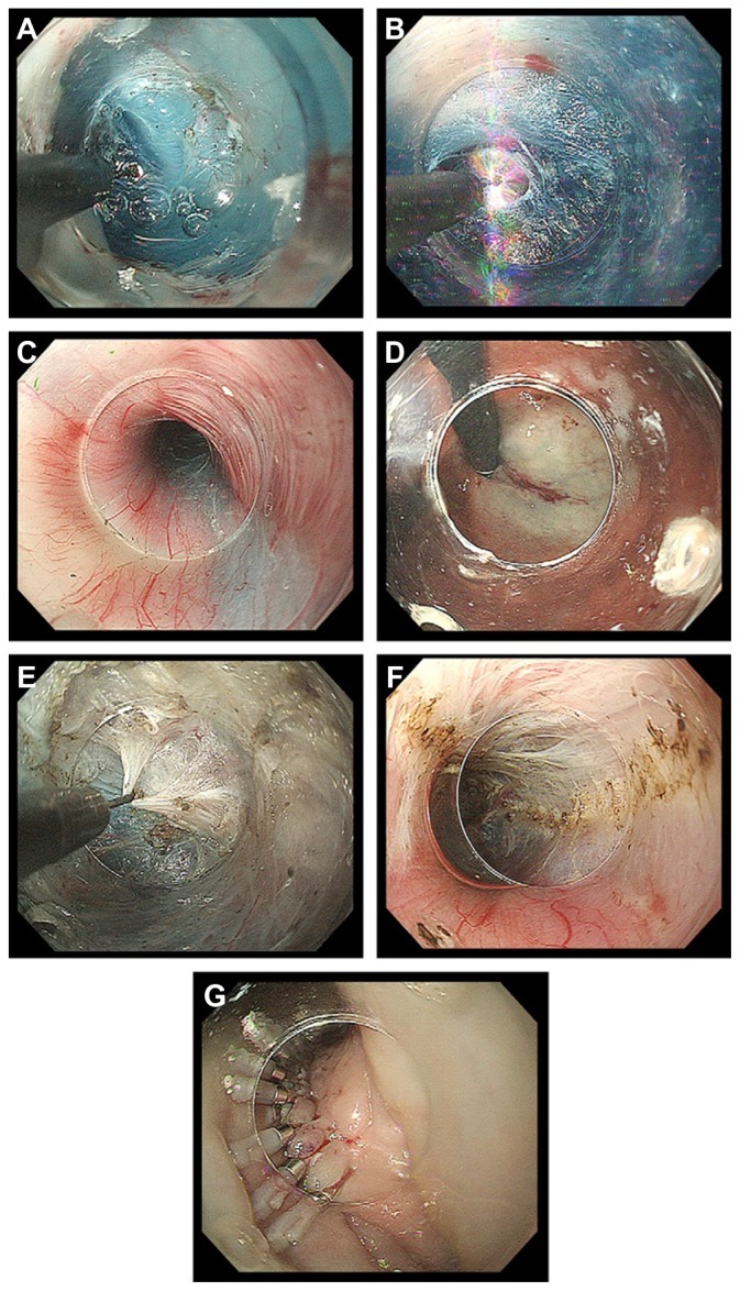
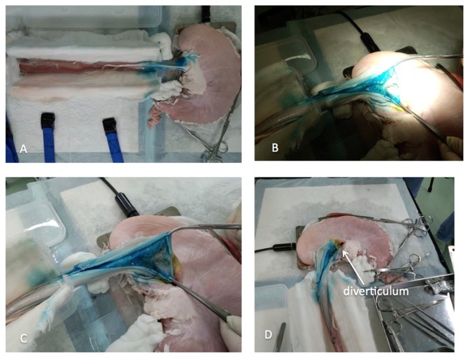
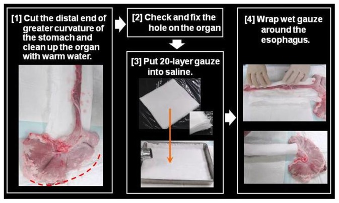
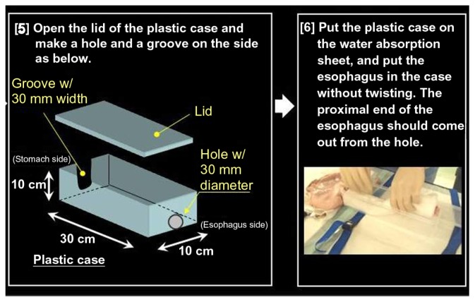
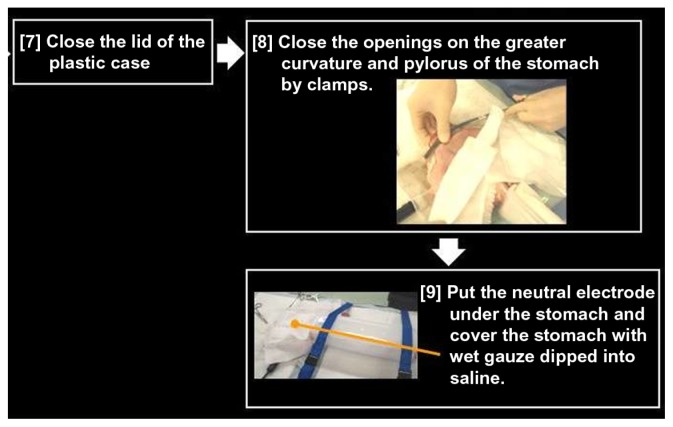
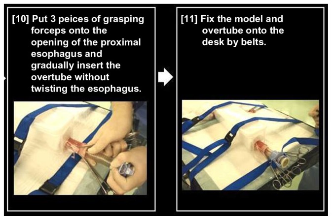
Peroral endoscopic myotomy (POEM) has been developed in the context of natural orifice transluminal endoscopic surgery (NOTES) as a minimally invasive endoscopic treatment for symptomatic esophageal achalasia, which is a chronic progressive benign disease with severe morbidity and difficult management. Since September 2008, POEM has been successfully performed in more than 200 consecutive patients with symptomatic achalasia at the Digestive Disease Center of Showa University, Northern Yokohama Hospital, Yokohama, Japan, with excellent short- and long-term results and absence of serious complications. International experience of POEM within clinical studies is also promising. According to these results, POEM is considered as a safe procedure that can be applied to all achalasia patients. However, the low incidence of achalasia (0.3%-1% per 100,000 population), in combination with the potential serious complications related to the technically demanding POEM procedure, has made training difficult. There is therefore an urgent need for an animal model for training to decrease the learning curve. Further, there are other ethical and training issues to address. The pig is the most appropriate animal model for training in POEM due to its anatomy being similar to that of humans. The porcine esophagus has the advantage of easy mobilization due to absence of tight junctions to surrounding organs. A non-survival porcine model would be a simple, inexpensive, and reproducible animal model for training in POEM, without the need for concern about complications. A possible training process might first involve observation of POEM performed by specialists, then training on non-survival and survival porcine models, followed by training in humans under specialist guidance and finally, performance of POEM in humans.

Keywords: Heller myotomy; non-survival; porcine esophagus; porcine organ model.


Figures

Figure 1
Figure 1
Classification of distal esophageal morphology in esophageal achalasia. Note: Cases from Showa University Northern Yokohama Hospital, Yokohama, Japan. Abbreviations: Sp, spindle type; Fk, flask type; Sig, sigmoid type.
Figure 2
Figure 2
Barium esophagogram before (left) and after (right) POEM in a case of classical achalasia, showing obvious opening of EGJ and smooth passage of contrast after POEM.
Figure 3
Figure 3
Barium esophagogram before (left) and after (right) peroral endoscopic myotomy (POEM) in a case of vigorous achalasia showing disappearance of the simultaneous body contractions, obvious opening of the esophagogastric junction and smooth passage of contrast after the POEM procedure.
Figure 4
Figure 4
Barium esophagogram of sigmoid type of achalasia before and after peroral endoscopic myotomy (POEM), showing significant reduction of esophageal dilatation and smooth passage of contrast from the esophagogastric junction.
Figure 5
Figure 5
(A) A distal small transparent “tapered” cap, securely attached at the tip of the endoscope, is used during submucosal dissection and myotomy. (B) Alternatively, a transparent oblique cap can be used; this is necessary for clipping the esophageal opening. (C) Triangle-tip knife.
Figure 6
Figure 6
(A) The CO2 insufflator (UCR; Olympus Medical Systems Corporation, Tokyo, Japan) used with a regular insufflating tube (MAJ-1742; Olympus) to maintain CO2 insufflation during endoscopy. (B) The air/water button of the endoscope should remain closed during peroral endoscopic myotomy. (C) High-frequency electrogenerator (VIO 300D; ERBE Elektromedizin GmbH, Tubingen, Germany). (D) The endoscopic trolley is shown situated at the right side of the intubated patient. Notes: The patient is in a supine position, with open abdomen and periodically checked during the procedure. Cut and coagulation pedals are fixed on metallic stairs on the right side of the patient.
Figure 7
Figure 7
(A) Longitudinal incision at the 2 o’clock position. (B) Dissection plane. (C) Palisade vessels within the submucosal layer at the esophagogastric junction. (D) The distal margin of the tunnel verified using a retroflexion view from within the gastric lumen. (E) The sharp tip of a triangle-tip knife is used to catch circular muscle bundles and then retract and dissect them toward esophageal lumen. (F) Longitudinal muscle is identified at the bottom of myotomy site. (G) Closure with endoscopic clips.
Figure 8
Figure 8
Non-survival organ model for training in peroral endoscopic myotomy (POEM). (A) Porcine esophagus and stomach are removed as one unit. POEM is performed following the same principles as in humans. At the end of the procedure, blue dye from the submucosal injection can be identified from the external side at the distal esophagus and esophagogastric junction. (B and C) After completion of POEM, circular myotomy is controlled by external longitudinal incision of the distal esophagus and gastric cardia. (D) A normal diverticulum identified at the gastric cardia after external longitudinal incision.
Figure 9
Figure 9
Preparing an organ model for training in peroral endoscopic myotomy. Notes: First, the distal end of gastric greater curvature is cut and the organ is cleaned with warm water. Following this, 20-layer wet gauzes are placed below the stomach and wrapped around the esophagus.
Figure 10
Figure 10
The organ model is put into a plastic case.
Figure 11
Figure 11
Preparing the pig organ model. Notes: Gastric greater curvature and pylorus are closed by clamps. The neutral electrode is placed under the stomach, which is covered with wet gauze dipped in saline.
Figure 12
Figure 12
Preparing the pig organ model. Notes: Three-prong grasping forceps are placed at the opening of the proximal esophagus and an overtube is gradually inserted without twisting the esophagus. Then, the organ model and overtube are fixed on the table with belts.

References

    1. Spechler SJ. UpToDate® [web site on the Internet] Waltham, MA: UpToDate, Inc; 2012. [Accessed May 22, 2012]. Clinical manifestations and diagnosis of achalasia. [updated Mar 31, 2011]. Available from: http://www.uptodate.com/contents/clinical-manifestations-and-diagnosis-o....
    1. Mikaeli J, Fazel A, Montazeri G, Yaghoobi M, Malekzadeh R. Randomized controlled trial comparing botulinum toxin injection to pneumatic dilatation for the treatment of achalasia. Aliment Pharmacol Ther. 2001;15(9):1389–1396. - PubMed
    1. Craft RO, Aguilar BE, Flahive C, et al. Outcomes of minimally invasive myotomy for the treatment of achalasia in the elderly. JSLS. 2010;14(3):342–347. - PMC - PubMed
    1. Mikaeli J, Islami F, Malekzadeh R. Achalasia: a review of Western and Iranian experiences. World J Gastroenterol. 2009;15(40):5000–5009. - PMC - PubMed
    1. Japanese Society for Esophageal Diseases. Guidelines for the Clinical and Pathologic Studies on Carcinoma of the Esophagus. 9th ed. Tokyo: Kanehara; 1999. - PubMed