

Multifocal Langerhans cell sarcoma involving epidermis: a case report and review
- PMID: 22889043
- PMCID: PMC3478178
- DOI: 10.1186/1746-1596-7-99
Multifocal Langerhans cell sarcoma involving epidermis: a case report and review
Abstract
Objective: To study the clinico-pathological characteristics of Langerhans cell sarcoma (LCS) which involving epidermis.
Methods: A case of primary multifocal LCS was analyzed in histopathology and immunophenotype.
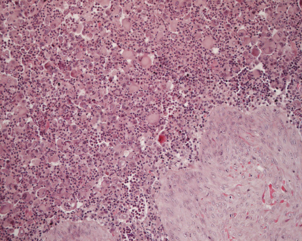
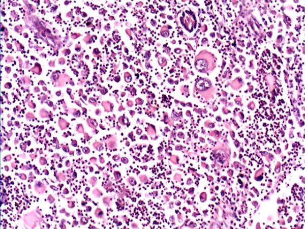
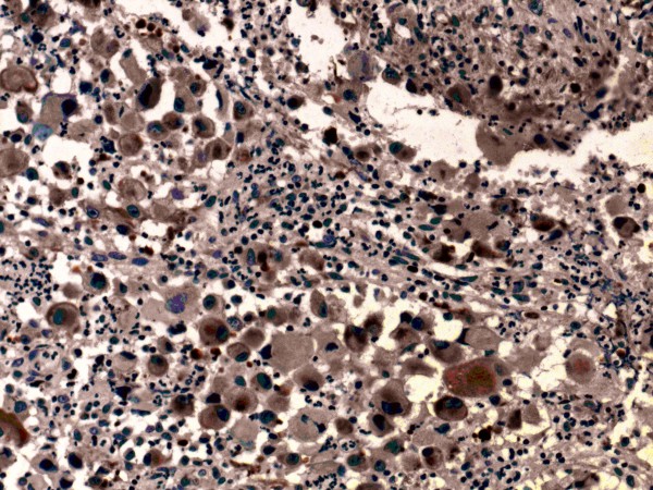
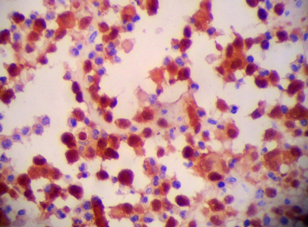
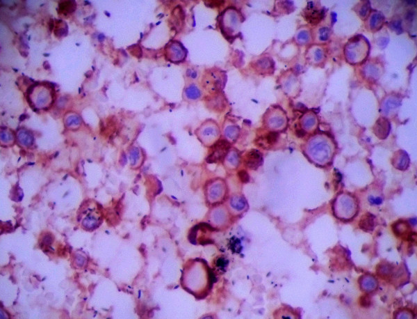
Results: A 41-year-old man with multifocal cutaneous LCS involving the inguina and waist was reported. Clinical and pathology data were available. Neoplastic cells with markedly malignant cytological features were observed. Tumor cells exhibited irregular shape with abundant and eosinophilic red staining cytoplasm; large, irregular-shaped, showing lobulated or dented nucleus and some cells with a longitudinal nuclear groove and prominent nucleoli. The tumor cells expressed CD1a, Langerin (CD207), S-100 protein, CD68 and vimentin, and did not express pan-T or B cell markers and epithelial markers. The patient died less than 1 year after diagnosis due to local recurrence and metastasis to the lung, despite the administration of local radiation and chemotherapy.
Conclusions: LCS is a tumor with markedly malignant cytological features that originates from Langerhans cells. Primary multifocal neoplasms involving epidermis is even rare. Accurate diagnosis is based on the histopathological and immunohistochemical of the tumor cells.
Virtual slide: The virtual slide(s) for this article can be found here: http://www.diagnosticpathology.diagnomx.eu/vs/1182345104754765.
Figures
References
-
- Jaffe E, Harris N, Stein H, In: World Health Organization classification of tumours: Pathology and genetics, tumours of haematopoietic and lymphoid tissues. Barnes L, Eveson JW, Reichart P, Sidransky D, editor. IARC Press, Lyon; 2008. Tumors derived from Langerhans cells; pp. 278–289.
-
- Shi-Yuan He X-LZ. 2 cases of Langerhans cell sarcoma misdiagnosed. J Guangxi Trad Chin Med University. 2000;17:93–95.
-
- Jing Chen J-YT, Ci PAN. et al. Langerhans cell sarcoma: the clinical features and diagnosis. J China Pediatr Blood Cancer. 2007;12:213–214.
-
- Lian YL, He HY, Liao SL. et al. Langerhans cell sarcoma of talus: report of a case. Zhonghua Bing Li Xue Za Zhi. 2006;35:697–698. - PubMed
Publication types
MeSH terms
Substances
LinkOut - more resources
Full Text Sources
Medical
