. 2012 Nov;18(11):890-902.
doi: 10.1089/ten.TEC.2011.0482. Epub 2012 Oct 1.

Immortalized functional endothelial progenitor cell lines from umbilical cord blood for vascular tissue engineering

Affiliations

Immortalized functional endothelial progenitor cell lines from umbilical cord blood for vascular tissue engineering

Praveen K Sobhan et al. Tissue Eng Part C Methods. 2012 Nov.

Abstract

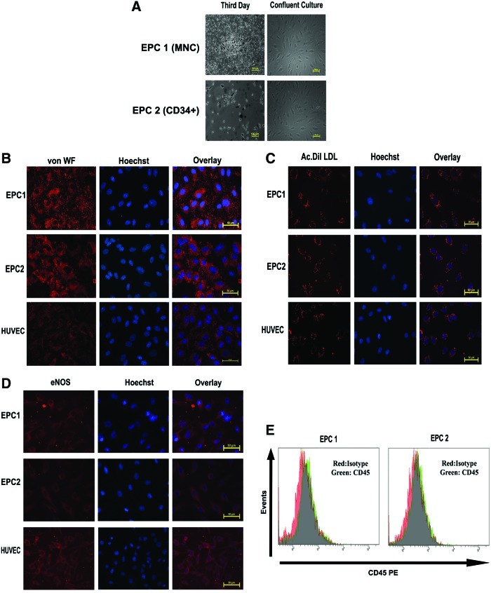
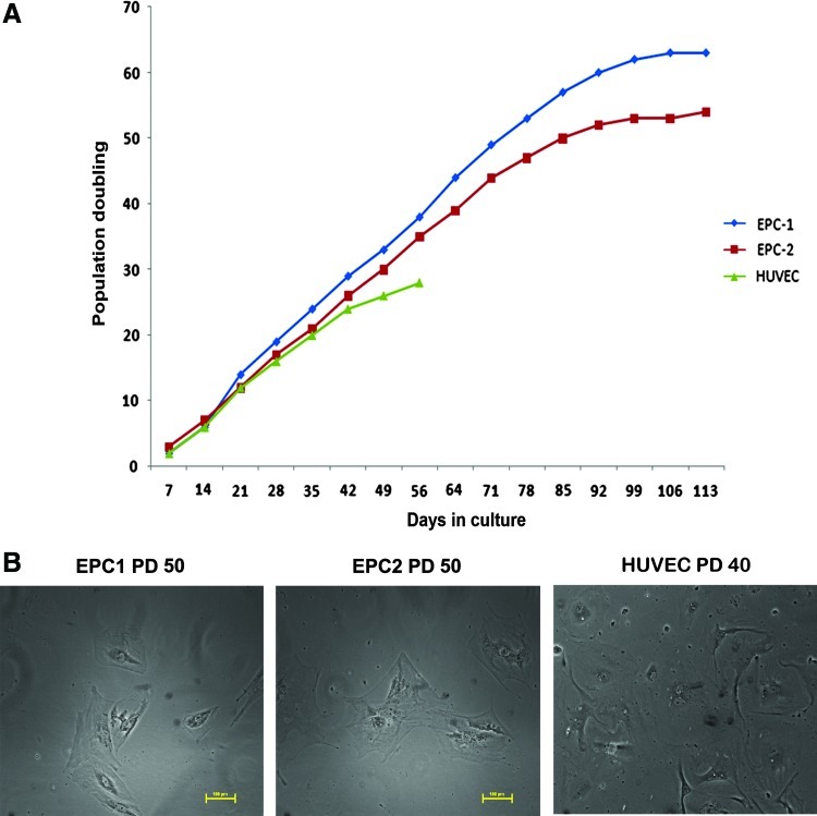
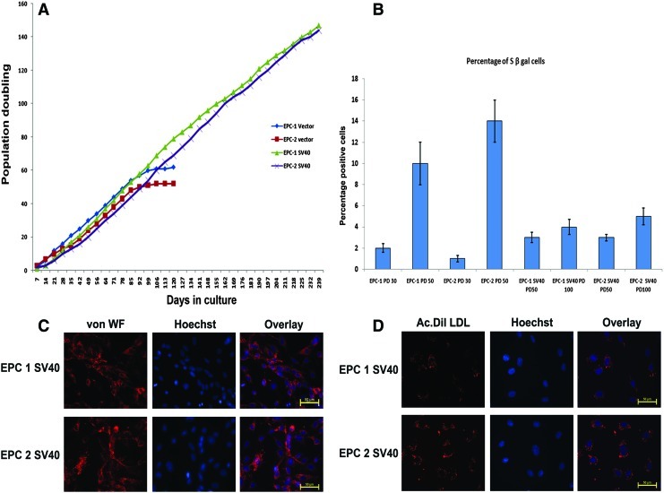
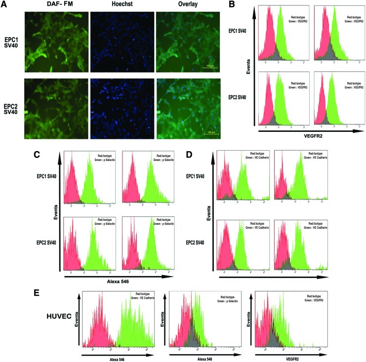
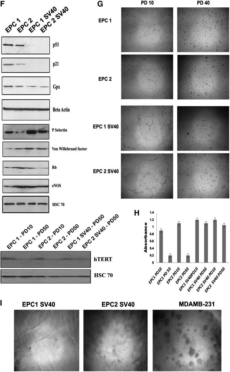
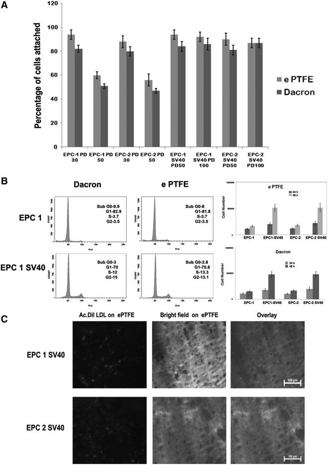
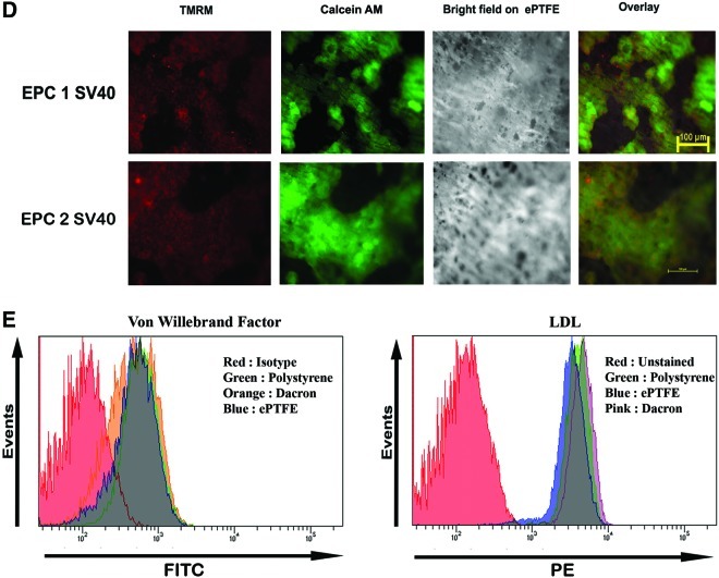
Endothelial progenitor cells (EPCs) play a significant role in multiple biological processes such as vascular homeostasis, regeneration, and tumor angiogenesis. This makes them a promising cell of choice for studying a variety of biological processes, toxicity assays, biomaterial-cell interaction studies, as well as in tissue-engineering applications. In this study, we report the generation of two clones of SV40-immortalized EPCs from umbilical cord blood. These cells retained most of the functional features of mature endothelial cells and showed no indication of senescence after repeated culture for more than 240 days. Extensive functional characterization of the immortalized cells by western blot, flow cytometry, and immunofluorescence studies substantiated that these cells retained their ability to synthesize nitric oxide, von Willebrand factor, P-Selectin etc. These cells achieved unlimited proliferation potential subsequent to inactivation of the cyclin-dependent kinase inhibitor p21, but failed to form colonies on soft agar. We also show their enhanced growth and survival on vascular biomaterials compared to parental cultures in late population doubling. These immortalized EPCs can be used as a cellular model system for studying the biology of these cells, gene manipulation experiments, cell-biomaterial interactions, as well as a variety of tissue-engineering applications.


Figures

FIG. 1.
FIG. 1.
(A) Mononuclear cells (MNCs) isolated from umbilical cord blood (upper panel) or CD34+ MACS-sorted cells were plated on endothelial cell growth medium-2 (EGM-2) on collagen-coated dishes. A representative image on 3rd day and confluent culture is shown (Mag. 10×). (B) The expanded endothelial progenitor cells (EPCs) emerged from MNC (EPC1) and CD34+ MACS-sorted cells (EPC2) grown on glass coverslips were fixed and stained with von Willebrand factor (von WF) primary antibody followed by Alexa-546-conjugated secondary antibody. The cells were also stained with Hoechst to visualize nuclei. Human umbilical cord vein endothelial cells (HUVECs) were employed as positive control. As seen, the cells from both cultures showed intense granular staining similar to HUVECs. (C) The indicated cells were incubated with Ac-Dil-LDL for 4 h and imaged with a fluorescent microscope after staining with Hoechst. Both EPC1 and EPC2 showed internalization of Ac-Dil-LDL to the same extent as HUVECs. (D) The indicated cells were stained with endothelial nitric oxide synthase antibody followed by Alexa 546 secondary antibody and imaged as described above. (E) Both EPC1 and EPC2 were incubated with CD45 PE antibody or isotope IgG, followed by FACS analysis. Both the cells showed negative expression for CD45. Color images available online at www.liebertpub.com/tec
FIG. 2.
FIG. 2.
(A) The growth kinetics of EPC1, EPC2, and HUVECs are shown using the population doubling (PD) calculated at each passage. (B) A representative image showing flattened senescent cells at PD60 in both EPC1 and EPC2, as well as HUVECs at PD40. Color images available online at www.liebertpub.com/tec
FIG. 3.
FIG. 3.
(A) The growth kinetics of the immortalized EPCs (EPC1 SV40 and EPC2 SV40) and respective vector-transfected cells are shown. (B) The percentage of senescent cells from the respective PD for different cells are shown after staining the cells for the S β gal stain as described (n=4). (C) The immortalized cells, EPC1 SV40 and EPC2 SV40, were grown on glass coverslips and stained for von WF as described. (D) The indicated cells were incubated with Ac-Dil-LDL and representative fluorescent images are shown. Color images available online at www.liebertpub.com/tec
FIG. 4.
FIG. 4.
(A) EPC1 SV40 and EPC2 SV40 cells were incubated with the intracellular nitric oxide sensor 4-amino-5-methylamino-2′,7′-difluorescein (DAF-FM). The DAF fluorescence was detected using an FITC filter. The cells were also stained with Hoechst to visualize the nuclei. (B) EPC1 SV40 and EPC2 SV40 cells were incubated with VEGFR-2 PE and analyzed by FACS. The red histogram indicates the respective isotype IgG. (C) The above panel of cells was fixed and incubated with a primary antibody against P-Selectin followed by Alexa 546 secondary antibody. The respective isotype IgG control is represented as red histogram. (D) The above panel of cell lines was processed for VE cadherin immunostaining as described above. (E) HUVECs stained with above panel of antibodies served as control. (F) EPC1, EPC2, and the respective immortalized clones were grown on 100-mm dishes. The protein was extracted from subconfluent cultures as described. About 80 μg of protein was resolved on SDS–PAGE, followed by western blotting using the antibody indicated. Beta-actin and HSC-70 served as loading control. (G) EPC1, EPC2, and the respective SV40-immortalized cells were grown on Matrigel-coated dishes. After 16 h, the tubules formed were visualized using 4× objective. (H) Telomerase activity was assayed with a TeloTAGGGTelomerase PCR ELISAPLUS detection kit as described. The absorbance values determined at 450 nm using a reference wavelength of ∼690 nm used for plotting the graph (n=3). (I) The immortalized EPC1, EPC2, and the breast cancer cell line MDAMB-231 were grown on soft-agar colony assay. The representative images are shown. Color images available online at www.liebertpub.com/tec
FIG. 4.
FIG. 4.
(A) EPC1 SV40 and EPC2 SV40 cells were incubated with the intracellular nitric oxide sensor 4-amino-5-methylamino-2′,7′-difluorescein (DAF-FM). The DAF fluorescence was detected using an FITC filter. The cells were also stained with Hoechst to visualize the nuclei. (B) EPC1 SV40 and EPC2 SV40 cells were incubated with VEGFR-2 PE and analyzed by FACS. The red histogram indicates the respective isotype IgG. (C) The above panel of cells was fixed and incubated with a primary antibody against P-Selectin followed by Alexa 546 secondary antibody. The respective isotype IgG control is represented as red histogram. (D) The above panel of cell lines was processed for VE cadherin immunostaining as described above. (E) HUVECs stained with above panel of antibodies served as control. (F) EPC1, EPC2, and the respective immortalized clones were grown on 100-mm dishes. The protein was extracted from subconfluent cultures as described. About 80 μg of protein was resolved on SDS–PAGE, followed by western blotting using the antibody indicated. Beta-actin and HSC-70 served as loading control. (G) EPC1, EPC2, and the respective SV40-immortalized cells were grown on Matrigel-coated dishes. After 16 h, the tubules formed were visualized using 4× objective. (H) Telomerase activity was assayed with a TeloTAGGGTelomerase PCR ELISAPLUS detection kit as described. The absorbance values determined at 450 nm using a reference wavelength of ∼690 nm used for plotting the graph (n=3). (I) The immortalized EPC1, EPC2, and the breast cancer cell line MDAMB-231 were grown on soft-agar colony assay. The representative images are shown. Color images available online at www.liebertpub.com/tec
FIG. 5.
FIG. 5.
(A) The EPC1 and EPC2 and the respective SV40-immortalized cells at the indicated PD were allowed to attach on to different surfaces. The percentage of cell attachment is shown (n=4). (B) The EPC1 vector-transfected and EPC1 SV40 cells at PD40 were allowed to grow for 24 h on Dacron and expanded polytetrafluoroethylene (ePTFE) fixed on 12-well plates. Then, the cells were trypsinized and analyzed for cell cycle status as described. The percentage of cells at each phase is also indicated. The cell proliferation analysis data for the indicated cells on Dacron and ePTFE are also shown (right). (C) The SV40-immortalized cells were grown on ePTFE for 24 h. Then, the cells were stained with Ac- Dil-LDL for 4 h. The epifluorescence image of internalized low-density lipoprotein (LDL) is shown. (D) The EPC1 SV40 cells and EPC2 SV40 cells were allowed to grow on ePTFE for 24 h. Then, the cells were stained with mitochondrial transmembrane potential-sensitive dye tetramethyl rhodamine methyl ester (TMRM) and calcium AM as described. Both TMRM and Calcium AM channels are shown. As seen, all the calcium-AM-positive cells retained granular red fluorescence, indicating maintenance of mitochondrial membrane potential. (E) The EPC1 SV40 cells were allowed to grow on Dacron, e PTFE, or tissue culture polystyrene for 24 h. Then, the cells were stained with von WF and analyzed by FACS (left). The same cells grown on same group of surfaces were also incubated with Ac-Dil-LDL and uptake was analyzed by FACS (right). Color images available online at www.liebertpub.com/tec
FIG. 5.
FIG. 5.
(A) The EPC1 and EPC2 and the respective SV40-immortalized cells at the indicated PD were allowed to attach on to different surfaces. The percentage of cell attachment is shown (n=4). (B) The EPC1 vector-transfected and EPC1 SV40 cells at PD40 were allowed to grow for 24 h on Dacron and expanded polytetrafluoroethylene (ePTFE) fixed on 12-well plates. Then, the cells were trypsinized and analyzed for cell cycle status as described. The percentage of cells at each phase is also indicated. The cell proliferation analysis data for the indicated cells on Dacron and ePTFE are also shown (right). (C) The SV40-immortalized cells were grown on ePTFE for 24 h. Then, the cells were stained with Ac- Dil-LDL for 4 h. The epifluorescence image of internalized low-density lipoprotein (LDL) is shown. (D) The EPC1 SV40 cells and EPC2 SV40 cells were allowed to grow on ePTFE for 24 h. Then, the cells were stained with mitochondrial transmembrane potential-sensitive dye tetramethyl rhodamine methyl ester (TMRM) and calcium AM as described. Both TMRM and Calcium AM channels are shown. As seen, all the calcium-AM-positive cells retained granular red fluorescence, indicating maintenance of mitochondrial membrane potential. (E) The EPC1 SV40 cells were allowed to grow on Dacron, e PTFE, or tissue culture polystyrene for 24 h. Then, the cells were stained with von WF and analyzed by FACS (left). The same cells grown on same group of surfaces were also incubated with Ac-Dil-LDL and uptake was analyzed by FACS (right). Color images available online at www.liebertpub.com/tec

Similar articles

Cited by

References

    1. Ahn J.B. Rha S.Y. Shin S.J. Jeung H.C. Kim T.S. Zhang X., et al. Circulating endothelial progenitor cells (EPC) for tumor vasculogenesis in gastric cancer patients. Cancer Lett. 2010;288:124. - PubMed
    1. Andreou I. Tousoulis D. Tentolouris C. Antoniades C. Stefanadis C. Potential role of endothelial progenitor cells in the pathophysiology of heart failure: clinical implications and perspectives. Atherosclerosis. 2006;189:247. - PubMed
    1. Arap W. Pasqualini R. Engineered embryonic endothelial progenitor cells as therapeutic Trojan horses. Cancer Cell. 2004;5:406. - PubMed
    1. Au P. Daheron L.M. Duda D.G. Cohen K.S. Tyrrell J.A. Lanning R.M., et al. Differential in vivo potential of endothelial progenitor cells from human umbilical cord blood and adult peripheral blood to form functional long-lasting vessels. Blood. 2008;111:1302. - PMC - PubMed
    1. Choi J.H. Hur J. Yoon C.H. Kim J.H. Lee C.S. Youn S.W., et al. Augmentation of therapeutic angiogenesis using genetically modified human endothelial progenitor cells with altered glycogen synthase kinase-3beta activity. J Biol Chem. 2004;279:49430. - PubMed

Publication types

Substances