Review
. 2012 Sep;249(1):158-75.
doi: 10.1111/j.1600-065X.2012.01146.x.

PAMPs and DAMPs: signal 0s that spur autophagy and immunity

Affiliations
Review

PAMPs and DAMPs: signal 0s that spur autophagy and immunity

Daolin Tang et al. Immunol Rev. 2012 Sep.

Abstract

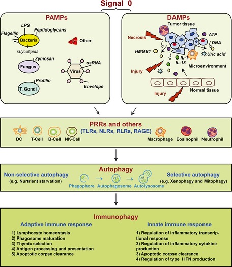
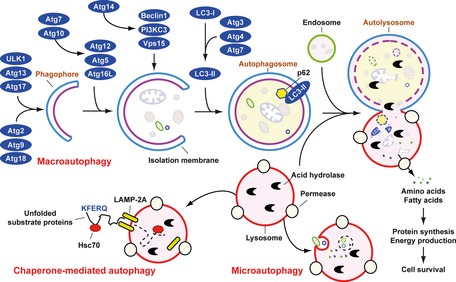
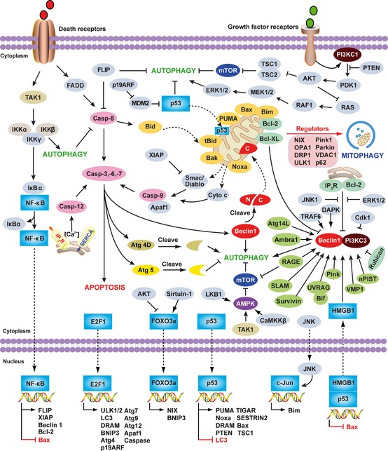
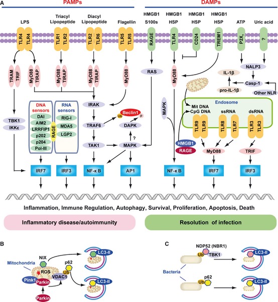
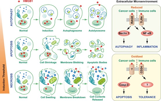
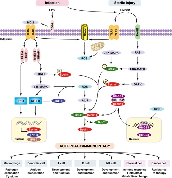
Pathogen-associated molecular pattern molecules (PAMPs) are derived from microorganisms and recognized by pattern recognition receptor (PRR)-bearing cells of the innate immune system as well as many epithelial cells. In contrast, damage-associated molecular pattern molecules (DAMPs) are cell-derived and initiate and perpetuate immunity in response to trauma, ischemia, and tissue damage, either in the absence or presence of pathogenic infection. Most PAMPs and DAMPs serve as so-called 'Signal 0s' that bind specific receptors [Toll-like receptors, NOD-like receptors, RIG-I-like receptors, AIM2-like receptors, and the receptor for advanced glycation end products (RAGE)] to promote autophagy. Autophagy, a conserved lysosomal degradation pathway, is a cell survival mechanism invoked in response to environmental and cellular stress. Autophagy is inferred to have been present in the last common eukaryotic ancestor and only to have been lost by some obligatory intracellular parasites. As such, autophagy represents a unifying biology, subserving survival and the earliest host defense strategies, predating apoptosis, within eukaryotes. Here, we review recent advances in our understanding of autophagic molecular mechanisms and functions in emergent immunity.


Figures

Figure 1
Figure 1
Signal 0s play critical roles in autophagy and immunity. Pathogen‐associated molecular patterns (PAMPs) and damage‐associated molecular patterns (DAMPs) serve as signal 0s, inducing autophagy and immunophagy in the emergent immune response before the later Signal 1 (antigenic peptide and major histocompatability molecules), Signal 2 (costimulatory molecules such as CD80 and CD86), both present on the surface of DCs recruited by the signal 0s. Signal 3 represents the DC provided IL‐6 family cytokine expression such as IL‐12 and IL‐23 which promote polarization of emergent T‐cell response. Signal 4 represents the integrin expression on DCs, defining the origin of the DCs and driving specialized molecules on T‐cells promoting T‐cell traffic to tissues. LPS, lipopolysaccharide; HMGB1, high mobility group box 1; ATP, adenosine‐5′‐triphosphate; PRRs, pattern recognition receptors; TLRs, Toll‐like receptors; NLRs, NOD‐like receptors; RLRs, RIG‐I‐like receptors; RAGE, receptor for advanced glycation end products.
Figure 2
Figure 2
Means by which autophagy delivers antigen into the autolysosome. Microautophagy refers to the sequestration of cytosolic components directly by lysosomes through invaginations within their limiting membrane. Chaperone‐mediated autophagy involves direct translocation of unfolded substrate proteins (KFERQ‐like motif) across the lysosome membrane through the action of a cytosolic and lysosomal chaperone heat shock cognate protein of 70 kDa (Hsc70), and the integral membrane receptor lysosome‐associated membrane protein type 2A (LAMP‐2A). In the case of macroautophagy, the cargo is sequestered within a unique double membrane cytosolic vesicle, an autophagosome. The autophagosome itself is formed by expansion of the phagophore. The autophagosome undergoes fusion with a late endosome or lysosome to form an autolysosome, in which the sequestered material is degraded. Degradation of membrane lipids and proteins by the autolysosome generates free fatty acids, nucleotides, and amino acids that can be reused by the cell to maintain mitochondrial ATP energy production, protein synthesis, and thereby promote cell survival. The molecular machinery of macroautophagy was largely discovered in yeast and the centrally important proteins referred to as autophagy‐related (ATG) proteins although some similar proteins in mammals have disparate names (Beclin‐1 = ATG6, LC3 = ATG8 for example).
Figure 3
Figure 3
Overview of the major signal transduction pathways that regulate autophagy and apoptosis. Common molecular regulators include gene products that affect both autophagy and apoptosis and the pathways that they influence. The process of apoptotic cell death is mediated by two central pathways: an extrinsic pathway involving cell surface receptors (the death receptor pathway), and an intrinsic pathway using mitochondria and the endoplasmic reticulum (the mitochondrial pathway). A third pathway is mediated by cytolytic T and NK cells delivering perforin/granzymes to promote apoptosis. CD95 (also called Fas or APO‐1) induces apoptosis by forming a death‐inducing signaling complex (DISC) at the receptor that contains FADD, caspase‐8, and the caspase‐8 regulator. Autophagy can degrade active caspase‐8. The FLICE inhibitory protein (FLIP), a caspase‐like molecule without proteolytic activity, protects cells from CD95‐induced apoptosis. FLIP can suppress autophagy. The intrinsic mitochondrial pathway is activated by diverse apoptotic signals such as DNA damage, growth factor deprivation, and oxidative stress. Cytoplasmic translocation of mitochondrial proteins [such as cytochrome c (cyt c) and Smac/DIABLO] lead to activation of the caspase cascade and initiate apoptosis. A pivotal event in the mitochondrial pathway is mitochondrial outer membrane permeabilization (MOMP), which is mainly regulated by Bcl‐2 family members. Some of the Bcl‐2 family members (e.g. Bcl‐2, and Bcl‐XL) are anti‐apoptotic, whereas others (e.g. Bax, tBid, Bad, Bim, PUMA, and Noxa) are pro‐apoptotic. Mitophagy is a well established mechanism necessary for elimination of dysfunctional mitochondria and regulation of mitochondrial quality in yeast or mammalian cells associated with cytosolic mediators such as NIX, Atg32, optic atrophy 1 (OPA1), dynamin‐related protein 1 (DRP1), unc‐51‐like kinase 1 (ULK1), Parkin, Pink1, voltage‐dependent anion channel 1 (VDAC1), p62/Sequestosome 1 (SQSTM1), mitofusin 1, and mitofusin 2. Beclin 1, the mammalian ortholog of yeast Atg6, plays a central role in autophagy. It interacts with several cofactors (e.g. Atg14L, UVRAG, Bif‐1, Rubicon, Ambra1, HMGB1, nPIST, VMP1, SLAM, IP 3R, PINK, and Survivin) to regulate the formation of Beclin 1‐PI3KC3 complexes, thereby inducing autophagy. In contrast, the BH3 domain of Beclin 1 is bound to and inhibited by Bcl‐2 or Bcl‐XL. This interaction can be disrupted by phosphorylation of Bcl‐2 and Beclin 1, or ubiquitination of Beclin 1. Interestingly, caspase‐mediated cleavage of Beclin 1 promotes cross‐talk between apoptosis and autophagy. Although apoptosis‐associated cleavage of Beclin 1 and Atg5 inactivates autophagy, the cleavage of Atg4D by caspase‐3 generates a fragment with increased autophagic activity. In the presence of growth factors, growth factor receptor signaling activates PI3KC1 at the plasma membrane. PI3KC1 activates the downstream target AKT, leading to activation of mammalian target of rapamycin (mTOR) by inhibiting the tuberous sclerosis complex 1/2 (TSC1–TSC2), which results in inhibition of autophagy. Overexpression of the phosphatase and tensin homolog (PTEN) gene, by an inducible promoter, antagonizes PI3KC1 to induce autophagy. RAS has a dual effect on autophagy. When it activates PI3KC1, autophagy is inhibited, but when it selectively activates the RAF1–mitogen‐activated protein kinase kinase (MEK)–extracellular signal‐regulated kinase (ERK) cascade, autophagy is stimulated. AMPK monitors the energy status of the cell by sensing the AMP:ATP ratio. Several upstream kinases, including liver kinase B1 (LKB1, which is activated by energy depletion), calcium/calmodulin kinase kinase‐β (CaMKKβ, which is activated by cytosolic Ca2+ levels), and TGFβ activated kinase‐1 (TAK‐1, which is also involved in IKK activation), can activate AMPK by phosphorylation. Activated AMPK promotes inhibition of mTOR kinase, which induces autophagy. NF‐κB and p53 play a double role in regulating autophagy in a transcription dependent and/or independent fashion. In contrast, E2F1 and FOXO3 positively regulate autophagy in a transcription dependent fashion. Nuclear HMGB1 inhibits the p53‐dependent transactivation from the Bax promoter. Accumulation of cytosolic HMGB1 sustains autophagy by liberating the Beclin 1 and PI3KC3 complexes. RAGE‐induced autophagy is associated with decreased phosphorylation of mTOR and increased Beclin1‐PI3KC3 interactions.
Figure 4
Figure 4
The role of TLR s, RLR s, and NLR s in PAMP and DAMP recognition. (A) Signaling pathways triggered by pathogen‐associated molecular pattern (PAMPs) and damage‐associated molecular pattern molecules (DAMPs). Lipopolysaccharide (LPS) activates both the myeloid differentiation factor 88 (MyD88)‐dependent and TIR‐domain‐containing adapter‐inducing interferon‐β (TRIF)‐dependent Toll‐like receptor 4 (TLR4) pathways. The MyD88‐dependent pathway is responsible for NF‐κB and mitogen‐activated protein kinase (MAPK) activation, which controls induction of proinflammatory cytokines. The TRIF‐dependent pathway activates IRF3 by TANK‐binding kinase 1 (TBK1)/IKKε, which is required for the induction of IFN‐inducible genes. TLR1‐TLR2 and TLR2‐TLR6 recognize bacterial triacylated lipopeptide or diacyl lipopeptide, respectively, and recruit TIR adapter protein (TIRAP) and MyD88 at the plasma membrane to activate the MyD88‐dependent pathway. TLR5 recognizes flagellin and activates the MyD88‐dependent pathway. TLR3, TLR7, TLR8, and TLR9 reside in the endosome and recognize dsRNA, ssRNA, CpG DNA, or mitochondrial DNA (Mit DNA), respectively. They recruit TRIF or MyD88 to activate the IRF3 or IRF7 pathway. All immunogenic nucleic acids bind indicated cytosolic DNA sensors or RNA sensors, including retinoid acid‐inducible gene I (RIG‐I)‐like receptors (RLRs), which are required for subsequent recognition by specific pattern recognition receptors to activate innate immune responses. DAMPs such as HMGB1, S100 proteins (S100s), and heat shock proteins (HSPs) recognize the receptor for advanced glycation end products (RAGE), TLR4, or triggering receptor expressed on myeloid cells‐1 (TREM‐1) and activate the MyD88‐MAPKNF‐κB pathway. HMGB1 and RAGE activate the TLR9‐MyD88 dependent pathway, which contributes to autoimmune pathogenesis. CD24 is a negative receptor to inhibit the DAMP‐induced TLR4 pathway. ATP binding of the P2X7 receptor and uric acid, as well as asbestos and alum, increase activation of caspase‐1 by the NALP3 inflammasome and other nucleotide‐binding oligomerization domain (NOD)‐like receptors (NLRs) to promote secretion of IL‐1β and IL‐18. PAMP and DAMP‐mediated signaling and induction of an innate immune response usually results in resolution of infection, but may also cause chronic inflammation or autoimmunity by altering various cell death and survival mechanisms. (B) Mitochondria in mammalian cells are removed by autophagy via the NIX adapter during developmental elimination of mitochondria, or via PTEN‐induced putative kinase 1 (Pink1) and Parkin‐mediated ubiquitination (Ub) of voltage‐dependent anion channel 1 (VDAC1), recognized by the adapter p62 for removal of stressed (e.g. depolarized or damaged) mitochondria. (C) Intracellular bacteria exposed to the cytosol are modified by ubiquitin (Ub) and recognized by the autophagic adapters p62 or nuclear dot protein 52 (NDP52)/TBK1 for sequestration into autophagosomes and subsequent elimination.
Figure 5
Figure 5
Cellular changes and HMGB 1 release observed with autophagy, apoptosis, and necrosis. The prototypical DAMP, high mobility group box 1 protein (HMGB1), is released with sustained autophagy, late apoptosis, and necrosis. Reducible HMGB1 binds to the receptor for advanced glycation end products (RAGE), inducing Beclin 1‐dependent autophagy, and binds both RAGE and Toll‐like receptor 2 (TLR2), TLR4, and TLR9, activating NF‐κB and promoting inflammation. In contrast, oxidized HMGB1 increases caspase 3 dependent apoptosis and tolerance by binding CD24 or other unknown receptors.
Figure 6
Figure 6
Signaling pathways triggered by LPS and HMGB 1 in autophagy/immunophagy. The inflammatory response, including the recruitment and migration of immune cells to the site of infection and release of cytokines, is mediated by lipopolysaccharide (LPS). LPS bound to LPS‐binding protein (LBP) is presented to CD14. CD14 maneuvers the LPSLBP complex to TLR4, and LPS, in combination with accessory molecule MD2, activates TLR4 signaling. LPS induces activation of the tumor necrosis factor receptor (TNFR)‐associated factor 6 (TRAF6)‐p38 MAPK pathway and induces the expression of TNF‐α and Beclin 1 by NF‐κB. NF‐κB inhibits TNF‐α‐induced autophagy. Multiple means to promote ROS production converge on the mitochondria or alternatively, NADPH oxidases such as NOX2 and NOX4. These in turn results in activation of autophagy through Atg4 activation, ultimately reducing the binding of Beclin 1 to Bcl‐2. TRIF‐dependent and/or MyD88‐dependent TLR4 pathway is required for LPS‐induced autophagy in macrophages. Moreover, TRAF6‐mediated ubiquitination of Beclin1 amplifies TLR4‐induced autophagy. HMGB1 links sterile injury and infection‐induced immunity. Stimuli that enhance reactive oxygen species promote cytosolic translocation of HMGB1 and thereby enhance autophagic flux. HMGB1 directly interacts with Beclin1, displacing Bcl‐2 requiring the cysteines at positions C23 and C45 within HMGB1. The HMGB1/RAGE interaction activates parallel signaling pathways, including ERK1/2 and NF‐ κB activation. Mitogen‐activated protein kinases (MAPKs), such as JNK1 and ERK1/2, also phosphorylate Bcl‐2 driving subsequent dissociation of the Beclin 1‐Bcl‐2 complex. Notably, DAPK phosphorylates Beclin 1, promoting the dissociation of Beclin 1 from Bcl‐2 like proteins, which in turn induces autophagy. Activation of phagocytic NADPH oxidase (NOX2) by HMGB1 requires TLR4 expression; the activation of other NADPH oxidases and interaction with TLRs is less clear. Thus PAMPs (LPS) and DAMPs (HMGB1) drive autophagy/immunophagy, regulating immune, stromal, and tumor cell functions.

References

    1. Janeway CA Jr. Approaching the asymptote? Evolution and revolution in immunology. Cold Spring Harb Symp Quant Biol 1989;54:1–13. - PubMed
    1. Janeway C. Immunogenicity signals 1,2,3… and 0. Immunol Today 1989;10:283–286. - PubMed
    1. Akira S, Hemmi H. Recognition of pathogen‐associated molecular patterns by TLR family. Immunol Lett 2003;85:85–95. - PubMed
    1. Matzinger P. Tolerance, danger, and the extended family. Annu Rev Immunol 1994;12:991–1045. - PubMed
    1. Rubartelli A, Lotze MT. Inside, outside, upside down: damage‐associated molecular‐pattern molecules (DAMPs) and redox. Trends Immunol 2007;28:429–436. - PubMed

Publication types

Substances