. 2012 Aug 13:9:160.
doi: 10.1186/1743-422X-9-160.

Increased cellular immune responses and CD4+ T-cell proliferation correlate with reduced plasma viral load in SIV challenged recombinant simian varicella virus - simian immunodeficiency virus (rSVV-SIV) vaccinated rhesus macaques

Affiliations

Increased cellular immune responses and CD4+ T-cell proliferation correlate with reduced plasma viral load in SIV challenged recombinant simian varicella virus - simian immunodeficiency virus (rSVV-SIV) vaccinated rhesus macaques

Bapi Pahar et al. Virol J. .

Abstract

Background: An effective AIDS vaccine remains one of the highest priorities in HIV-research. Our recent study showed that vaccination of rhesus macaques with recombinant simian varicella virus (rSVV) vector - simian immunodeficiency virus (SIV) envelope and gag genes, induced neutralizing antibodies and cellular immune responses to SIV and also significantly reduced plasma viral loads following intravenous pathogenic challenge with SIVMAC251/CX1.

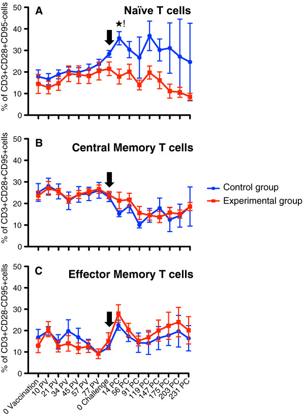
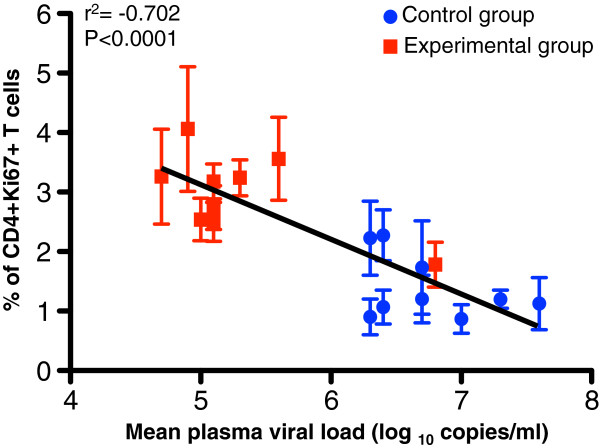
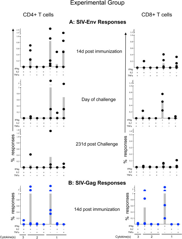
Findings: The purpose of this study was to define cellular immunological correlates of protection in rSVV-SIV vaccinated and SIV challenged animals. Immunofluorescent staining and multifunctional assessment of SIV-specific T-cell responses were evaluated in both Experimental and Control vaccinated animal groups. Significant increases in the proliferating CD4+ T-cell population and polyfunctional T-cell responses were observed in all Experimental-vaccinated animals compared with the Control-vaccinated animals.

Conclusions: Increased CD4+ T-cell proliferation was significantly and inversely correlated with plasma viral load. Increased SIV-specific polyfunctional cytokine responses and increased proliferation of CD4+ T-cell may be crucial to control plasma viral loads in vaccinated and SIVMAC251/CX1 challenged macaques.


Figures

Figure 1
Figure 1
Percentages of (A) naïve (CD28 + CD95-), (B) central memory (CD28 + CD95+) and (C) effector memory (CD28-CD95+) CD3+ T-cells in peripheral blood of both Experimental (EG) and Control group (CG) of macaques. Note that naïve T-cell population showed progressive increase in both groups following immunization. A significant increase in naïve T-cell population was observed in animals from CG group compared to EG after SIV challenge. Following challenge there was increased and decreased expansion of effector and central memory T-cell population respectively in both EG and CGs of animals. Cells were gated through CD3+ “bright” T lymphocytes and at least 20,000 events were collected by gating on lymphocytes. Percentages of respective cell population represent cells out of total lymphocytes. PV and PC denote post vaccination and post challenge time points, in days, respectively. Black arrow shows the time of SIVMAC251 challenge. Asterisk (*) indicates a significant difference between EG (n = 5) and CG (n = 4) for the specified cell subset using the Student’s t-test, with a significance level of p < 0.05.
Figure 2
Figure 2
Peripheral blood mononuclear cell proliferation assessed by %Ki67 expression is shown in CD3+CD4+ (A) and CD3+CD8+ (B) cells. Both CD4+ and CD8+ T-cell proliferation increased following SIVMAC251 infection between 14 and 42 days of infection, but CD4+ T-cells in Experimental group (EG) maintained higher proliferation rates compared to Control group (CG). In contrast, proliferation in CD8+ T-cells both EGs and CGs returned to baseline thereafter. Cells were gated through CD3+ “bright” T lymphocytes and at least 20,000 events were collected by gating on lymphocytes. Asterisk (*) indicates significant differences between EG (n = 5) and CG (n = 4) for the specified cell subsets using the Student’s t-test, with a significance level of p < 0.05.
Figure 3
Figure 3
An inverse correlation is shown between the CD3+CD4+ T-cell proliferation responses and plasma viral load in Experimental group (red squares, n = 5) and Control groups (blue circles, n = 4) of macaques following SIVMAC251challenge for all timepoints from d14 and d231. Note that the lower viral loads demonstrated in the EG showed higher % of CD4+Ki67+ T-cells while the CG that had much higher viral loads had a lower % of CD4+Ki67+ T-cells. Interestingly, the one exception in EG overall trend was the high viral load at the d14 peak, yet the correlation held and at that point, the % of CD4+Ki67+ T-cells was low. Linear regression analysis of proliferating CD4+ T-cells and mean plasma viral load from all macaques was calculated using GraphPad Prism (version 5.0d, GraphPad software, CA).
Figure 4
Figure 4
Intracellular cytokine responses measured against SIV-Env (A) and SIV-Gag (B) antigens in Experimental vaccinated macaques were shown. PBMC were unstimulated (medium control) or stimulated for 6 h with different SIV-Env and/or SIV-Gag peptide pools at 14d post immunization, day of challenge and 231d post challenge time points. Cells were gated on singlets, lymphocytes, followed by live cells and then on CD3+ T-cells and subsequently on CD3+CD4+ and CD3+CD8+ T-cell subsets. CD3+CD4+ or CD3+CD8+ T-cells were further analyzed for the presence of IFNγ, TNFα and /or IL2 positive cells by using Flowjo, and SPICE software. Increased polyfunctional responses were detected both in CD4 and CD8 T-cells, however the antigen specific CD4 responses were higher compared to CD8 specific responses. SIV-Gag specific responses were also higher compared to SIV-Env antigens. Individual animal responses are depicted by each dot and gray bars represent mean values of respective responses from all animals (n = 5). Positive symbols represent cells staining positive for a cytokine response, and minus symbols represent cells staining negative for a cytokine response. The presence of three different cytokine producing cells, two different cytokine producing cells and single cytokine producing cells are denoted under the bottom-most graphs (left to right) for each CD4 and CD8 cells as 3, 2 and 1 cytokine(s) respectively. The criterion for a positive cytokine response was a two-fold increase in frequency for that specific antigen and cytokine above the medium control culture. All values were subtracted from medium control before the analysis.

References

    1. Pahar B, Lackner AA, Piatak M Jr, Lifson JD, Wang X, Das A, Ling B, Montefiori DC, Veazey RS. Control of viremia and maintenance of intestinal CD4(+) memory T-cells in SHIV(162P3) infected macaques after pathogenic SIV(MAC251) challenge. Virology. 2009;387:273–284. - PMC - PubMed
    1. Koff WC, Johnson PR, Watkins DI, Burton DR, Lifson JD, Hasenkrug KJ, McDermott AB, Schultz A, Zamb TJ, Boyle R, Desrosiers RC. HIV vaccine design: insights from live attenuated SIV vaccines. Nat Immunol. 2006;7:19–23. - PubMed
    1. Marthas ML, Miller CJ, Sutjipto S, Higgins J, Torten J, Lohman BL, Unger RE, Ramos RA, Kiyono H, McGhee JR. et al.Efficacy of live-attenuated and whole-inactivated simian immunodeficiency virus vaccines against vaginal challenge with virulent SIV. J Med Primatol. 1992;21:99–107. - PubMed
    1. Rerks-Ngarm S, Pitisuttithum P, Nitayaphan S, Kaewkungwal J, Chiu J, Paris R, Premsri N, Namwat C, de Souza M, Adams E. et al.Vaccination with ALVAC and AIDSVAX to prevent HIV-1 infection in Thailand. N Engl J Med. 2009;361:2209–2220. - PubMed
    1. Barouch DH, Liu J, Li H, Maxfield LF, Abbink P, Lynch DM, Iampietro MJ, Sanmiguel A, Seaman MS, Ferrari G. et al.Vaccine protection against acquisition of neutralization-resistant SIV challenges in rhesus monkeys. Nature. 2012;482:89–93. - PMC - PubMed

Publication types

MeSH terms