. 2012 Sep 27;120(13):2658-68.
doi: 10.1182/blood-2011-05-355396. Epub 2012 Aug 13.

Effects of plerixafor in combination with BCR-ABL kinase inhibition in a murine model of CML

Affiliations

Effects of plerixafor in combination with BCR-ABL kinase inhibition in a murine model of CML

Anupriya Agarwal et al. Blood. .

Abstract

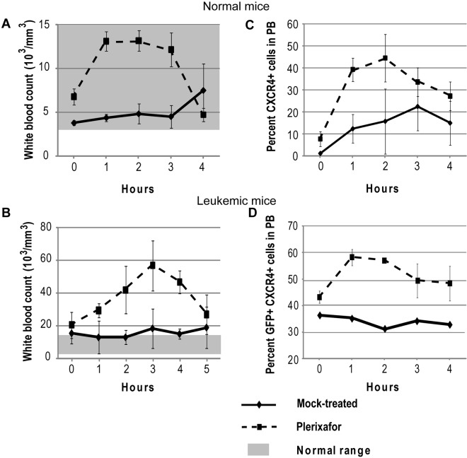
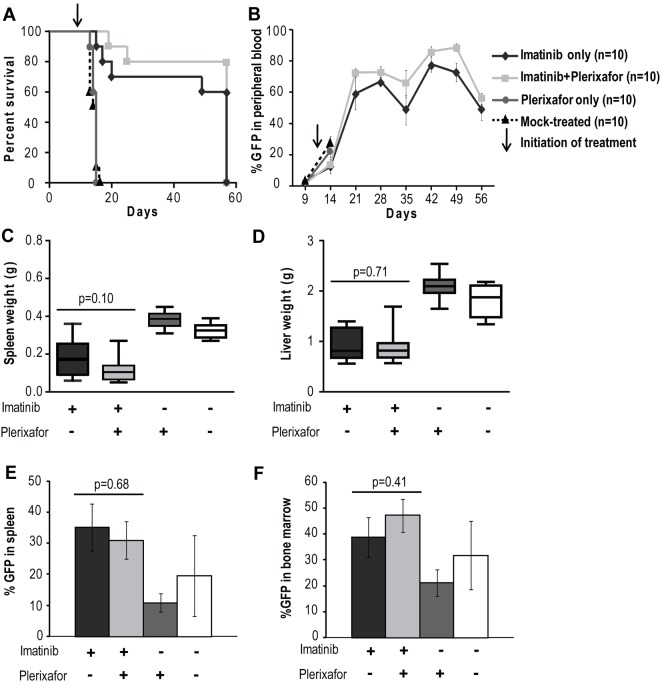
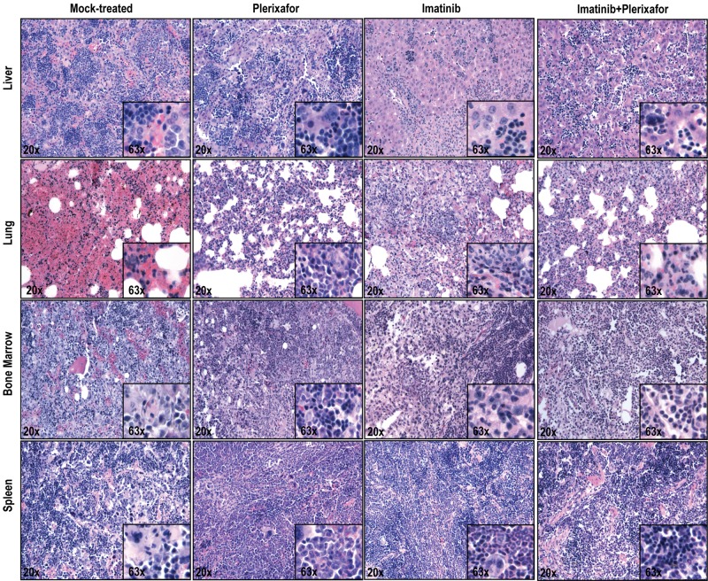
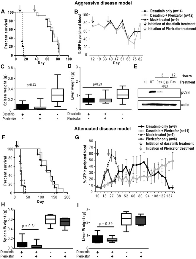
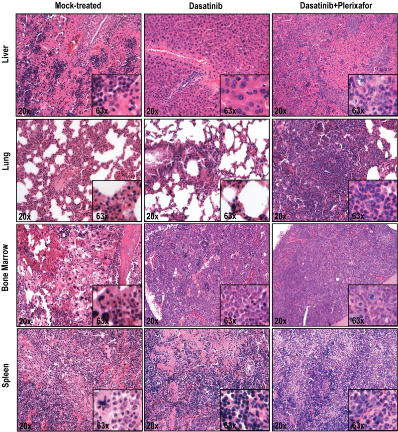
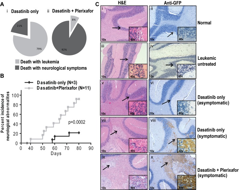
Sequestration in the bone marrow niche may allow leukemic stem cells to evade exposure to drugs. Because the CXCR4/SDF-1 axis is an important mechanism of leukemic stem cell interaction with marrow stroma, we tested whether plerixafor, an antagonist of CXCR4, may dislodge chronic myeloid leukemia (CML) cells from the niche, sensitizing them to tyrosine kinase inhibitors. We initially treated mice with retrovirally induced CML-like disease with imatinib plus plerixafor. Plerixafor mobilized CXCR4(+) cells, but no difference was observed in leukemia burden, possibly reflecting insufficient disease control by imatinib. In a second series of experiments, we tested the combination of plerixafor with dasatinib in the same as well as an attenuated CML model. Despite much improved leukemia control, plerixafor failed to reduce leukemia burden over dasatinib alone. In addition, mice receiving plerixafor had an increased incidence of neurologic symptoms in association with CNS infiltration by BCR-ABL-expressing cells. We conclude that plerixafor is ineffective in reducing leukemia burden in this model but promotes CNS infiltration. Beneficial effects of combining tyrosine kinase inhibitors with plerixafor may be observed in a situation of minimal residual disease, but caution is warranted when disease control is incomplete.


Figures

Figure 1
Figure 1
Plerixafor mobilizes CXCR4+ cells into the peripheral blood. Normal (A,C) and leukemic (B,D) mice were treated with a single dose of plerixafor, and WBC and CXCR4 expression in peripheral blood was measured over time. (A-B) Peripheral blood WBC count of normal and leukemic mice treated with plerixafor. (C-D) CXCR4 expression in peripheral blood of normal and leukemic mice treated with plerixafor.
Figure 2
Figure 2
Addition of plerixafor to imatinib does not improve survival or decrease disease burden of leukemic mice. (A) Kaplan-Meier survival analysis of mock-treated, plerixafor only, imatinib only, or imatinib + plerixafor treatment groups (P = .3228, log-rank test). The experiment was terminated at day 57. (B) FACS analysis for GFP to assess the proportion of leukemia cells in the peripheral blood. (C-D) Spleen and liver weights measured at necropsy. (E-F) Percentage GFP+ cells in spleen and bone marrow measured by FACS.
Figure 3
Figure 3
Histopathologic examination of imatinib and imatinib + plerixafor treated mice shows no significant differences. Spleen, liver, lung, and bone marrow were stained with hematoxylin and eosin.
Figure 4
Figure 4
Addition of plerixafor to dasatinib does not improve survival or decrease disease burden of leukemic mice. Experiment was performed with both aggressive (A-E) and attenuated disease models (F-I). (A,F) Kaplan-Meier survival analysis of mice treated with dasatinib only or in combination with plerixafor (P = .2934, log-rank test). (B,G) FACS analysis for GFP+ cells to assess the proportion of leukemia cells in the peripheral blood. (C-D,H-I) Spleen and liver weight measured at necropsy of leukemic mice. (E) pCrkL levels measured by Western blot analysis in bone marrow cell lysates of dasatinib (Das) and dasatinib + plerixafor (Das + PLX) treated mice after indicated time of treatment compared with normal mice (NL) and untreated leukemic mice (UT).
Figure 5
Figure 5
Histopathologic analysis of mice treated with dasatinib + plerixafor shows slightly increased infiltration of leukemic cells in all organs compared with dasatinib only treatment. Spleen, liver, lung, and bone marrow sections were stained with hematoxylin and eosin.
Figure 6
Figure 6
Plerixafor combination with dasatinib increases neurologic symptoms of leukemic mice. (A) Percentage incidence of neurologic symptoms in dasatinib only and combination group (i-ii) after excluding 5 mice from each group, which were taken off treatment at day 68. (B) Incidence of neurologic symptoms, from first occurrence of symptoms to death in symptomatic mice. Significant difference was observed between dasatinib only and dasatinib + plerixafor groups (P = .0002, log-rank test) in the occurrence of neurologic symptoms. (C) Hematoxylin and eosin (left panel) and anti-GFP immunohistochemistry (right panel) on brain sections. Arrow indicates the area of the brain, which is shown in higher magnification normal brain structure (i-vi) and leukemic infiltrates (vii-x).

References

    1. Deininger MW, Goldman JM, Melo JV. The molecular biology of chronic myeloid leukemia. Blood. 2000;96(10):3343–3356. - PubMed
    1. Druker BJ, Guilhot F, O'Brien SG, et al. Five-year follow-up of patients receiving imatinib for chronic myeloid leukemia. N Engl J Med. 2006;355(23):2408–2417. - PubMed
    1. Hughes TP, Kaeda J, Branford S, et al. Frequency of major molecular responses to imatinib or interferon alfa plus cytarabine in newly diagnosed chronic myeloid leukemia. N Engl J Med. 2003;349(15):1423–1432. - PubMed
    1. Rousselot P, Huguet F, Rea D, et al. Imatinib mesylate discontinuation in patients with chronic myelogenous leukemia in complete molecular remission for more than 2 years. Blood. 2007;109(1):58–60. - PubMed
    1. Graham SM, Jorgensen HG, Allan E, et al. Primitive, quiescent, Philadelphia-positive stem cells from patients with chronic myeloid leukemia are insensitive to STI571 in vitro. Blood. 2002;99(1):319–325. - PubMed

Publication types

MeSH terms