

Evidence for ape and human specializations in geniculostriate projections from VGLUT2 immunohistochemistry
- PMID: 22889767
- PMCID: PMC3503454
- DOI: 10.1159/000341135
Evidence for ape and human specializations in geniculostriate projections from VGLUT2 immunohistochemistry
Abstract
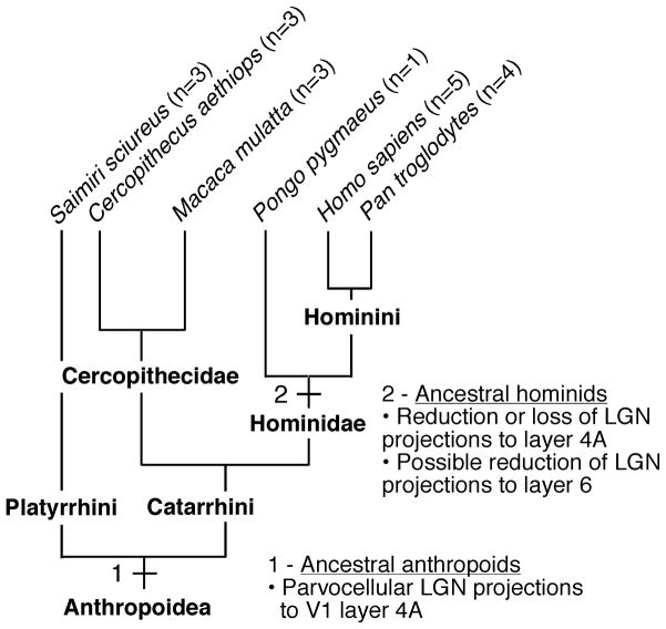
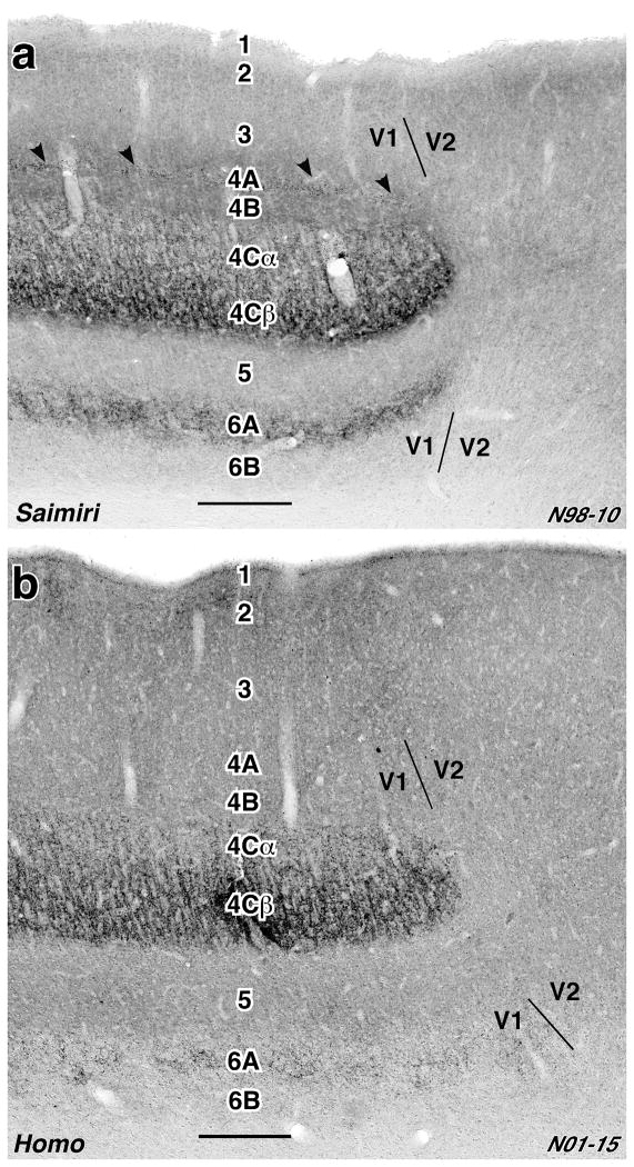
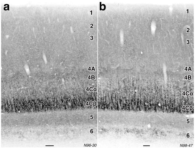
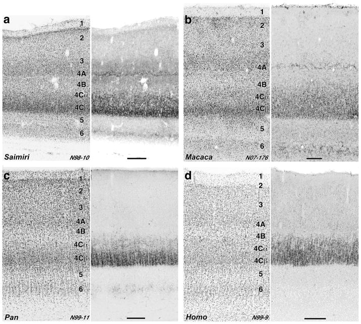
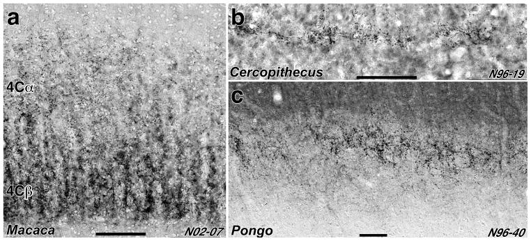
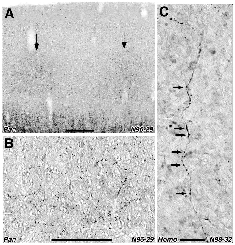
Vesicular glutamate transporters (VGLUTs) reuptake glutamate into synaptic vesicles at excitatory synapses. VGLUT2 is localized in the cortical terminals of neuronal somas located in the main sensory nuclei of the thalamus. Thus, immunolabeling of cortex with antibodies to VGLUT2 can reveal geniculostriate terminal distributions in species in which connectivity cannot be studied with tract-tracing techniques, permitting broader comparative studies of cortical specializations. Here, we used VGLUT2 immunohistochemistry to compare the organization of geniculostriate afferents in primary visual cortex in hominid primates (humans, chimpanzees, and an orangutan), Old World monkeys (rhesus macaques and vervets), and New World monkeys (squirrel monkeys). The New and Old World monkeys had a broad, dense band of terminal-like labeling in cortical layer 4C, a narrow band of labeling in layer 4A, and additional labeling in layers 2/3 and 6, consistent with results from conventional tract-tracing studies in these species. By contrast, although the hominid primates had a prominent layer 4C band, labeling of layer 4A was sparse or absent. Labeling was also present in layers 2/3 and 6, although labeling of layer 6 was weaker in hominids and possibly more individually variable than in Old and New World monkeys. These findings are consistent with previous observations from cytochrome oxidase histochemistry and a very small number of connectivity studies, suggesting that the projections from the parvocellular layers of the lateral geniculate nucleus to layer 4A were strongly reduced or eliminated in humans and apes following their evolutionary divergence from the other anthropoid primates.
Copyright © 2012 S. Karger AG, Basel.
Figures

Similar articles
-
Human-specific organization of primary visual cortex: alternating compartments of dense Cat-301 and calbindin immunoreactivity in layer 4A.Cereb Cortex. 2002 Jul;12(7):671-91. doi: 10.1093/cercor/12.7.671. Cereb Cortex. 2002. PMID: 12050080
-
Distribution of vesicular glutamate transporter 2 (VGluT2) in the primary visual cortex of the macaque and human.J Comp Neurol. 2013 Jan 1;521(1):130-51. doi: 10.1002/cne.23165. J Comp Neurol. 2013. PMID: 22684983 Free PMC article.
-
Distinctive compartmental organization of human primary visual cortex.Proc Natl Acad Sci U S A. 1999 Sep 28;96(20):11601-6. doi: 10.1073/pnas.96.20.11601. Proc Natl Acad Sci U S A. 1999. PMID: 10500223 Free PMC article.
-
Visual system of a naturally microphthalmic mammal: the blind mole rat, Spalax ehrenbergi.J Comp Neurol. 1993 Feb 15;328(3):313-50. doi: 10.1002/cne.903280302. J Comp Neurol. 1993. PMID: 8440785 Review.
-
Laminar organization of geniculostriate projections. A common organizational plan based on layers rather than individual functional classes.Brain Behav Evol. 1988;32(3):187-92. doi: 10.1159/000116546. Brain Behav Evol. 1988. PMID: 3058267 Review.
Cited by
-
Oxytocin- and arginine vasopressin-containing fibers in the cortex of humans, chimpanzees, and rhesus macaques.Am J Primatol. 2018 Oct;80(10):e22875. doi: 10.1002/ajp.22875. Epub 2018 May 24. Am J Primatol. 2018. PMID: 29797339 Free PMC article.
-
Cytochrome oxidase "blobs": a call for more anatomy.Brain Struct Funct. 2021 Dec;226(9):2793-2806. doi: 10.1007/s00429-021-02360-2. Epub 2021 Aug 12. Brain Struct Funct. 2021. PMID: 34382115 Free PMC article. Review.
-
The postnatal development of MT, V1, LGN, pulvinar and SC in prosimian galagos (Otolemur garnettii).J Comp Neurol. 2020 Dec 1;528(17):3075-3094. doi: 10.1002/cne.24885. Epub 2020 Feb 24. J Comp Neurol. 2020. PMID: 32067231 Free PMC article.
-
Major Feedforward Thalamic Input Into Layer 4C of Primary Visual Cortex in Primate.Cereb Cortex. 2019 Jan 1;29(1):134-149. doi: 10.1093/cercor/bhx311. Cereb Cortex. 2019. PMID: 29190326 Free PMC article.
-
Differential maturation of vesicular glutamate and GABA transporter expression in the mouse auditory forebrain during the first weeks of hearing.Brain Struct Funct. 2016 Jun;221(5):2619-73. doi: 10.1007/s00429-015-1062-3. Epub 2015 Jul 10. Brain Struct Funct. 2016. PMID: 26159773 Free PMC article.
References
-
- Barroso-Chinea P, Castle M, Aymerich MS, Perez-Manso M, Erro E, Tunon T, Lanciego JL. Expression of the mRNAs encoding for the vesicular glutamate transporters 1 and 2 in the rat thalamus. J Comp Neurol. 2007;501:703–715. - PubMed
-
- Billings-Gagliardi S, Chan-Palay V, Palay SL. A review of lamination in area 17 of the visual cortex Macaca mulatta. J Neurocytol. 1974;3:619–629. - PubMed
-
- Blümcke I, Hof PR, Morrison JH, Celio MR. Distribution of parvalbumin immunoreactivity in the visual cortex of Old World monkeys and humans. J Comp Neurol. 1990;301:417–432. - PubMed
Publication types
MeSH terms
Substances
Grants and funding
LinkOut - more resources
Full Text Sources
Research Materials
Miscellaneous
