. 2012 Oct;181(4):1250-70.
doi: 10.1016/j.ajpath.2012.06.036. Epub 2012 Aug 11.

A RHAMM mimetic peptide blocks hyaluronan signaling and reduces inflammation and fibrogenesis in excisional skin wounds

Affiliations

A RHAMM mimetic peptide blocks hyaluronan signaling and reduces inflammation and fibrogenesis in excisional skin wounds

Cornelia Tolg et al. Am J Pathol. 2012 Oct.

Abstract

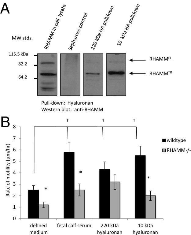
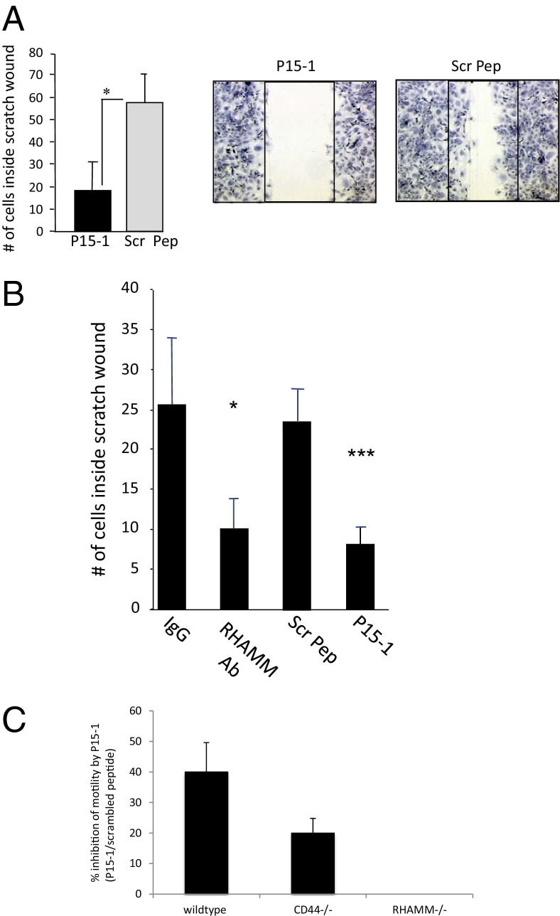
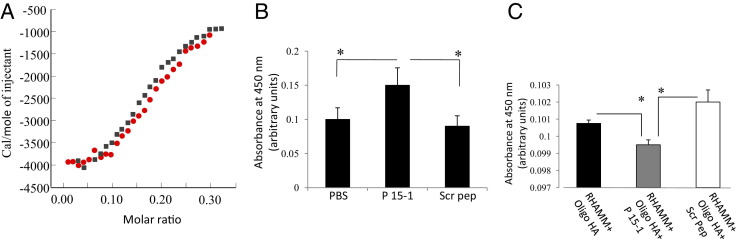
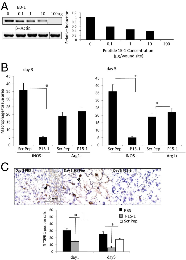
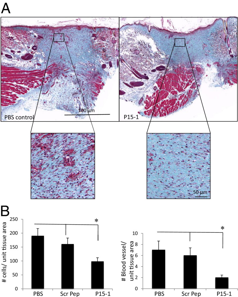
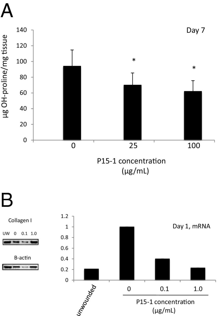
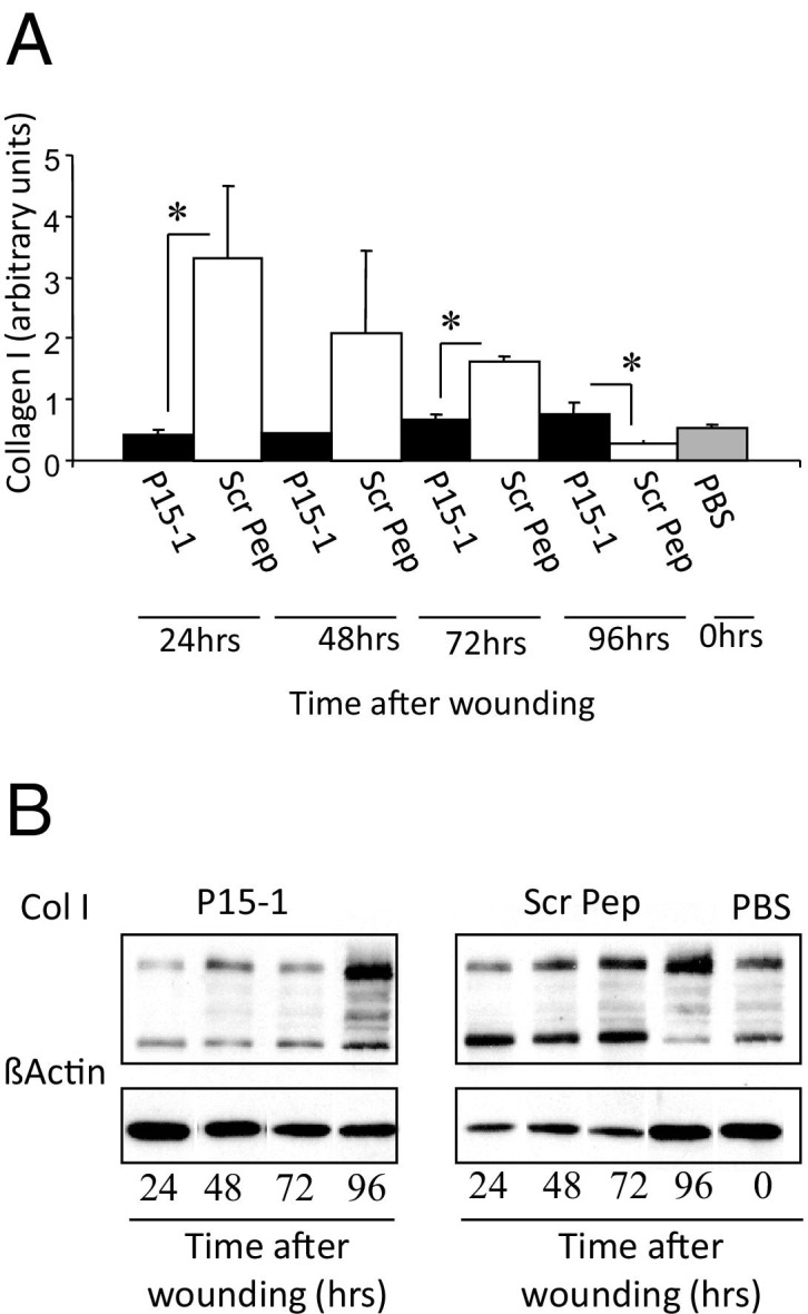
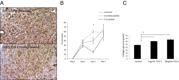
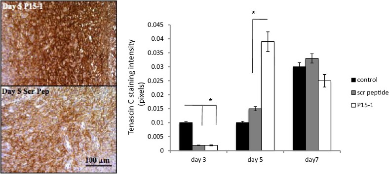
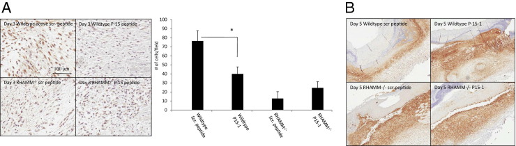
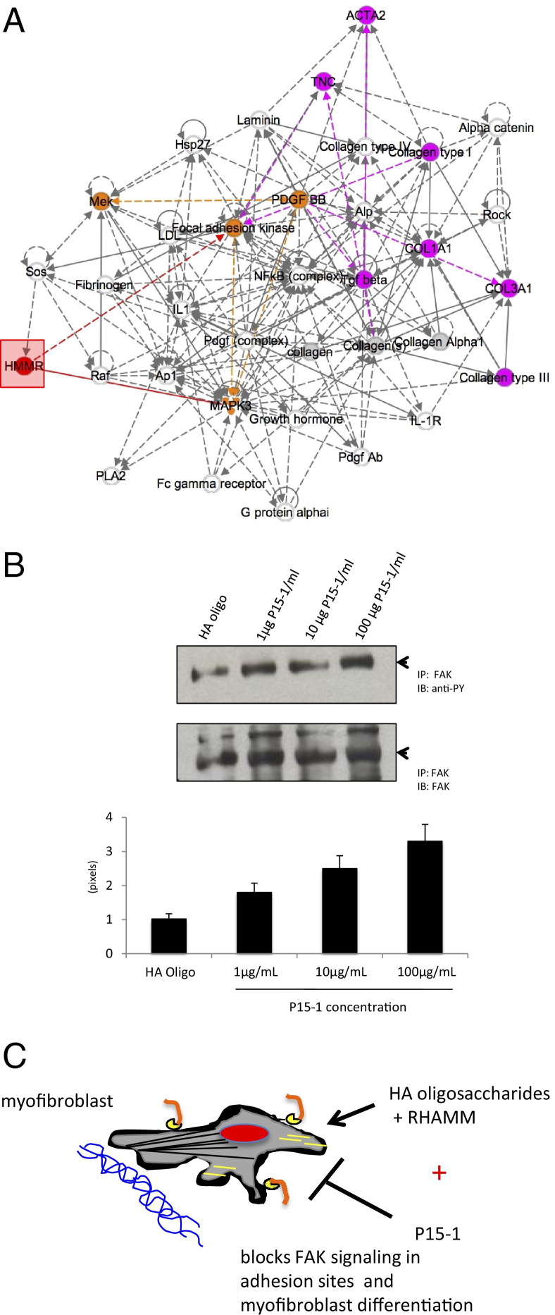
Hyaluronan is activated by fragmentation and controls inflammation and fibroplasia during wound repair and diseases (eg, cancer). Hyaluronan-binding peptides were identified that modify fibrogenesis during skin wound repair. Peptides were selected from 7- to 15mer phage display libraries by panning with hyaluronan-Sepharose beads and assayed for their ability to block fibroblast migration in response to hyaluronan oligosaccharides (10 kDa). A 15mer peptide (P15-1), with homology to receptor for hyaluronan mediated motility (RHAMM) hyaluronan binding sequences, was the most effective inhibitor. P15-1 bound to 10-kDa hyaluronan with an affinity of K(d) = 10(-7) and appeared to specifically mimic RHAMM since it significantly reduced binding of hyaluronan oligosaccharides to recombinant RHAMM but not to recombinant CD44 or TLR2,4, and altered wound repair in wild-type but not RHAMM(-/-) mice. One topical application of P15-1 to full-thickness excisional rat wounds significantly reduced wound macrophage number, fibroblast number, and blood vessel density compared to scrambled, negative control peptides. Wound collagen 1, transforming growth factor β-1, and α-smooth muscle actin were reduced, whereas tenascin C was increased, suggesting that P15-1 promoted a form of scarless healing. Signaling/microarray analyses showed that P15-1 blocks RHAMM-regulated focal adhesion kinase pathways in fibroblasts. These results identify a new class of reagents that attenuate proinflammatory, fibrotic repair by blocking hyaluronan oligosaccharide signaling.


Figures

Figure 1
Figure 1
A motogenic response to HA fragments requires RHAMM. A: RHAMM binds to 220-kDa and 10-kDa HA. Lysates from RHAMM overexpressing mesenchymal cells were incubated with 220-kDa and 10-kDa HA coupled to Sepharose beads. Proteins bound to beads were resolved on SDS-PAGE, and RHAMM was detected by Western blot analysis. Arrows indicate bands that represent full-length RHAMM (RHAMMFL) and a processed (truncated) RHAMMTR that is commonly expressed after an injury. B: Wild-type and RHAMM−/− MEFs were seeded sparsely on tissue culture plastic and serum starved in defined medium. Cell migration was stimulated with 10% FCS (positive control), 220-kDa HA, or 10-kDa HA and quantified by real-time cinemicrography. Values represent mean ± SD of 50 cells. *P < 0.001 between wild-type and RHAMM−/− cells, P < 0.001 versus defined medium.
Figure 2
Figure 2
P15-1 blocks HA oligosaccharide–induced migration in RHAMM-expressing fibroblasts. A: Confluent monolayers of rat dermal fibroblasts were scratch wounded using disposable, plastic Pipetman tips (Gilson, Middleton, WI). Cells were incubated with culture medium containing 500 ng of HA oligosaccharides (10 kDa, sized 30mer HA) plus either 50 μg/mL P15-1 or scrambled control peptide (Scr Pep). Twenty-four hours later, cell migration was quantified by counting cells that migrated into the scratch wound. P15-1 significantly reduces the number of cells that have migrated into the wound in comparison to scrambled peptide controls. Values represent the mean ± SD of three culture wells. ***P < 0.001. B: Confluent monolayers of RHAMM-expressing MEF were scratch wounded using pipette tips. Cells were incubated with medium containing anti-RHAMM Ab (30 μg/mL), P15-1 (30 μg/mL), or Scr Pep (30 μg/mL). In the control experiments, cells were incubated with medium alone. After 11 to 14 hours, cells that migrated into the scratch wound were counted. P15-1 significantly decreased (***P < 0.001) migration of RHAMM-expressing MEF, and to the same extent as RHAMM-blocking Ab (*P < 0.05). Scrambled control peptide and Isotype-matched IgG control (IgG) had no effect. Values represent mean ± SD of three culture wells. C: Effect of P15-1 and scrambled control peptide on scratch wound–induced migration of wild-type, CD44−/−, and RHAMM−/− MEFs. Peptide treatment and cell migration quantification were performed as described in B. The graph shows that whereas the migration of wild-type and CD44−/− MEF were significantly inhibited by P15-1, it had no effect on migration of RHAMM−/− MEF. Values represent mean ± SD of nine culture wells.
Figure 3
Figure 3
Peptide 15-1 binds to HA oligosaccharides and blocks their interaction with recombinant RHAMM protein. A: The graph shows quantification of the interaction between P15-1 and HA oligosaccharides by ITC. P15-1 bound to HA oligosaccharides (avg. 10 kDa) with a binding constant of kDa = 10−7 mol/L. A 0.9 mmol/L solution of HA was injected into MES (pH 6.0) containing 0.6 mmol/L peptide 15-1, and the energy released on interaction with P15-1 was determined by ITC. Binding curves of two independent experiments (red or black) are depicted. B: HA oligosaccharide:P15-1 interactions were confirmed with ELISA. ELISA plates were coated with HA oligosaccharides (1 mg/mL), followed by incubation with either biotinylated P15-1 or Scr Pep (50 μg/mL). PBS alone was used as control. Bound biotinylated peptides were detected by streptavidin-horseradish peroxidase. Peptide 15-1 binds to HA oligosaccharides. Values represent the mean ± SD of three ELISA wells. C: ELISA plates were coated with 1 μg/mL recombinant RHAMM followed by incubation with either 10 μg/mL 30mer HA or HA oligosaccharides (10 kDa) together with P15-1 or Scr Pep (1 μg/mL). Bound HA was detected with biotinylated aggrecan followed by streptavidin-horseradish peroxidase. P15-1 reduces RHAMM/HA binding. Values represent the mean ± SD of n = 3. *P < 0.001.
Figure 4
Figure 4
HA is fragmented in excisional wounds. Agarose gel electrophoretic analysis of molecular weight distribution for HA isolated from full-thickness excisional wounds 0 to 14 days (D) after wounding. HA was visualized in the electrophoretic gels by Stains-All dye. Mainly high molecular weight HA (3050 to 4570 kDa) was present at day 0, but a broad distribution of HA sizes appeared by day 3, and were prominently observed at day 7. HA molecular weight distribution was measured by densitometry of agarose gels, and results are normalized to the weight of tissue used for HA isolation. Arrow indicates bands representing 10-kDa Oligo-HA, used for experimental analyses.
Figure 5
Figure 5
Peptide 15-1 reduces macrophage influx into excisional skin wounds and TGFβ1 production. A: Full-thickness excisional wounds were covered with either vehicle alone or vehicle containing different concentrations (100 ng/mL to 100 μg/wound site) of P15-1. Wound tissue was isolated 24 hours later. Expression of the macrophage-specific marker ED-1 was analyzed by PCR. Amplification of β-actin transcripts was used as loading control. Amplification products were separated on an agarose gel. The ED-1 amplification product was quantified by densitometry and corrected for equal loading by calculating the ratio ED-1:β-actin control. P15-1 decreased the amount of wound ED-1 in a dose-dependent manner. Values are mean ± SD. n = 3. B: Full-thickness excisional skin wounds were treated with either P15-1 or scrambled peptide (Scr Pep; 50 μg/wound site). Wound sections were stained with iNos- or Arg1-specific Ab. Positive cells were counted. P15-1 treatment reduced infiltration of proinflammatory (iNos+) macrophages into wounds. All graphs represent results of n = 3 wounds, five areas/wound. Values are mean ± SD. n = 15. *P < 0.001. C: Full-thickness excisional wounds were treated with vehicle alone, P15-1 (50 μg/wound site), or scrambled peptide (50 μg/wound site). Day 1 and day 3 wound sections were stained with TGFβ1-specific Ab as described in Materials and Methods. Microscopic images were taken at an original magnification of ×60, and TGFβ1+ cells (arrows) were counted. The number of TGFβ1+ cells is reduced in P15-1–treated wounds at days 1 and 3 after wounding. Graphs represent mean ± SD. n = 30. *P < 0.01.
Figure 6
Figure 6
Peptide 15-1 reduces fibroblast density and blood vessel density in granulation tissue. A: Full-thickness day 7 excisional wounds were treated with collagen vehicle alone (PBS control), scrambled peptide in collagen vehicle (image not shown), or P15-1 in collagen vehicle (P15-1, 50 μg/wound site). Cross sections of day 7 wounds were stained with Masson's trichrome, allowing differentiation of collagen (blue), muscle, and cells. Angiogenesis and fibrogenesis was reduced in P15-1–treated wounds at day 7. B: Granulation tissue fibroblasts and blood vessels were counted in wound sections. P15-1 significantly decreased the numbers of fibroblasts and blood vessels in granulation tissue (day 7) compared to both PBS and scrambled peptide (scr pep) controls. The effects of scrambled peptide on fibroblasts and blood vessels were not significantly different from vehicle control. Graphs represent the mean ± SD. n = 6 wounds. *P < 0.001.
Figure 7
Figure 7
P15-1 inhibits collagen I accumulation within wounds. A: Full-thickness excisional wounds were treated with collagen vehicle alone (0 μg/mL P15-1 concentration) or P15-1 + collage vehicle (25, 100 μg/wound site). At day 7, wounds were isolated, and the OH-proline content was analyzed as described in Materials and Methods. P15-1 significantly reduced wound collagen at day 7 after wounding at both concentrations (*P < 0.01). Each column represents the mean ± SD. n = 6. B: Full-thickness excisional wounds were treated with either collagen vehicle alone or P15-1 in collagen vehicle (100 ng/mL to 1 μg/wound site). Wound tissue mRNA was isolated 24 hours after wounding, and collagen I transcripts were amplified by RT PCR. Amplification of β-actin transcripts was used as loading control. Amplification products were separated on an agarose gel and quantified by densitometry. The collagen I amplification products were corrected for equal loading by calculating the ratio collagen I/β-actin control. Collagen I expression in control wounds were set to one. P15-1 decreased collagen I mRNA expression 24 hours after wounding. The values represent typical results that were replicated three separate times.
Figure 8
Figure 8
Peptide 15-1 reduces in vitro collagen production of cultured dermal fibroblasts. Monolayers of rat dermal fibroblasts were scratched multiple times using a plastic comb. A: Cells were incubated for the indicated times in medium containing either peptide 15-1 (30 μg/mL) or scrambled control peptide (Scr Pep; 30 μg/mL). B: Collagen I production was quantified by Western blot analysis. β-actin was used as loading control. Protein band intensities were determined by densitometry, and the ratio of Col I/β-actin was calculated. P15-1 detectably reduced collagen I protein levels at 24, 48, and 72 hours after its addition. Values represent the mean ± SD. n = 3. *P < 0.001.
Figure 9
Figure 9
P15-1 reduces wound contraction and smooth muscle actin in day 5 wounds. A: Full-thickness excisional wounds were treated with collagen vehicle alone or collagen vehicle plus P15-1 peptide. Wounds were harvested at day 5 post injury, and cross sections were stained with smooth muscle actin–specific Ab. A: Panel depicts representative images of collagen vehicle–treated control wounds and P15-1 in collagen vehicle–treated wounds. Smooth muscle actin staining was reduced in P15-1–treated wounds. B: Wound contraction was reduced in P15-1–treated wounds at day 5 after wounding. Full-thickness excisional wounds were either treated with collagen vehicle plus P15-1, collagen vehicle plus scrambled control peptide, or left untreated. Wound contraction was measured by tracing wound edges on days 3, 5, and 7 after wounding and quantification of the wound area by image analysis. The graph depicts percentage wound contraction. n = 6 wounds/treatment. C: P15-1 inhibits collagen gel contraction in culture. Collagen I gel contraction by human foreskin fibroblasts was analyzed as described in Materials and Methods. Graph depicts the gel area ± SD of n = 4 replicates/experiment. *P < 0.01.
Figure 10
Figure 10
Peptide 15-1 increases wound tenascin C protein. Full-thickness excisional wounds were covered with collagen vehicle alone (image not shown), P15-1 in collagen gel (50 μg/wound site), or scrambled control peptide in collagen gel (50 μg/wound site). Sections were stained with tenascin C–specific Ab as described in Materials and Methods. Microscopic images were taken, and tenascin C staining intensity was quantified by image analysis using Image J. P15-1 increased tenascin C protein expression in wounds at day 5 after injury. Graphs represent the mean ± SD. n = 6 wounds. *P < 0.001.
Figure 11
Figure 11
P15-1 does not affect TGFβ1 or tenascin C levels in RHAMM−/− wounds. A and B: Full-thickness excisional wounds of wild-type and RHAMM−/− mice were treated with collagen vehicle plus either P15-1 or scrambled control peptide. A: Cross sections of day 3 wounds were stained with TGFβ1-specific Ab. The graph depicts the number of positively stained cells per microscopic field ± SD. n = 4. *P < 0.001. B: Cross sections of day 5 wounds were stained with tenascin C–specific Ab. A representative image of n = 4 is shown.
Figure 12
Figure 12
RHAMM-regulated signaling pathways blocked by P15-1. A: Protein or mRNA expression of genes, which are subject to regulation by both RHAMM and P15-1 (see Materials and Methods), were identified by performing immunohistochemistry of wound beds or microarray analysis of RHAMM transfected versus parental fibroblasts, and P15-1–treated versus scrambled peptide–treated RHAMM-expressing fibroblasts. Genes that were altered in their expression by P15-1 (but not by negative controls), as well as by RHAMM transfection, were identified and analyzed for functional networks/canonical signaling pathways using Ingenuity Pathway Analysis (Ingenuity Systems, Redwood City, CA). Analyses identified the top functional network (shown in A) as one associated with connective tissue disorders, dermatological diseases, and inflammation. The most significant molecular and cellular functions predicted by these associations included cell cycle (P < 3.7 × 10⁻⁷) and cell-to-cell signaling and interaction (P < 3.9 × 10⁻⁷). The top canonical signaling pathway was FAK (focal adhesion kinase) signaling (P = 2.5 × 10⁻⁷) as shown in A. Genes that have previously been linked to RHAMM (red box, identified as HMMR) are marked in orange, and those that have been shown to be directly or indirectly modified through an action on signaling pathways are marked in solid (direct) or dashed (indirect) red lines. Genes identified in the present study to be regulated by RHAMM and P15-1 on the FAK signaling pathway are pink, and their relationships to each other are indicated by solid or dashed pink lines. B: Based on the analysis in A, the consequence of P15-1 on FAK tyrosine phosphorylation status was assessed following exposure to HA oligosaccharides (10 kDa) as described in Materials and Methods. The control, scrambled peptide, resulted in light phosphorylation of FAK, whereas by contrast, P15-1 treatment strongly and significantly increased FAK phosphorylation at all doses tested (P < 0.001). This was sustained over time and was associated with inability of focal adhesions, which contain large signaling complexes linked to the microenvironment, to turnover and signal appropriately (data not shown). C: Literature searches of the FAK pathway shown in A regulate myofibroblast differentiation within wounds (see Discussion). The consequences of RHAMM, HA oligosaccharides and blocking of these interactions by P15-1 are modeled in C. HA oligosaccharides bind to cell surface RHAMM, which in turn regulates signaling in focal adhesions through FAK, thus contributing to myofibroblast differentiation/migration. P15-1 is proposed to compete for RHAMM in binding to HA oligosaccharides thus blocking appropriate RHAMM signaling through FAK/ERK1. This aberrant signaling block by P15-1 results in a downstream decrease in Tgfβ1, Acta2, Col1, and Col3 expression and an increase in Tnc expression with a consequent loss of myofibroblast phenotype.

References

    1. Veiseh M., Turley E.A. Hyaluronan metabolism in remodeling extracellular matrix: probes for imaging and therapy of breast cancer. Integr Biol (Camb) 2011;3:304–315. - PubMed
    1. Toole B.P., Slomiany M.G. Hyaluronan: a constitutive regulator of chemoresistance and malignancy in cancer cells. Semin Cancer Biol. 2008;18:244–250. - PMC - PubMed
    1. Jiang D., Liang J., Noble P.W. Hyaluronan in tissue injury and repair. Annu Rev Cell Dev Biol. 2007;23:435–461. - PubMed
    1. Jiang D., Liang J., Noble P.W. Hyaluronan as an immune regulator in human diseases. Physiol Rev. 2011;91:221–264. - PMC - PubMed
    1. Volpi N., Schiller J., Stern R., Soltes L. Role, metabolism, chemical modifications and applications of hyaluronan. Curr Med Chem. 2009;16:1718–1745. - PubMed

Publication types

MeSH terms