Review
. 2012 Sep 19;367(1602):2517-28.
doi: 10.1098/rstb.2012.0054.

Evolution of the eukaryotic protein kinases as dynamic molecular switches

Affiliations
Review

Evolution of the eukaryotic protein kinases as dynamic molecular switches

Susan S Taylor et al. Philos Trans R Soc Lond B Biol Sci. .

Abstract

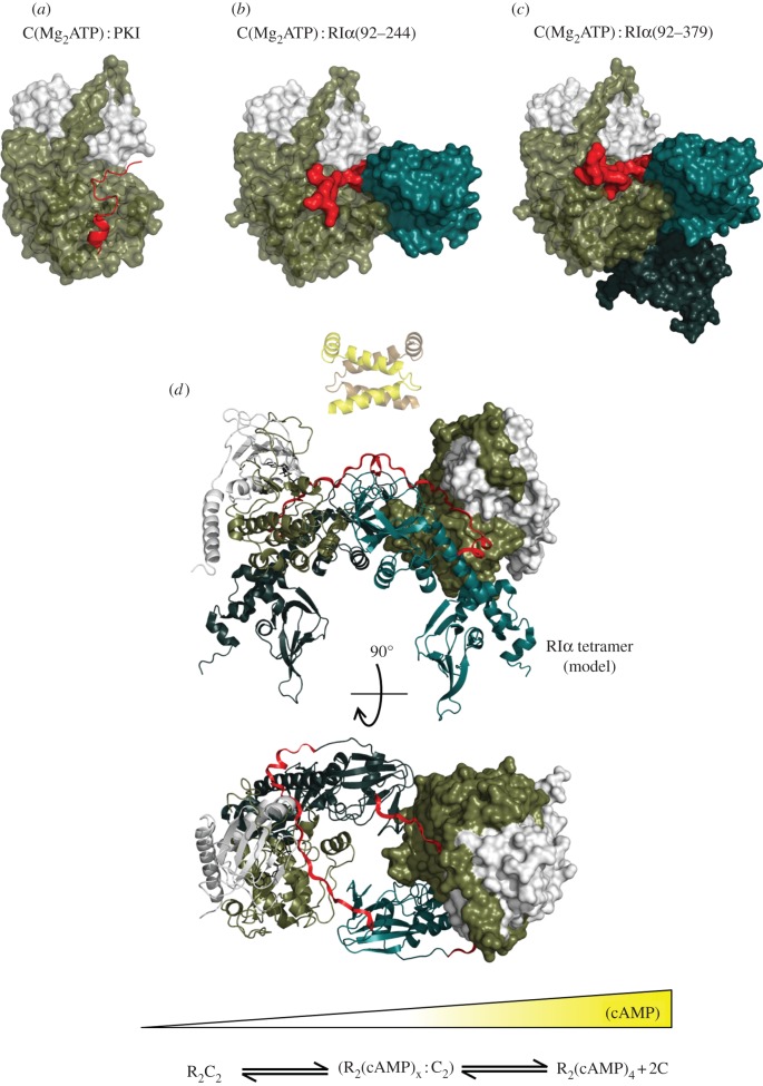
Protein kinases have evolved in eukaryotes to be highly dynamic molecular switches that regulate a plethora of biological processes. Two motifs, a dynamic activation segment and a GHI helical subdomain, distinguish the eukaryotic protein kinases (EPKs) from the more primitive eukaryotic-like kinases. The EPKs are themselves highly regulated, typically by phosphorylation, and this allows them to be rapidly turned on and off. The EPKs have a novel hydrophobic architecture that is typically regulated by the dynamic assembly of two hydrophobic spines that is usually mediated by the phosphorylation of an activation loop phosphate. Cyclic AMP-dependent protein kinase (protein kinase A (PKA)) is used as a prototype to exemplify these features of the PKA superfamily. Specificity in PKA signalling is achieved in large part by packaging the enzyme as inactive tetrameric holoenzymes with regulatory subunits that then are localized to macromolecular complexes in close proximity to dedicated substrates by targeting scaffold proteins. In this way, the cell creates discrete foci that most likely represent the physiological environment for cyclic AMP-mediated signalling.


Figures

Figure 1.
Figure 1.
Conserved core of the eukaryotic protein kinases. The bottom panels (ce) highlight functional motifs in the N-lobe (a) and the C-lobe (b) using PKA as a prototype for the EPK family. Helices are shown in red; β-strands in teal. (a) The N-lobe contains five β-strands and a large αC-helix. (b) The C-lobe is mostly helical with a large activation segment. A four-stranded β-sheet rests on the helical core and forms one surface of the active site cleft. ATP is bound in the cleft between the two lobes. (c) The phosphates of ATP are positioned by a conserved glycine-rich loop between the β1- and β2-strands. (d) Conserved residues Lys72 from the β3-strand, Glu91 from the αC-helix, and Asp164 from the DFG motif in the activation segment where Mg2+ ions are show as purple balls. (e) The catalytic loop also contains a set of catalytically important residues: Asp166, Lys168, Asn171.
Figure 2.
Figure 2.
Hydrophobic spines define the internal architecture of the EPKs. (a) Two hydrophobic spines span the two lobes of the kinase core and provide a firm but flexible connection between the N- and C-lobes. (b) The regulatory spine (R-spine) contains four residues from different kinase subdomains and is anchored to the αF-helix by conserved Asp220. The catalytic spine (C-spine) is completed by ATP. (c,d) In the inactive state, the R-spine is typically disassembled. Disassembly of the R-spine can be achieved in different ways: by movement of the αC-helix like in cyclin-dependent kinase 2 (CDK2) (c) or by movement of the activation segment like in insulin receptor kinase (IRK) (d).
Figure 3.
Figure 3.
Activation segment and the helical GHI subdomain distinguish EPKs from ELKs. The activation segment (shown in red) that joins the DFG motif to the αF-helix is shown on the left side of the two top panels while the helical GHI subdomains that follow the αF-helix are shown on the right and indicated in teal. (a) PKA, a prototype for EPKs, is compared with a eukaryote-like kinase: (b) choline kinase. (c) The extended activation segment (red), which is typically regulated by phosphorylation, is a unique feature of the EPKs. The GHI helical subdomain is also conserved in EPKs and functions as a docking site and, most likely, as an allosteric link to the active site in EPKs. While the ELKs also have helical subdomains following the F-helix, these helical regions are not conserved with respect to each other or to the EPKs (shown in teal). The GHI subdomain and the activation segment are bound to each other and to the αF-helix via a set of conserved hydrophobic interactions (shown as transparent surfaces) and a buried salt bridge Glu208–Arg280.
Figure 4.
Figure 4.
Tails and linkers. Flanking regions wrap around the conserved kinase core in different ways for the various kinases but typically, the kinase core alone is not sufficient for optimal activity. These tails provide stability and allosteric mechanisms for regulation. Three EPK examples are shown: (a) protein kinase A (PKA), (b) casein kinase II (CK2) and (c) ERK2. Each kinase core is displayed as a ribbon. The N-terminal tails are shown as a teal surface while the C-terminal tails are in red.
Figure 5.
Figure 5.
The activation loops and the C-tails are regulated by phosphorylation. (a) Shows how the tails from the three different kinases shown in figure 4 wrap around the core in different ways but fill the same space. (b) The C-tails are a conserved feature of the AGC subfamily of EPKs. (c) The C-tail of most AGC kinases, exemplified here by PKC ζ (teal), are assembled into an active conformation by phosphorylation at a turn motif and at the hydrophobic motif near the C-terminus. The C-tail of PKA (red) ends with the HF motif and lacks the final phosphorylation site.
Figure 6.
Figure 6.
Phosphorylation of the activation segment drives the assembly of the R-spine. (a) The fully phosphorylated active PKA catalytic subunit shows how phosphorylation of Thr197 creates the contiguous R-spine that spans both lobes. (b) The activation loop phosphate interacts with five different subdomains. (c) In the absence of phosphorylation the R-spine is broken and the C-helix is pushed away from the active site.
Figure 7.
Figure 7.
The catalytic subunit of PKA serves as a scaffold that interacts with multiple regulatory proteins. On the left (a) is a representation of the catalytic subunit bound to a peptide from the heat-stable protein kinase inhibitor (PKI) shown in red [22]. The middle (b) shows the catalytic subunit bound to a deletion mutant of RIα that contains a single nucleotide-binding domain (CNB-A) [57]. On the right (c) is the catalytic subunit bound to a deletion mutant of RIα that contains both single nucleotide-binding domains (CNB-A and CNB-B) [58]. The catalytic subunit is shown as a space-filling model with the residues from 1 to 126 in ivory and residues from 127 to 350 in tan. The R-subunits are also shown as space-filling models. The inhibitor site that docks to the catalytic site is shown in red. The CNB-A domain is in turquoise and the CNB-B domain is in dark teal. Panel (d) shows a model of the tetrameric RIα holoenzyme based on the crystal structure of a complex of a deletion mutant of the RIα subunit that contains an extended linker segment [59]. The dimerization domain shown in yellow is joined to the tetramer by a flexible linker. Rotation of the tetramer by 90°, minus the D/D domain shows how the N-linker of each heterodimer is docked onto the surface of the R-subunit CNB-A domain in the opposite heterodimer. This view also indicates the symmetry that is achieved in the tetramer. At the bottom is a gradient of cAMP, which regulates the activation and conformational state of the PKA holoenzyme.

Similar articles

Cited by

References

    1. Krebs E. G., Fischer E. H. 1956. The phosphorylase-B to phosphorylase-A converting enzyme of rabbit skeletal muscle. Biochim. Biophys. Acta 20, 150–157 10.1016/0006-3002(56)90273-6 (doi:10.1016/0006-3002(56)90273-6) - DOI - DOI - PubMed
    1. Krebs E. G., Graves D. J., Fischer E. H. 1959. Factors affecting the activity of muscle phosphorylase B kinase. J. Biol. Chem. 234, 2867–2873 - PubMed
    1. Manning G., Whyte D. B., Martinez R., Hunter T., Sudarsanam S. 2002. The protein kinase complement of the human genome. Science 298, 1912–1934 10.1126/science.1075762 (doi:10.1126/science.1075762) - DOI - DOI - PubMed
    1. Taylor S. S., Kornev A. P. 2011. Protein kinases: evolution of dynamic regulatory proteins. Trends Biochem. Sci. 36, 65–77 10.1016/j.tibs.2010.09.006 (doi:10.1016/j.tibs.2010.09.006) - DOI - DOI - PMC - PubMed
    1. Davoren P. R., Sutherland E. W. 1963. The cellular location of adenyl cyclase in the pigeon erythrocyte. J. Biol. Chem. 238, 3016–3023 - PubMed

Publication types