. 2012 Sep 19;367(1602):2584-93.
doi: 10.1098/rstb.2012.0076.

Mutational properties of amino acid residues: implications for evolvability of phosphorylatable residues

Affiliations

Mutational properties of amino acid residues: implications for evolvability of phosphorylatable residues

Pau Creixell et al. Philos Trans R Soc Lond B Biol Sci. .

Erratum in

  • Philos Trans R Soc Lond B Biol Sci. 2012 Nov 5;367(1602):3058

Abstract

As François Jacob pointed out over 30 years ago, evolution is a tinkering process, and, as such, relies on the genetic diversity produced by mutation subsequently shaped by Darwinian selection. However, there is one implicit assumption that is made when studying this tinkering process; it is typically assumed that all amino acid residues are equally likely to mutate or to result from a mutation. Here, by reconstructing ancestral sequences and computing mutational probabilities for all the amino acid residues, we refute this assumption and show extensive inequalities between different residues in terms of their mutational activity. Moreover, we highlight the importance of the genetic code and physico-chemical properties of the amino acid residues as likely causes of these inequalities and uncover serine as a mutational hot spot. Finally, we explore the consequences that these different mutational properties have on phosphorylation site evolution, showing that a higher degree of evolvability exists for phosphorylated threonine and, to a lesser extent, serine in comparison with tyrosine residues. As exemplified by the suppression of serine's mutational activity in phosphorylation sites, our results suggest that the cell can fine-tune the mutational activities of amino acid residues when they reside in functional protein regions.


Figures

Figure 1.
Figure 1.
Creative methodologies and evolution. As an analogy to protein evolution in the hunt for new protein function, we have illustrated different strategies to design a radio. (a) As Jacob described several years ago, nature does not evolve by creating de novo protein function from a blank canvas resembling a free designer who can build a radio using some predefined instructions and any imaginable radio parts. (b) Instead, nature is more of an innovator who tinkers with existing proteins before finding new protein function by a process of mutation and selection. Following with our analogy, the tinkerer does not generate a radio from scratch, but it tinkers with existing devices by combining and substituting pieces, and the best design is selected for. (c) In this study, we extend this concept by highlighting the fact that the sources and targets of mutations cannot be chosen arbitrarily, but instead some amino acid substitutions will be more likely than others (different probabilities). Unlike in (b), where different substitution probabilities are not considered, tinkering with the loudspeaker in the radio is more likely to lead to some radio parts than others.
Figure 2.
Figure 2.
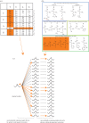
Exploring evolutionary mutational targets. (a) In this codon table, we have highlighted amino acid residues that are close to (one nucleotide mutation away from) methionine in mutational space. (b) In this table of physico-chemical properties of different amino acid residues, we have highlighted amino acid residues that are close (similar) to methionine in physico-chemical space (adapted from www.wikipedia.org). (c) Combining mutational and physico-chemical space allows rationalization of why some mutational paths (amino acid substitutions) are more frequent than others. Here, we have highlighted in orange the most preferred mutational paths owing to short mutational distance (first arrow) and short physico-chemical distance (second arrow). Residue conservation has been illustrated as a loop, and it should be considered as another possible mutational path with very short mutational and physico-chemical distance.
Figure 3.
Figure 3.
Exploring evolutionary mutational targets. (a) The relative contribution of the genetic code (by disfavouring amino acid residue substitutions that require several nucleotide mutations) and the physico-chemical properties (by disfavouring amino acid residue substitutions between dissimilar residues) to mutation will vary over evolutionary time. The restrictions imposed by the genetic code will have higher influence when comparing short-evolutionary distances, whereas the physico-chemical properties of amino acid residues will have a constant influence, because selection against radical changes in physico-chemical space will always be applied before a mutation becomes fixed. (b) Graphical representation of the phylogenetic tree whose ancestral sequences (L1, L2, L3, L4, L5, L6 and L7) we have reconstructed as described in §7. (c) Here, we confirm the principle described in (a), by comparing mutational targets of methionine between L1 and human and between L7 and human and showing that in shorter evolutionary distances (L1: red), methionine tends to mutate only to residues that are one nucleotide mutation away, while for longer times (L7: black), more targets are possible.
Figure 4.
Figure 4.
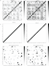
A comparison of amino acid substitution matrices. (a) Representation of a normalized version of the BLOSUM62 matrix. (b) Representation of a normalized version of the PAM70 matrix. (c) Representation of our L1 and L7 MUTA matrices, including versions without conservation (bottom) in order to better visualize the non-conservative amino acid substitutions.
Figure 5.
Figure 5.
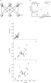
Mutability–targetability plots. (a) Toy model to represent three mutable objects and how they can evolve, with each letter representing one element at the ancestral (bottom) or target (top) sequence and each arrow representing the frequency of every possible mutational path. (b) Any mutable system, such as the one represented in (a), can be represented in a mutability–targetability plot, an xy scatter plot where each element (e.g. A–C) is located in a precise coordinate depending on their mutational properties, i.e. how often it mutates (mutability) and how often it is the result of a mutation from another residue (targetability). Depending on their location, we can consider the mutable elements fast or slow evolving (high mutability and high targetability or low mutability and low targetability, respectively) or likely to increase or decrease in frequency (low mutability and high targetability or high mutability and low targetability, respectively). (c) Mutability–targetability plots computed for all the amino acid residues at different evolutionary distance (L2, L5 and L7). In order to avoid frequency-related biases, we normalized all mutation frequencies before computing mutabilities and targetabilities for each amino acid (for more information refer to §7).
Figure 6.
Figure 6.
Phosphorylation site evolution. (a) As a direct consequence of our previous results, our hypothesis was that phosphorylation at serine, with the highest mutability and targetability rates, would evolve faster than phosphorylation at threonine, which has slightly lower but still relatively high mutability and targetability rates, and much faster than tyrosine, with very low mutability and targetability rates. (b) For each phosphorylatable amino acid residue, we have computed the fraction of phosphorylated residue (in black) that is conserved (same amino acid) in the ancestor L7. For comparison, we have computed conservation fraction for the residue, regardless of phosphorylated state (white) and conditional on whether the residue resides in a disordered (cyan both phosphorylated and unphosphorylated residues and blue only phosphorylated residues) or ordered protein region (orange both phosphorylated and unphosphorylated residues and red phosphorylated). Statistical significance (*p < 0.05) was assessed by Fisher's exact test.

References

    1. Jacob F. 1977. Evolution and tinkering. Science 196, 1161–1166 10.1126/science.860134 (doi:10.1126/science.860134) - DOI - DOI - PubMed
    1. Pastor-Satorras R., Smith E., Solé R. V. 2003. Evolving protein interaction networks through gene duplication. J. Theor. Biol. 222, 199–210 10.1016/S0022-5193(03)00028-6 (doi:10.1016/S0022-5193(03)00028-6) - DOI - DOI - PubMed
    1. Force A., Lynch M., Pickett F. B., Amores A., Yan Y. L., Postlethwait J. 1999. Preservation of duplicate genes by complementary, degenerative mutations. Genetics 151, 1531–1545 - PMC - PubMed
    1. Dayhoff M. O., Schwartz R., Orcutt B. C. 1978. A model of evolutionary change in proteins. In Atlas of protein sequence and structure, vol. 5, suppl. 3 (ed. Dayhoff M. O.), pp. 345–352 Washington, DC: National Biomedical Research Foundation
    1. Henikoff S., Henikoff J. G. 1992. Amino acid substitution matrices from protein blocks. Proc. Natl Acad. Sci. USA 89, 109 15–109 19. 10.1073/pnas.89.22.10915 (doi:10.1073/pnas.89.22.10915) - DOI - DOI - PMC - PubMed

Publication types

MeSH terms