. 2012 Oct 28;14(40):13853-60.
doi: 10.1039/c2cp41386h. Epub 2012 Aug 14.

On rate limitations of electron transfer in the photosynthetic cytochrome b6f complex

Affiliations

On rate limitations of electron transfer in the photosynthetic cytochrome b6f complex

S Saif Hasan et al. Phys Chem Chem Phys. .

Abstract

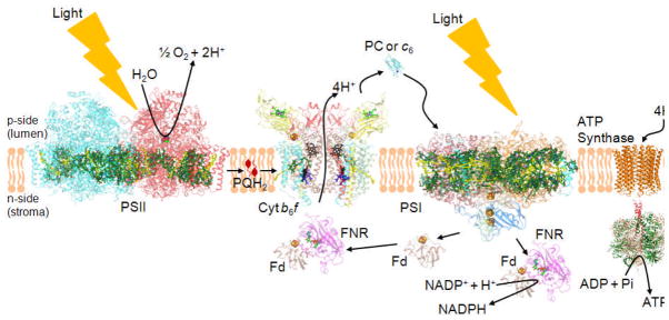
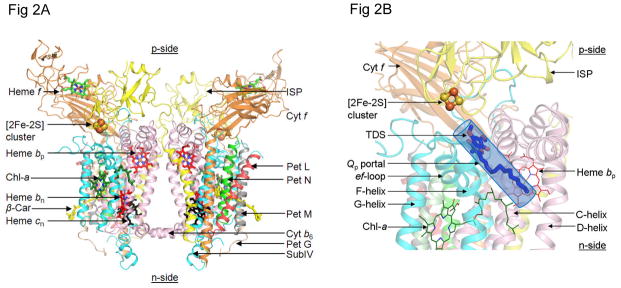
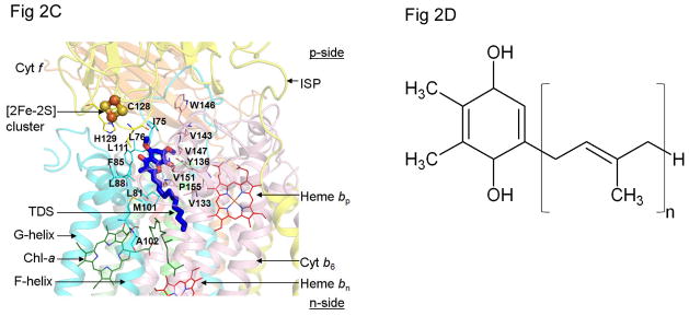
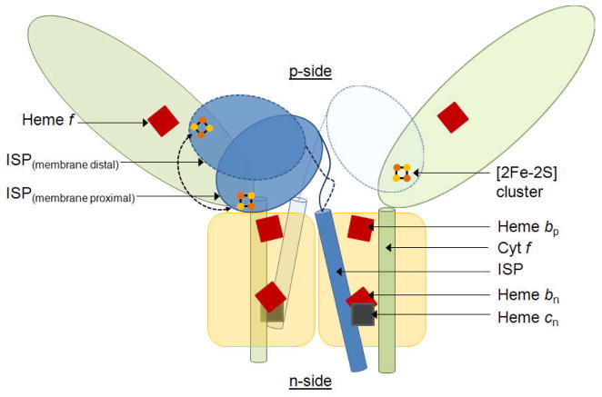
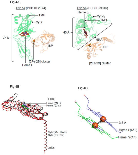
Considering information in the crystal structures of the cytochrome b(6)f complex relevant to the rate-limiting step in oxygenic photosynthesis, it is enigmatic that electron transport in the complex is not limited by the large distance, approximately 26 Å, between the iron-sulfur cluster (ISP) and its electron acceptor, cytochrome f. This enigma has been explained for the respiratory bc(1) complex by a crystal structure with a greatly shortened cluster-heme c(1) distance, leading to a concept of ISP dynamics in which the ISP soluble domain undergoes a translation-rotation conformation change and oscillates between positions relatively close to the cyt c(1) heme and a membrane-proximal position close to the ubiquinol electron-proton donor. Comparison of cytochrome b(6)f structures shows a variation in cytochrome f heme position that suggests the possibility of flexibility and motion of the extended cytochrome f structure that could entail a transient decrease in cluster-heme f distance. The dependence of cyt f turnover on lumen viscosity is consistent with a role of ISP - cyt f dynamics in determination of the rate-limiting step under conditions of low light intensity. Under conditions of low light intensity and proton electrochemical gradient present, for example, under a leaf canopy, it is proposed that a rate limitation of electron transport in the b(6)f complex may also arise from steric constraints in the entry/exit portal for passage of the plastoquinol and -quinone to/from its oxidation site proximal to the iron-sulfur cluster.


Figures

Figure 1
Figure 1. The electron transport chain of oxygenic photosynthesis
Formation of the trans-membrane Δμ̃H+, by the linear electron transport chain, which extends from the H2O oxidation site to that for reduction of NADP+, by H+ translocation in the PSII reaction center and cytochrome b6f complex is shown, along with the utilization of this gradient by the ATP synthase. The rate-limitation in the linear ETC is associated with the oxidation of plastoquinol (PQH2; see PQ in lipid bilayer) by the b6f complex, which involves the structure-based entry/exit of the quinol-quinone to/from the [2Fe-2S] cluster of the iron-sulfur protein, and electron/proton transfer from the quinol to the cluster. Cyt b6f (PDB ID 2E74), cytochrome b6f; Fd (PDB ID 1EWY), ferredoxin; FNR (PDB ID 1EWY), ferredoxin-NADP+-reductase; PC (PDB ID 2Q5B), plastocyanin; PSII (PDB ID 3ARC) and PSI (PDB ID 1JB0), photosynthetic reaction center complexes.
Figure 2
Figure 2
(A) Crystal structure of dimeric cytochrome b6f complex from the cyanobacterium, M. laminosus [(4) (PDB ID 2E74)], drawn in ribbon format, showing the eight subunits of the complex and five redox prosthetic groups, hemes f, bp, bn, the unique heme cn (59, 60) and the [2Fe-2S] cluster present in each monomer. The two other prosthetic groups in each monomer, are a single chlorophyll a molecule ligated to two H2O molecules (4) and, separated by a distance of 14 Å in each monomer, a single β-carotene molecule (61, 1, 2). Color code: cytochrome f TMH (orange), cytochrome b6 (pink), Iron-Sulfur protein (yellow), subunit IV (cyan), small subunits with single TMH are PetG (wheat), PetL (red), PetM (gray) and PetN (green). (B) p-side quinol/quinone binding site and 11 × 12 Å p-side entry/exit portal (Qp portal) for plastoquinol/plastoquinone; [2Fe-2S] cluster is shown as orange and yellow spheres, orange for Fe, S for sulfur; (C) Quinone/quinol binding site inferred from the crystal structure obtained in the presence of the p-side quinone-analogue inhibitor, tridecyl-stigmatellin (TDS, blue and red) (4) [n. b., at the high concentrations used for crystallization, the TDS was found to also bind in an n-side quinone-binding niche]. C and D helices belong to cyt b6 (pink) and F and G- helices to subunit IV (cyan). (D) Plastoquinol substrate of cyt b6f; number of isoprenoid groups in extended tail of the plastoquinol molecule, n=9.
Figure 2
Figure 2
(A) Crystal structure of dimeric cytochrome b6f complex from the cyanobacterium, M. laminosus [(4) (PDB ID 2E74)], drawn in ribbon format, showing the eight subunits of the complex and five redox prosthetic groups, hemes f, bp, bn, the unique heme cn (59, 60) and the [2Fe-2S] cluster present in each monomer. The two other prosthetic groups in each monomer, are a single chlorophyll a molecule ligated to two H2O molecules (4) and, separated by a distance of 14 Å in each monomer, a single β-carotene molecule (61, 1, 2). Color code: cytochrome f TMH (orange), cytochrome b6 (pink), Iron-Sulfur protein (yellow), subunit IV (cyan), small subunits with single TMH are PetG (wheat), PetL (red), PetM (gray) and PetN (green). (B) p-side quinol/quinone binding site and 11 × 12 Å p-side entry/exit portal (Qp portal) for plastoquinol/plastoquinone; [2Fe-2S] cluster is shown as orange and yellow spheres, orange for Fe, S for sulfur; (C) Quinone/quinol binding site inferred from the crystal structure obtained in the presence of the p-side quinone-analogue inhibitor, tridecyl-stigmatellin (TDS, blue and red) (4) [n. b., at the high concentrations used for crystallization, the TDS was found to also bind in an n-side quinone-binding niche]. C and D helices belong to cyt b6 (pink) and F and G- helices to subunit IV (cyan). (D) Plastoquinol substrate of cyt b6f; number of isoprenoid groups in extended tail of the plastoquinol molecule, n=9.
Fig. 3
Fig. 3
Motion of ISP soluble domain required for productive electron transfer in the cytochrome b6f complex. Crystal structures of the cyt b6f complex show the [2Fe-2S] cluster of the ISP soluble domain at a distance of 25.9 Å – 27.2 Å from heme f (Table 3) in a membrane proximal position. Electron transfer cannot proceed at a millisecond time scale over such a large distance. The ISP soluble domain, and possibly the luminal domain of cytochrome f as well, must undergo a motion to position the [2Fe-2S] cluster in proximity (16–18 Å) to heme f in a membrane distal position. Color scheme: Cyt f, light green; ISP, blue; Cyt b6, subIV, Pet G, L, M, N, yellow; Hemes f, bp, bn, red; heme cn, gray; [2Fe-2S] cluster, brown-yellow.
Fig. 4
Fig. 4
Structures of cytochrome f with a displaced heme position. (A) The cyt f soluble domain (left) consists of a 75 Å long extended β sheet structure which is larger than the cyt c1 soluble domain (right). In this restricted space, the cyt b6f ISP soluble domain does not have the same conformational freedom as the cyt bc1 ISP soluble domain. Hence, the ISP of cyt b6f may undergo motion on a smaller scale as demonstrated by the relative insensitivity of the ISP hinge to mutations that restrict mobility (51). (B) For physiologically relevant electron transfer from the [2Fe-2S] cluster of the ISP soluble domain to heme f in cyt b6f, the [2Fe-2S] cluster-heme f distance must be 16 Å – 18 Å. This may involve a small scale movement of the cyt f soluble domain as seen between the cyt b6f crystal structures obtained from M. laminosus (PDB ID 2E74, black) and C. reinhardtii (PDB ID 1Q90, red). Structural superposition performed in PyMol between conserved residues of the cyt f trans-membrane helix (PDB ID 2E74, chain D, residues 258–277; PDB ID 1Q90, chain A, residues 255–274) using a Cα pair fitting algorithm (RMSD=0.84 Å). (C) In the superposed structure of cyt f from M. laminosus and C. reinhardtii, the heme f Fe atoms are separated by 3.8 Å. (M. l., Mastigocladus laminosus, C. r. Chlamydomonas reinhardtii).
Figure 5
Figure 5
The p-side quinone entry/exit portal. (A) Amino acid sequences of segment of F-helix of subunit IV of cytochrome b6f complex showing conservation of proline residues at positions 105 and 112. (B) Schematic of trans-membrane F- and C-helices that define the p-side entry/exit portal for plastoquinol-plastoquinone transfer proximal to [2Fe-2S] cluster in the b6f complex. Two proline residues, 105 and 112, cause a “bent” conformation of the wild-type trans-membrane F-helix of subunit IV relative to the trans-membrane C-helix of the cyt b subunit.
Figure 6
Figure 6
Anticipated effects on the time course of cytochrome f reduction, measured by the absorbance change at 556 nm, the alpha-band peak of the ‘reduced-oxidized’ difference spectrum vs. 540 nm as a reference of the different genetic constructs that are predicted to change the size of the PQH2/PQ portal and thereby the rate-limiting step in the linear electron transport chain that can be assayed by the time course of cytochrome f reduction. (A) Introduction of additional proline residues designed to increase the portal size would increase the rate, i. e., decrease the half-time, of cyt f reduction, as seen in the trend from the blue → red → green → yellow traces that describe the time course of reduction after cytochrome f is oxidized by the red (μsec) light flash. (B) Change of Pro105 and Pro112 to Ala would cause a decrease in the rate of cyt f reduction (red, green traces) relative to the wild type (blue), with the effect predicted to be greater if both Pro→Ala changes are made (yellow trace).

References

    1. Kurisu G, Zhang H, Smith JL, Cramer WA. Structure of the cytochrome b6f complex of oxygenic photosynthesis: tuning the cavity. Science (New York, NY. 2003;302:1009–1014. - PubMed
    1. Stroebel D, Choquet Y, Popot J-L, Picot D. An atypical heam in the cytochrome b6f complex. Nature. 2003;426:413–418. - PubMed
    1. Yan J, Kurisu G, Cramer WA. Intraprotein transfer of the quinone analogue inhibitor 2,5-dibromo-3-methyl-6-isopropyl-p-benzoquinone in the cytochrome b6f complex. Proceedings of the National Academy of Sciences of the United States of America. 2006;103:69–74. - PMC - PubMed
    1. Yamashita E, Zhang H, Cramer WA. Structure of the cytochrome b6f complex: quinone analogue inhibitors as ligands of heme cn. J Mol Biol. 2007;370:39–52. - PMC - PubMed
    1. Baniulis D, Yamashita E, Whitelegge JP, Zatsman AI, Hendrich MP, Hasan SS, Ryan CM, Cramer WA. Structure-Function, Stability, and Chemical Modification of the Cyanobacterial Cytochrome b6f Complex from Nostoc sp. PCC 7120. J Biol Chem. 2009;284:9861–9869. - PMC - PubMed

Publication types

Substances