. 2012;30(4):815-26.
doi: 10.1159/000341460. Epub 2012 Aug 10.

Oxidative stress triggers Ca-dependent lysosome trafficking and activation of acid sphingomyelinase

Affiliations

Oxidative stress triggers Ca-dependent lysosome trafficking and activation of acid sphingomyelinase

Xiang Li et al. Cell Physiol Biochem. 2012.

Abstract

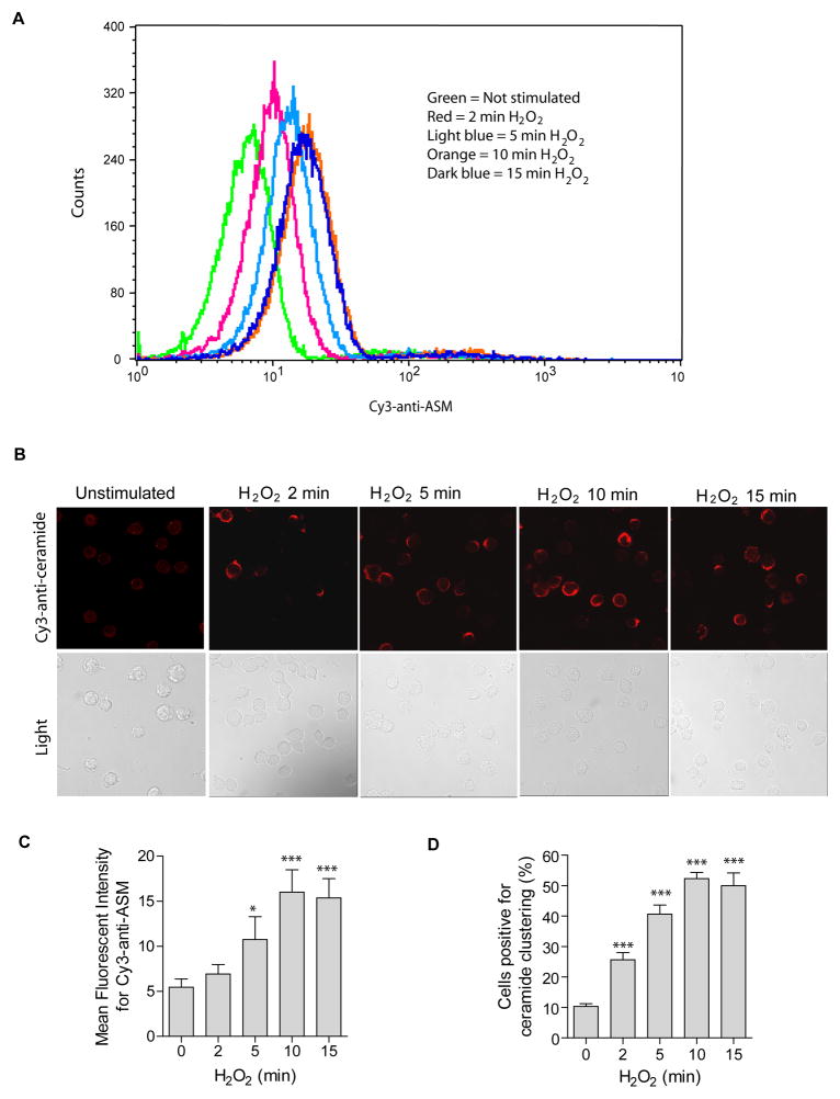
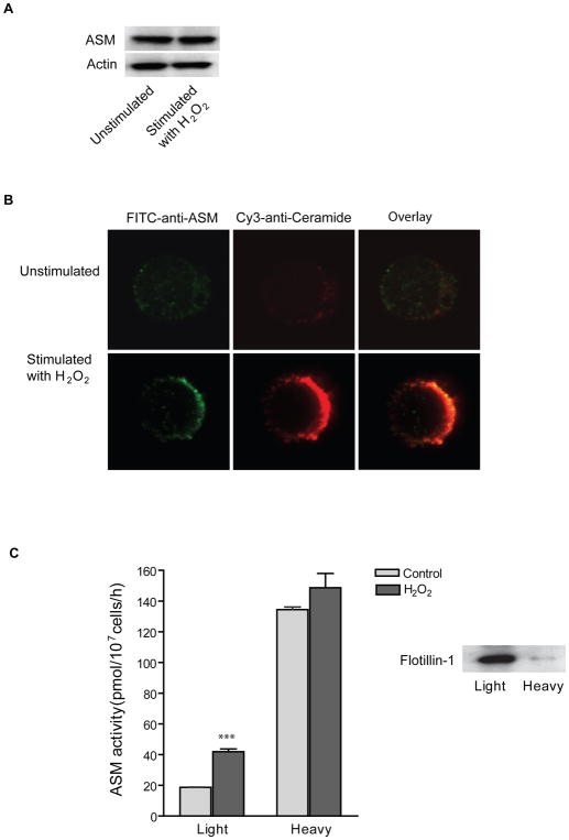
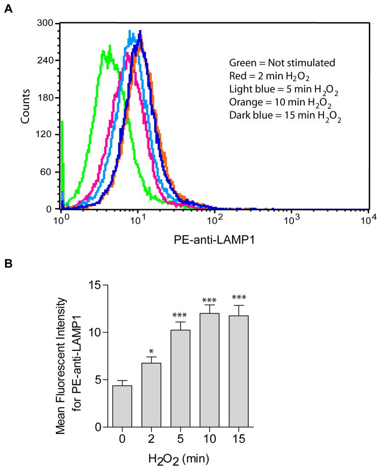
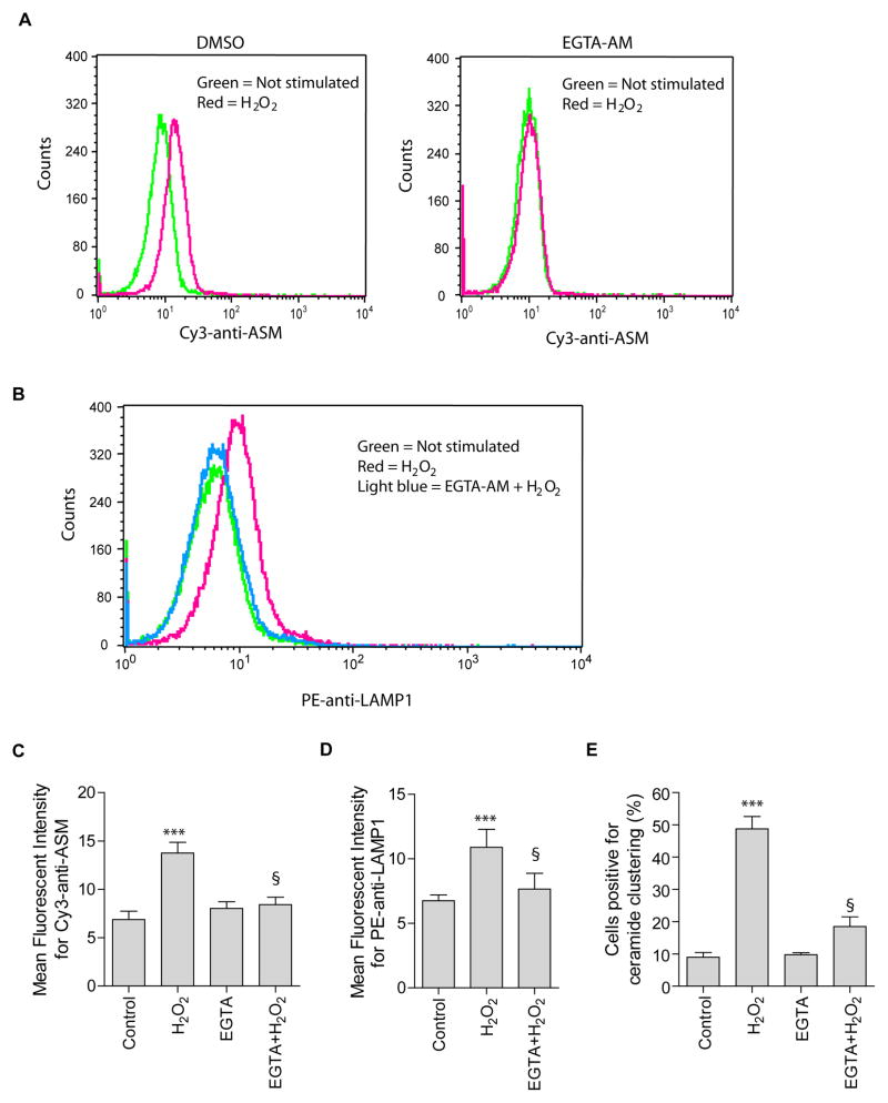
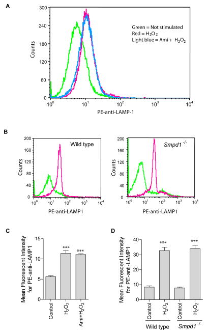
Recent studies demonstrate that rapid translocation of the acid sphingomyelinase (ASM), a lysosomal hydrolase, to the outer leaflet of the cell membrane and concomitant release of ceramide constitute a common cellular signaling cascade to various stimuli including CD95 ligation, UV-irradiation, bacterial and viral infections. Reactive oxygen species (ROS) were shown to play a crucial role in regulating this signaling cascade at least for some bacterial infections and UV-irradiation. However, the precise role of ROS for regulation of ASM is unknown. Here, by confocal microscopy and flow cytometry analysis, we demonstrate that hydrogen peroxide (H(2)O(2)), a primary form of ROS in mammalian cells, induces very rapid translocation of ASM and formation of ceramide-enriched membrane platforms in the plasma membrane of Jurkat T cells. In parallel, H(2)O(2) triggers lysosome trafficking and fusion with the plasma membrane, i.e. lysosome exocytosis, as detected by exposure of a lysosome-associated protein, LAMP1. Depletion of intracellular Ca(2+) by cell permeable EGTA-AM inhibits H(2)O(2)-induced lysosome exocytosis, ASM translocation and formation of ceramide-enriched platforms. Pharmacological inhibition or genetic deficiency of ASM did not affect H(2)O(2)-induced lysosome exocytosis. These results indicate that ROS-induced membrane translocation of ASM is mediated by exocytosis of lysosomes, which is dependent on intracellular Ca(2+) release.


Figures

Fig. 1
Fig. 1. Oxidative stress induces ASM externalization and formation of ceramide-enriched membrane platforms
(A) Oxidative stress by H2O2 (1 mM) induces a translocation of ASM onto the cell surface. Surface ASM was detected by flow cytometry analysis of cells incubated with Cy3-labeled goat anti-ASM antibodies. Shown is a representative flow cytometry analysis from four independent experiments. (B) H2O2 (1 mM)-induced ASM translocation correlates with rapid clustering of ceramide in the membrane and formation of ceramide-enriched membrane platforms. Representative confocal fluorescence microscopy and light images from three independent experiments are shown. (C) Displayed are the summarized data showing the mean fluorescence intensity for Cy3-anti-ASM staining from four independent experiments. (D) The quantitative analysis of the data from four independent experiments, which each included analysis of 200 cells/time point, shows the formation of ceramide-enriched membrane platforms in a large proportion of the cells. Displayed is the percentage of cells with a ceramide-enriched membrane platform at indicated time points. Panel C and D show the mean ± S.D. Significant differences between treated and non-treated controls were determined by t-test and are indicated by *** (P<0.001, t-test) or * (P<0.05, t-test), respectively.
Fig. 2
Fig. 2. Oxidative stress induces ASM translocation and activation of ASM onto membrane lipid rafts
(A) Western blot analysis of ASM and Actin in Jurkat cells stimulated with or without H2O2 (10 min, 1 mM). The experiments were repeated three times with similar results. (B) Confocal microscopy of Jurkat cells revealed a translocation of ASM onto the surface of the cell membrane and a co-localization of ASM with ceramide-enriched membrane platforms. Shown is a representative results from three independent experiments. (C) ASM activity in DRM fractions is increased upon H2O2 stimulation. Low density membrane fractions were collected and designated as DRMs (Light). High density membrane fractions were collected in parallel and designated as none-DRM fraction (Heavy). The DRM maker protein flotillin-1 was detected by Western blot analysis. Shown is the mean ± S.D. of three independent experiments. Significant differences between treated and non-treated controls were determined by t-test and are indicated by ***, respectively (P<0.001, t-test).
Fig. 3
Fig. 3. H2O2induces lysosome excytosis
The translocation of LAMP1 onto the cell surface served as marker for lysosome fusion with the plasma membrane and was detected by flow cytometry analysis using PE-conjugated goat anti-LAMP1. (A) Shown is a representative flow cytometry analysis from four independent experiments. (B) Summarized data showing the mean fluorescence intensity for PE-anti-LAMP1 staining (n=4). Shown are the mean ± SD. Significant differences between treated and non-treated controls were determined by t-test and are indicated by *** (P<0.001, t-test) or * (P<0.05, t-test), respectively.
Fig. 4
Fig. 4. H2O2-induced lysosomal excytosis and ASM externalization are Ca2+-dependent
(A, B) Jurkat cells were stimulate with or without H2O2 (1 mM, 10 min) in the absence (DMSO) or presence of EGTA-AM (100 μM) in a Ca2+-free PBS buffer. H2O2-induced ASM externalization and lysosomal excytosis were detected by flow cytometry analysis using Cy3-conjugated anti-ASM (A) and PE-conjugated goat anti-LAMP1 (B). Shown is a representative flow cytometry analysis from four independent experiments. (C) and (D) summarized data of four independent studies and show the mean fluorescence intensity for Cy3-anti-ASM or PE-anti-LAMP1 staining. (E) Attenuation of H2O2-induced ceramide-enriched membrane platforms by EGTA-AM. Displayed is the percentages of cells with a ceramide-enriched membrane platform in the cells treated with 1 mM H2O2 for 10 min (n = 4). Panel C–E show the mean ± S.D. Significant differences were determined by t-test (***, H2O2 vs. Control, P<0.001; §, EGTA+H2O2 vs. H2O2, P<0.001).
Fig. 5
Fig. 5. Effect of ASM inhibition or deficiency on H2O2-induced lysosomal exocytosis
(A) Shown is a representative flow cytometry analysis diagram using PE-conjugated goat anti-LAMP1 antibodies in Jurkat cells with or without ASM inhibitor amitriptyline (10 μM) (n = 4). (B) Spleen T cells were isolated from wild-type and Asm deficient (Smpd1−/−) mice, stimulated without (green) or with H2O2 (red) and stained with PE-conjugated goat anti-LAMP1 antibodies. Shown are representative flow cytometry analyses from 3 independent experiments. Summarized data from 3 independent studies show the mean fluorescence intensity for PE-anti-LAMP1 staining in Jurkat cells (C) or spleen T cells (D). Displayed is the mean ± S.D. Significant differences between treated and non-treated controls were determined by t-test and are indicated by *** (P<0.001, t-test).

Similar articles

Cited by

References

    1. Ferlinz K, Hurwitz R, Vielhaber G, Suzuki K, Sandhoff K. Occurrence of two molecular forms of human acid sphingomyelinase. Biochem J. 1994;301 (Pt 3):855–862. - PMC - PubMed
    1. Levade T, Jaffrezou JP. Signalling sphingomyelinases: Which, where, how and why? Biochim Biophys Acta. 1999;1438:1–17. - PubMed
    1. Huynh KK, Eskelinen EL, Scott CC, Malevanets A, Saftig P, Grinstein S. Lamp proteins are required for fusion of lysosomes with phagosomes. Embo J. 2007;26:313–324. - PMC - PubMed
    1. McGovern MM, Pohl-Worgall T, Deckelbaum RJ, Simpson W, Mendelson D, Desnick RJ, Schuchman EH, Wasserstein MP. Lipid abnormalities in children with types a and b niemann pick disease. J Pediatr. 2004;145:77–81. - PubMed
    1. McGovern MM, Aron A, Brodie SE, Desnick RJ, Wasserstein MP. Natural history of type a niemann-pick disease: Possible endpoints for therapeutic trials. Neurology. 2006;66:228–232. - PubMed

Publication types